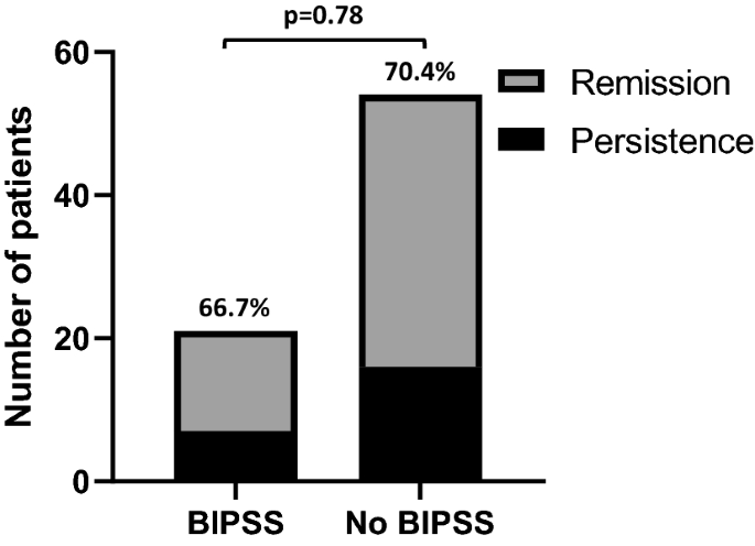
Jump to content

MaryO

~Chief Cushie~
  • Posts

    8,082
  • Joined

  • Last visited

  • Days Won

    556

Posts posted by MaryO

  1. https://doi.org/10.1016/j.aace.2021.11.005Get rights and content
    Under a Creative Commons license
    open access


    Highlights


    Due to the physiologic rise of ACTH during pregnancy, unstimulated ACTH levels may not be an accurate marker to differentiate between adrenal and ACTH-independent Cushing's syndrome.

    The desmopressin stimulation test can be done during pregnancy to investigate the etiology of Cushing's syndrome.

    Non-gadolinium enhanced pituitary imaging may not detect pituitary adenoma, which is the most common cause of Cushing's disease. Contrast-enhanced pituitary magnetic resonance imaging should be considered in pregnant women with ACTH-dependent Cushing's syndrome.

    Due to increase maternal and fetal morbidities in active Cushing's syndrome, prompt diagnosis and appropriate treatment are essential. The treatment of choice is transsphenoidal surgery during the second trimester, preferably at a high-volume pituitary center.

    There were significantly lower rates of fetal complications in women with active Cushing's syndrome than a cured disease, including low birth weight.

    Abstract

    Objective

    The hypothalamic-pituitary-adrenal axis stimulation during pregnancy complicates the investigation of Cushing's syndrome. Our objective is to present a pregnant patient with Cushing syndrome caused by pituitary tumor in which the desmopressin stimulation test helped in the diagnosis and led to appropriate management.

    Case report

    A 27-year-old woman with 9-week gestation presented with proximal myopathy for 2 months. She had high blood pressure, wide abdominal purplish striae, and proximal myopathy. Her past medical history revealed hypertension and dysglycemia for 1 year. The 8 AM cortisol was 32.4 μg/dL (5-18), late-night salivary cortisol at 11 PM was 0.7 μg/dL (<0.4), and the mean 24-hour urinary free cortisol was 237.6 μg/day (21.0-143.0). The mean ACTH concentrations at 8 AM were 44.0 pg/mL (0-46.0). Non-gadolinium enhanced pituitary magnetic resonance imaging (MRI) reported no obvious lesion. The desmopressin stimulation test showed a 70% increase in ACTH levels from baseline after desmopressin administration. The pituitary MRI with gadolinium showed an 8x8x7-mm pituitary adenoma. Transsphenoidal surgery with tumor removal was done, which showed ACTH-positive tumor cells. After the surgery, the patient carried on the pregnancy uneventfully.

    Discussion

    During pregnancy, the ACTH level may not be an accurate marker to help in the differential diagnosis of Cushing's syndrome. Moreover, non-gadolinium pituitary imaging may not detect small pituitary lesions.

    Conclusion

    In the present Case, the desmopressin stimulation test suggested the diagnosis of Cushing's disease, which subsequently led to successful treatment. This suggested that the desmopressin test may serve as a useful test to diagnose Cushing's disease in pregnant individuals.

    Keywords

    Cushing's disease
    Cushing's syndrome
    desmopressin stimulation test
    pregnancy

    Introduction

    Pregnancy rarely occurs during the course of Cushing's syndrome (CS).1,2 Given the increase in maternal and fetal morbidities in women with active CS, early diagnosis and treatment of CS are essential.2

    The diagnosis of CS using the usual diagnostic tests is challenging due to stimulation of the hypothalamic-pituitary-adrenal axis during pregnancy. The physiologic rise of ACTH from the 7th week of pregnancy also complicates the investigation for the etiology of CS.1 The concern of gadolinium use during pregnancy can affect the sensitivity in detecting small pituitary lesions in ACTH-dependent CS if using non-gadolinium pituitary imaging. Desmopressin is a vasopressin analog selective for V2 receptors. The desmopressin stimulation test has been proposed as a useful procedure for the differential diagnosis of CS.3 Desmopressin stimulates the increase in ACTH and cortisol in patients with CS caused by pituitary tumor or Cushing's disease (CD) but not in the majority of normal, obese subjects and patients with adrenal CS or ectopic ACTH syndrome.3,4 However, there were limited data on the desmopressin stimulation test during pregnancy.

    Here we present the 27-year-old woman with CS in which the desmopressin stimulation test helped in the diagnosis of CD and led to successful treatment.

    Case presentation

    A 27-year-old woman with 9-week gestation was referred from the orthopedic department to evaluate CS. She presented with proximal myopathy for 2 months. On physical examination, she had Cushingoid appearance, wide purplish striae, bruising, and proximal muscle weakness. Her blood pressure was 160/100 mmHg, and her body mass index was 32.2 kg/m2. Her past medical history revealed that she had hypertension, dyslipidemia, and impaired fasting glucose for 1 year without taking any medication. She also gained 20 kg in the past 2 years. The 8 AM cortisol (chemiluminescent immunometric assay, Immulite/Siemens) was 32.4 μg/dL (normal , 5.0-18.0), late-night salivary cortisol at 11 PM (electrochemiluminescence immunoassay, Roche Cobas) was 0.7 μg/dL (normal, <0.4), and the mean 24-hour urinary free cortisol (UFC) (radioimmunoassay, Immulite/Siemens) was 237.6 μg/day (normal, 21.0-143.0). ACTH concentrations at 8 AM (chemiluminescent immunometric assay, Immulite/Siemens) were 48.4 and 39.6 pg/mL (normal, 0-46.0) (Table 1). At 12 weeks of gestation, non-gadolinium enhanced pituitary magnetic resonance imaging (MRI) reported a mild bulging contour of the right lateral aspect of the pituitary gland without an obvious abnormal lesion (Figure 2A). The desmopressin stimulation test was then carried out at 14 weeks of gestation. Serial blood samples for ACTH and cortisol were obtained basally (at 8 AM) and at 15, 30, 45, and 60 minutes after the intravenous administration of 10 μg of desmopressin. The results were shown in Table 2. Compared with baseline, ACTH levels increased from 34.7 to 58.9 pg/mL (70%) at 15 minutes after desmopressin administration (a ≥35% increase in ACTH levels was considered an indication of CD in non-pregnant individuals)3 (Figure 1). The pituitary MRI with gadolinium revealed an 8x8x7-mm circumscribed lesion with heterogeneous iso- to hyperintensity on T2W in the right inferolateral aspect of the anterior pituitary lobe. The lesion had a delayed enhancement compared to normal pituitary tissue (Figure 2B). Non-contrast MRI adrenal glands showed bilateral normal adrenal glands without mass or nodule. Other abdominal organs were unremarkable. Regarding comorbidities, she had hypertension and gestational diabetes mellitus (GDM). The HbA1c level was 5.7% (39 mmol/mol). Using a two-step strategy, GDM was diagnosed at 12 weeks of gestation. Hypertension and GDM were controlled with 750 mg of methyldopa and 50 units of insulin per day, respectively.

    Table 1. Laboratory investigations of the present Case

    Variable At 9 weeks of gestation
    8 AM cortisol, μg/dL (5.0-18.0) 32.4
    Salivary cortisol (11 PM , <0.4 μg/dL) 0.7
    UFC (21.0-143.0 μg/day) 183.5 and 291.6
    ACTH, pg/mL (8 AM, 0-46.0) 48.4 and 39.6
    DHEAS (8 AM, 35.0-430.0 μg/dL) 378.0
    PAC (upright position, 8 AM), ng/dL 5.2
    PRA (upright position, 8 AM), ng/mL/hr 2.1
    Potassium, mmol/L 3.6


    UFC, urinary free cortisol; ACTH, adrenocorticotrophic hormone; DHEAS, dehydroepiandrosterone sulphate; PAC, plasma aldosterone concentration; PRA, plasma renin activity.

    1-s2.0-S2376060521001231-gr2.jpg
    1. Download : Download high-res image (130KB)
    2. Download : Download full-size image

    Figure 2. Pituitary imaging of the present Case. (A) A non-gadolinium MRI of the pituitary gland at 12 weeks of gestation showing a mild bulging contour of the right lateral aspect of the pituitary gland without an obvious abnormal lesion (B) An MRI of the pituitary gland with gadolinium at 14 weeks of gestation showing an 8x8x7-mm circumscribed lesion with heterogeneous iso- to hyperintensity on T2W in the right inferolateral aspect of the anterior pituitary lobe. The lesion had a delayed enhancement compared to normal pituitary tissue.

    Table 2. Desmopressin stimulation test results performing at 14 weeks of gestation

    Time 0 min 15 min 30 min 45 min 60 min
    ACTH (pg/mL) 34.7 58.9 57.4 49.9 38.2
    Cortisol (μg/dL) 30.6 30.2 29.7 29.6 31.0


    ACTH, adrenocorticotrophic hormone

    1-s2.0-S2376060521001231-gr1.jpg
    1. Download : Download high-res image (76KB)
    2. Download : Download full-size image

    Figure 1. Percentage of ACTH increase after desmopressin administration (time 0 min).

    Transsphenoidal surgery with tumor removal was performed at 18 weeks of gestation. Pathological findings showed a 1.3x1.0x0.3 cm of tissue with segments of the pituitary gland and tumor. There were monomorphous round nuclei, stippled chromatin, indistinct nucleoli, and pale eosinophilic cytoplasm cells. These cells were reactive with ACTH and showed loss of reticulin framework, unlike the normal pituitary gland. The next day after the surgery, her 8 AM cortisol was 6.0 μg/dL. Hydrocortisone supplement was started and continued throughout pregnancy. Antihypertensives were discontinued, and the insulin dosages decreased to less than 20 units per day. At 38 weeks of gestation, she gave birth to a 2300-gm male newborn (small for gestational age). Dysglycemia and hypertension resolved after the delivery. One year after the first child's delivery, the patient had a spontaneous pregnancy without GDM or hypertension. The 8 AM cortisol was 3.9 μg/dL, and hydrocortisone replacement was continued. The patient successfully delivered a term 3300-gm male infant without fetal or maternal complications. Two years after the first transsphenoidal surgery, a 1-μg cosyntropin stimulation test was performed, the basal cortisol was 11.7 μg/dL, and the peak serum cortisol was 23.8 μg/dL. Steroid replacement was withdrawn.

    Discussion

    Herein we present a 27-year-old woman who was evaluated during her first pregnancy for clinical and laboratory features suggestive of CD. Her morning serum and late-night salivary cortisol concentrations were elevated in addition to non-suppressed ACTH, but a definitive diagnosis was not obtained by a non-gadolinium pituitary MRI. The diagnosis of CD was suggested, however, by the results of a desmopressin stimulation test. The pituitary MRI with gadolinium was proceeded and revealed a pituitary lesion greater than 6 mm.

    The prevalence of pregnancy is low due to reduced fertility in CS. To date, there have been less than 300 pregnant patients with CS reported in the literature.2 In pregnancy, the most frequent etiology of CS is adrenal CS (60%), followed by ACTH-producing pituitary adenomas or CD (35%), and very rarely ectopic ACTH (<5%).1 In contrast, CD is the most common cause of CS in non-pregnant people (approximately 70 percent). The clinical diagnosis of CS during pregnancy may be missed due to overlapping features between pregnancy and CS. However, wide purplish cutaneous striae and proximal myopathy are signs with high discrimination index when CS is suspected.5 These signs are not present in normal pregnancy.

    In this present Case, CS was diagnosed with apparent clinical features of CS in addition to an elevated UFC and late-night salivary cortisol. The patient denied taking any supplements and her 8 AM cortisol was not suppressed and therefore did not suggest an etiology of exogenous steroid use. Pregnant women without CS may have elevated UFC and late-night salivary cortisol due to increased total and free plasma cortisol from the first trimester until the end of pregnancy.6 This results from an elevated concentration of cortisol transport protein and the increase in placental ACTH and CRH. According to the current guideline, UFC is the recommended test when CS is suspected during pregnancy.5 Since UFC increases during the second trimester, it may not be a reliable marker after the first trimester of pregnancy unless the level is clearly increased (up to 2- to 3-fold the upper limit of normal values).1 Late-night salivary cortisol is also one of the useful tests to diagnose CS during pregnancy because the circadian rhythm of cortisol is preserved in normal pregnancy. Furthermore, it is not influenced by the changes in the binding proteins.7 However, the previous study has shown that late-night salivary cortisol increased progressively throughout pregnancy. When compared with non-pregnant women, median values of late-night salivary cortisol in pregnant women were 1.1, 1.4, and 2.1 times higher in the first, second, and third trimesters respectively. The cutoff values for late-night salivary cortisol on each gestational trimester were: first trimester 0.255 μg/dL, second trimester 0.260 μg/dL, and third trimester 0.285 μg/dL. The respective sensitivities and specificities in each trimester were: first trimester 92 and 100%, second trimester 84 and 98%, and third trimester 80 and 93%.8

    Given the non-suppressed ACTH levels after the 7th week of gestation, we were not able to summarize whether the etiology was adrenal CS or ACTH-dependent CS which could be either CD or ectopic ACTH syndrome. In non-pregnant individuals, ACTH suppression usually identifies adrenal CS. However, in pregnancy, ACTH levels were non-suppressed in half of those with adrenal CS due to continued stimulation of maternal hypothalamic-pituitary-adrenal axis by placental CRH.1 Therefore, using the ACTH thresholds in general populations can lead to misdiagnosis when investigating the etiology of CS in pregnant individuals. The hypothalamic-pituitary-adrenal axis response to exogenous glucocorticoids is blunted in pregnant women. Following an overnight dexamethasone administration, pregnant women without CS may have non-suppressed plasma cortisol and UFC.6 In non-pregnant individuals with CS, the high-dose dexamethasone suppression test identify CD with a sensitivity of 82% and a specificity of 50%.4 During pregnancy, the high-dose dexamethasone suppression test failed to identify almost half of the patients with CD.1 Inferior petrosal sinus sampling is usually avoided due to the risk of excessive radiation exposure. Since the non-gadolinium MRI also showed no obvious pituitary lesion in the present Case, in addition to the limitation of the high-dose dexamethasone suppression test and inferior petrosal sinus sampling in pregnancy, we used desmopressin stimulation to help in the investigation of CD since desmopressin can stimulate an ACTH response in a considerable proportion of patients with CD but not in most patients with adrenal CS or ectopic ACTH syndrome.3,4

    Desmopressin has been assigned to pregnancy category B by the US Food and Drug Administration (FDA). In the most recent guideline update on the diagnosis and management of CD, the desmopressin stimulation test can be used to differentiate ectopic CS and CD in patients with normal or high ACTH and have no adenoma or equivocal results of pituitary MRI. However, the guideline did not mention the use of this test in pregnant individuals.9 The literature regarding the use of desmopressin stimulation tests in pregnancy is limited. We were able to identify one study in a pregnant patient with active CS, who was surgically confirmed as CD, in which the desmopressin stimulation test was performed at 10 weeks of gestation and after the delivery. Compared with age-matched healthy non-pregnant women, there were different responses of cortisol and ACTH after desmopressin administration in a pregnant patient with active CS.10 The ACTH peaks after the administration of desmopressin were higher in the pregnant patient. CRH stimulation test was also performed in the pregnant patient with CD. Desmopressin stimulated ACTH values during pregnancy and after the delivery were not significantly different, while the CRH stimulated ACTH values were significantly higher when the test was performed after the delivery. The authors did not mention optimal cutoff values for these diagnostic tests.10 In non-pregnant individuals, the ACTH increase of more than 35% at 15 minutes after the desmopressin administration gave the sensitivity of 84% and the specificity of 43% in the diagnosis of CD.3 Another recent study in ACTH-dependent CS showed that the threshold increase in the ACTH level after desmopressin stimulation of 45% identified CD with a sensitivity of 91% and a specificity of 75%.4 Using the non-pregnant cutoff values for the desmopressin stimulation test, the diagnosis of CD was made in our patient who was later surgically confirmed as CD.

    Pituitary microadenomas were the cause of CD in almost 90% of non-pregnant individuals.11 In pregnant women with CD, pituitary microadenomas were also reported to be more common than macroadenomas.1,12 Almost 40% of pituitary microadenomas in CD were invisible or poorly visible in non-contrast MRI, in which contrast-enhanced MRI detected them.13 In the Case series from Lindsay et al., the non-contrast MRI could not correctly identify pituitary adenomas in 38% of pregnant patients with available data.1 The same case series reported a pregnant patient having normal pituitary MRI and was later surgically confirmed as having CD from a 3x3 adenoma with positive staining for ACTH. In the present case, a mild bulging contour of the pituitary gland, although without an obvious abnormal lesion, in addition to desmopressin test results, suggested the need for contrast-enhanced pituitary MRI. Gadolinium contrast is FDA pregnancy category C since it is water-soluble and can cross the placenta into the fetus and amniotic fluid.14 However, since a non-gadolinium MRI may not detect pituitary microadenoma even in patients with normal imaging results,1,15 we suggested physicians consider pituitary MRI with gadolinium as initial imaging in pregnant patients with clinical suspicion of CD.

    Prompt diagnosis and treatment of CS are essential due to a higher rate of fetal loss in active CS patients without treatment than those who received either medical or surgical treatment. There are significantly lower rates of various fetal complications, including low birth weight, in women with active CS than in cured CS.2 Although medical and surgical treatment were not compared as prognostic factors for complications, experts recommend transsphenoidal surgery in the second trimester as the treatment of choice for CD in pregnancy.1,15 Medical treatment should be the second choice when surgery cannot be carried out or late diagnosis is made.

    Conclusion

    In the present Case, the results from the desmopressin stimulation test and the pituitary MRI with gadolinium suggested the diagnosis of CD, which subsequently led to successful treatment. This suggested that the desmopressin test may serve as a useful test to diagnose CD even in the context of pregnancy.

    Conflicts of Interest

    None of the authors have any potential conflicts of interest associated with this research.


    References


    Funding Statement


    This research did not receive any specific grant from funding agencies in the public, commercial, or not-for-profit sectors.


    Acknowledgements


    The authors would like to thank you all the colleagues in the Division of Endocrinology and Metabolism, Department of Medicine, Faculty of medicine, Chulalongkorn University for all the support.

    © 2021 Published by Elsevier Inc. on behalf of the AACE.
     
    • Like 1
  2. This article is based on reporting that features expert sources.

    U.S. News & World Report

    Adrenal Fatigue: Is It Real?

    You may have heard of so-called 'adrenal fatigue,' supposedly caused by ongoing emotional stress. Or you might have come across adrenal support supplements sold online to treat it. But if someone suggests you have the controversial, unproven condition, seek a second opinion, experts say. And if someone tries to sell you dietary supplements or other treatments for adrenal fatigue, be safe and save your money.


    Tired man sitting at desk in modern office

    (GETTY IMAGES)


    Physicians tend to talk about 'reaching' or 'making' a medical diagnosis. However, when it comes to adrenal fatigue, endocrinologists – doctors who specialize in diseases involving hormone-secreting glands like the adrenals – sometimes use language such as 'perpetrating a diagnosis,' 'misdiagnosis,' 'made-up diagnosis,' 'a fallacy' and 'nonsense.'

    About 20 years ago, the term "adrenal fatigue" was coined by Dr. James Wilson, a chiropractor. Since then, certain practitioners and marketers have promoted the notion that chronic stress somehow slows or shuts down the adrenal glands, causing excessive fatigue.

    "The phenomenon emerged from the world of integrative medicine and naturopathic medicine," says Dr. James Findling, a professor of medicine and director of the Community Endocrinology Center and Clinics at the Medical College of Wisconsin. "It has no scientific basis, and there's no merit to it as a clinical diagnosis."


    An online search of medical billing code sets in the latest version of the International Classification of Diseases, or the ICD-10, does not yield a diagnostic code for 'adrenal fatigue' among the 331 diagnoses related either to fatigue or adrenal conditions or procedures.

    In a March 2020 position statement, the American Association of Clinical Endocrinologists and American College of Endocrinology addressed the use of adrenal supplements "to treat common nonspecific symptoms due to 'adrenal fatigue,' an entity that has not been recognized as a legitimate diagnosis."


    The position statement warned of known and unknown health risks of off-label use and misuse of hormones and supplements in patients without an established endocrine diagnosis, as well as unnecessary costs to patients and the overall health care system.

    Study after study has refuted the legitimacy of adrenal fatigue as a medical diagnosis. An August 2016 systematic review combined and analyzed data from 58 studies on adrenal fatigue including more than 10,000 participants. The conclusion in a nutshell: "Adrenal fatigue does not exist," according to review authors in the journal BMC Endocrine Disorders.

    Adrenal Action

    You have two adrenal glands in your body. These small triangular glands, one on top of each kidney, produce essential hormones such as aldosterone, cortisol and male sex hormones such as DHEA and testosterone.

    Cortisol helps regulate metabolism: How your body uses fat, protein and carbohydrates from food, and cortisol increases blood sugar as needed. It also plays a role in controlling blood pressure, preventing inflammation and regulating your sleep/wake cycle.

    As your body responds to stress, cortisol increases. This response starts with signals between two sections in the brain: The hypothalamus and the pituitary gland, which act together to release a hormone that stimulates the adrenal glands to make cortisol. This interactive unit is called the hypothalamic pituitary adrenal axis.

    While some health conditions really do affect the body's cortisol-making ability, adrenal fatigue isn't among them.

    "There's no evidence to support that adrenal fatigue is an actual medical condition," says Dr. Mary Vouyiouklis Kellis, a staff endocrinologist at Cleveland Clinic. "There's no stress connection in the sense that someone's adrenal glands will all of a sudden just stop producing cortisol because they're so inundated with emotional stress."

    If anything, adrenal glands are workhorses that rise to the occasion when chronic stress occurs. "The last thing in the body that's going to fatigue are your adrenal glands," says Dr. William F. Young Jr., an endocrinology clinical professor and professor of medicine in the Mayo Clinic College of Medicine at Mayo Clinic in Rochester, Minnesota. "Adrenal glands are built for stress – that's what they do. Adrenal glands don't fatigue. This is made up – it's a fallacy."

    The idea of adrenal glands crumbling under stress is "ridiculous," Findling agrees. "In reality, if you take a person and subject them to chronic stress, the adrenal glands don't shut down at all," Findling says. "They keep making cortisol – it's a stress hormone. In fact, the adrenal glands are just like the Energizer Bunny – they just keep going. They don't stop."

    Home cortisol tests that allow consumers to check their own levels can be misleading, Findling says. "Some providers who make this (adrenal fatigue) diagnosis, provide patients with testing equipment for doing saliva cortisol levels throughout the day," he says. "And then, regardless of what the results are, they perpetrate this diagnosis of adrenal fatigue."

    Saliva cortisol is a legitimate test that's frequently used in diagnosing Cushing's syndrome, or overactive adrenal glands, Findling notes. However, he says, a practitioner pursuing an adrenal fatigue diagnosis could game the system. "What they do is: They shape a very narrow normal range, so narrow, in fact, that no normal human subject could have all their saliva cortisol (levels) within that range throughout the course of the day," he says. "Then they convince the poor patients that they have adrenal fatigue phenomena and put them on some kind of adrenal support."


    Loaded Supplements

    How do you know what you're actually getting if you buy a dietary supplement marketed for adrenal fatigue or 'adrenal support' use? To find out, researchers purchased 12 such supplements over the counter in the U.S.

    Laboratory tests revealed that all supplements contained a small amount of thyroid hormone and most contained at least one steroid hormone, according to the study published in the March 2018 issue of Mayo Clinic Proceedings. "These results may highlight potential risks for hidden ingredients in unregulated supplements," the authors concluded.


    Supplements containing thyroid hormones or steroids can interact with a patient's prescribed medications or have other side effects.

    "Some people just assume they have adrenal fatigue because they looked it up online when they felt tired and they ultimately buy these over-the-counter supplements that can be very dangerous at times," Vouyiouklis Kellis says. "Some of them contain animal (ingredients), like bovine adrenal extract. That can suppress the pituitary axis. So, as a result, your body stops making its own cortisol or starts making less of it, and as a result, you can actually worsen the condition rather than make it better."

    Any form of steroid from outside the body, whether a prescription drug like prednisone or extract from cows' adrenal glands, "can shut off the pituitary," Vouyiouklis Kellis explains. "Because it's signaling to the pituitary like: Hey, you don't need to stimulate the adrenals to make cortisol, because this patient is taking it already. So, as a result, the body ultimately doesn't produce as much. And, so, if you rapidly withdraw that steroid or just all of a sudden decide not to take it anymore, then you can have this acute response of low cortisol."

    Some adrenal support products, such as herbal-only supplements, may be harmless. However, they're unlikely to relieve chronic fatigue.


    Fatigue: No Easy Answers

    If you're suffering from ongoing fatigue, it's frustrating. And you're not alone. "I have fatigue," Young Jr. says. "Go to the lobby any given day and say, 'Raise your hand if you have fatigue.' Most of the people are going to raise their hands. It's a common human symptom and people would like an easy answer for it. Usually there's not an easy answer. I think 'adrenal fatigue' is attractive because it's like: Aha, here's the answer."

    There aren't that many causes of endocrine-related fatigue, Young Jr. notes. "Hypothyroidism – when the thyroid gland is not working – is one." Addison's disease, or adrenal insufficiency, can also lead to fatigue among a variety of other symptoms. Established adrenal conditions – like adrenal insufficiency – need to be treated.

    "In adrenal insufficiency, there is an intrinsic problem in the adrenal gland's inability to produce cortisol," Vouyiouklis Kellis explains. "That can either be a primary problem in the adrenal gland or an issue with the pituitary gland not being able to stimulate the adrenal to make cortisol."


    Issues can arise even with necessary medications. "For example, very commonly, people are put on steroids for various reasons: allergies, ear, nose and throat problems," Vouyiouklis Kellis says. "And with the withdrawal of the steroids, they can ultimately have adrenal insufficiency, or decrease in cortisol."

    Opioid medications for pain also result in adrenal sufficiency, Vouyiouklis Kellis says, adding that this particular side effect is rarely discussed. People with a history of autoimmune disease can also be at higher risk for adrenal insufficiency.

    Common symptoms of adrenal insufficiency include:

    • Fatigue.
    • Weight loss.
    • Decreased appetite.
    • Salt cravings.
    • Low blood pressure.
    • Abdominal pain.
    • Nausea, vomiting or diarrhea.
    • Muscle weakness.
    • Hyperpigmentation (darkening of the skin).
    • Irritability.

    Medical tests for adrenal insufficiency start with blood cortisol levels, and tests for the ACTH hormone that stimulates the pituitary gland.

    "If the person does not have adrenal insufficiency and they're still fatigued, it's important to get to the bottom of it," Vouyiouklis Kellis says. Untreated sleep apnea often turns out to be the actual cause, she notes.

    "It's very important to tease out what's going on," Vouyiouklis Kellis emphasizes. "It can be multifactorial – multiple things contributing to the patient's feeling of fatigue." The blood condition anemia – a lack of healthy red blood cells – is another potential cause.

    "If you are fatigued, do not treat yourself," Vouyiouklis Kellis says. "Please seek a physician or a primary care provider for evaluation, because you don't want to go misdiagnosed or undiagnosed. It's very important to rule out actual causes that would be contributing to symptoms rather than ordering supplements online or seeking an alternative route like self-treating rather than being evaluated first."

    SOURCES

    The U.S. News Health team delivers accurate information about health, nutrition and fitness, as well as in-depth medical condition guides. All of our stories rely on multiple, independent sources and experts in the field, such as medical doctors and licensed nutritionists. To learn more about how we keep our content accurate and trustworthy, read our editorial guidelines.

    James Findling, MD

    Findling is a professor of medicine and director of the Community Endocrinology Center and Clinics at the Medical College of Wisconsin.

    Mary Vouyiouklis Kellis, MD

    Vouyiouklis Kellis is a staff endocrinologist at Cleveland Clinic.

    William F. Young Jr., MD

    Young Jr. is an endocrinology clinical professor and professor of medicine in the Mayo Clinic College of Medicine at Mayo Clinic in Rochester, Minnesota

    From https://health.usnews.com/health-care/patient-advice/articles/adrenal-fatigue-is-it-real?

  3. 24c7ae85-e04e-e21a-9ea2-ad244cae1798.png


    The CAHmelia clinical trials are exploring a new investigational treatment for classic CAH.


    CAHmelia 203 and CAHmelia 204 are clinical trials to test tildacerfont in adults with classic CAH, which may offer you and your loved ones hope of a brighter future – one where you may not have to choose between symptom management and long-term health.


    Tildacerfont is a new type of oral, once-daily investigational treatment – one that is not a steroid – that is currently being tested in adults with classic CAH. By reducing the amount of androgens your body makes, tildacerfont may improve your classic CAH symptoms.


    This investigational treatment will not replace your steroid treatment but may allow you to manage your disease with lower amounts of steroids at normal or near-normal doses.


    Who can take part in this trial? You may be able to take part if you:

    • Are at least 18 years of age
    • Have a confirmed diagnosis of classic CAH due to 21-OH deficiency
    • Have been on the same daily dose of steroids (GCs and/or mineralocorticoids) for at least 1 month before starting the trial

    Both trials are now open for enrollment.


    Tildacerfont is an investigational treatment not authorized for use in people outside the clinical trial.


    For more information, go to: clarahealth.com/studies/cahmelia


    • Like 1
  4. Dexamethasone suppression test measures whether adrenocorticotrophic hormone (ACTH) secretion by the pituitary can be suppressed.

    How the Test is Performed

    During this test, you will receive dexamethasone. This is a strong man-made (synthetic) glucocorticoid medicine. Afterward, your blood is drawn so that the cortisol level in your blood can be measured.

    There are two different types of dexamethasone suppression tests: low dose and high dose. Each type can either be done in an overnight (common) or standard (3-day) method (rare). There are different processes that may be used for either test. Examples of these are described below.

    Common:

    • Low-dose overnight -- You will get 1 milligram (mg) of dexamethasone at 11 p.m., and a health care provider will draw your blood the next morning at 8 a.m. for a cortisol measurement.
    • High-dose overnight -- The provider will measure your cortisol on the morning of the test. Then you will receive 8 mg of dexamethasone at 11 p.m. Your blood is drawn the next morning at 8 a.m. for a cortisol measurement.

    Rare:

    • Standard low-dose -- Urine is collected over 3 days (stored in 24-hour collection containers) to measure cortisol. On day 2, you will get a low dose (0.5 mg) of dexamethasone by mouth every 6 hours for 48 hours.
    • Standard high-dose -- Urine is collected over 3 days (stored in 24-hour collection containers) for measurement of cortisol. On day 2, you will receive a high dose (2 mg) of dexamethasone by mouth every 6 hours for 48 hours.

    Read and follow the instructions carefully. The most common cause of an abnormal test result is when instructions are not followed.

    How to Prepare for the Test

    The provider may tell you to stop taking certain medicines that can affect the test, including:

    • Antibiotics
    • Anti-seizure drugs
    • Medicines that contain corticosteroids, such as hydrocortisone, prednisone
    • Estrogen
    • Oral birth control (contraceptives)
    • Water pills (diuretics)

    How the Test will Feel

    When the needle is inserted to draw blood, some people feel moderate pain. Others feel only a prick or stinging. Afterward, there may be some throbbing or slight bruising. This soon goes away.

    Why the Test is Performed

    This test is done when the provider suspects that your body is producing too much cortisol. It is done to help diagnose Cushing syndrome and identify the cause.

    The low-dose test can help tell whether your body is producing too much ACTH. The high-dose test can help determine whether the problem is in the pituitary gland (Cushing disease) or from a different site in the body (ectopic).

    Dexamethasone is a man-made (synthetic) steroid that binds to the same receptor as cortisol. Dexamethasone reduces ACTH release in normal people. Therefore, taking dexamethasone should reduce ACTH level and lead to a decreased cortisol level.

    If your pituitary gland produces too much ACTH, you will have an abnormal response to the low-dose test. But you can have a normal response to the high-dose test.

    Normal Results

    Cortisol level should decrease after you receive dexamethasone.

    Low dose:

    • Overnight -- 8 a.m. plasma cortisol lower than 1.8 micrograms per deciliter (mcg/dL) or 50 nanomoles per liter (nmol/L)
    • Standard -- Urinary free cortisol on day 3 lower than 10 micrograms per day (mcg/day) or 280 nmol/L

    High dose:

    • Overnight -- greater than 50% reduction in plasma cortisol
    • Standard -- greater than 90% reduction in urinary free cortisol

    Normal value ranges may vary slightly among different laboratories. Some labs use different measurements or may test different specimens. Talk to your doctor about the meaning of your specific test results.

    What Abnormal Results Mean

    An abnormal response to the low-dose test may mean that you have abnormal release of cortisol (Cushing syndrome). This could be due to:

    The high-dose test can help tell a pituitary cause (Cushing disease) from other causes. An ACTH blood test may also help identify the cause of high cortisol.

    Abnormal results vary based on the condition causing the problem.

    Cushing syndrome caused by an adrenal tumor:

    • Low-dose test -- no decrease in blood cortisol
    • ACTH level -- low
    • In most cases, the high-dose test is not needed

    Ectopic Cushing syndrome:

    • Low-dose test -- no decrease in blood cortisol
    • ACTH level -- high
    • High-dose test -- no decrease in blood cortisol

    Cushing syndrome caused by a pituitary tumor (Cushing disease)

    • Low-dose test -- no decrease in blood cortisol
    • High-dose test -- expected decrease in blood cortisol

    False test results can occur due to many reasons, including different medicines, obesity, depression, and stress. False results are more common in women than men.

    Most often, the dexamethasone level in the blood is measured in the morning along with the cortisol level. For the test result to be considered accurate, the dexamethasone level should be higher than 200 nanograms per deciliter (ng/dL) or 4.5 nanomoles per liter (nmol/L). Dexamethasone levels that are lower can cause a false-positive test result.

    Risks

    There is little risk involved with having your blood taken. Veins and arteries vary in size from one patient to another, and from one side of the body to the other. Taking blood from some people may be more difficult than from others.

    Other risks associated with having blood drawn are slight, but may include:

    • Excessive bleeding
    • Fainting or feeling lightheaded
    • Multiple punctures to locate veins
    • Hematoma (blood accumulating under the skin)
    • Infection (a slight risk any time the skin is broken)

    Alternative Names

    DST; ACTH suppression test; Cortisol suppression test

    References

    Chernecky CC, Berger BJ. Dexamethasone suppression test - diagnostic. In: Chernecky CC, Berger BJ, eds. Laboratory Tests and Diagnostic Procedures. 6th ed. St Louis, MO: Elsevier Saunders; 2013:437-438.

    Guber HA, Oprea M, Russell YX. Evaluation of endocrine function. In: McPherson RA, Pincus MR, eds. Henry's Clinical Diagnosis and Management by Laboratory Methods. 24th ed. St Louis, MO: Elsevier; 2022:chap 25.

    Newell-Price JDC, Auchus RJ. The adrenal cortex. In: Melmed S, Auchus RJ, Goldfine AB, Koenig RJ, Rosen CJ, eds. Williams Textbook of Endocrinology. 14th ed. Philadelphia, PA: Elsevier; 2020:chap 15.

    Review Date 5/13/2021

    Updated by: Brent Wisse, MD, Board Certified in Metabolism/Endocrinology, Seattle, WA. Also reviewed by David Zieve, MD, MHA, Medical Director, Brenda Conaway, Editorial Director, and the A.D.A.M. Editorial team.

    From https://medlineplus.gov/ency/article/003694.htm

  5. Abstract

    Purpose

    Dynamic testing represents the mainstay in the differential diagnosis of ACTH-dependent Cushing’s syndrome. However, in case of undetectable or detectable lesion < 6 mm on MRI, bilateral inferior petrosal sinus sampling (BIPSS) is suggested by current guidelines. Aim of this study was to analyze the performance of CRH, desmopressin and high-dose dexamethasone suppression test (HDDST) in the differential diagnosis of ACTH-dependent Cushing’s syndrome as well as the impact of invasive and noninvasive tests on surgical outcome in patients affected by Cushing’s disease (CD).

    Methods

    Retrospective analysis on 148 patients with CD and 26 patients with ectopic ACTH syndrome.

    Results

    Among CD patients, negative MRI/lesion < 6 mm was detected in 97 patients (Group A); 29 had a 6–10 mm lesion (Group B) and 22 a macroadenoma (Group C). A positive response to CRH test, HDSST and desmopressin test was recorded in 89.4%, 91·4% and 70.1% of cases, respectively. Concordant positive response to both CRH/HDDST and CRH/desmopressin tests showed a positive predictive value of 100% for the diagnosis of CD. Among Group A patients with concordant CRH test and HDDST, no difference in surgical outcome was found between patients who performed BIPSS and those who did not (66.6% vs 70.4%, p = 0.78).

    Conclusions

    CRH, desmopressin test and HDDST have high accuracy in the differential diagnosis of ACTH-dependent CS. In patients with microadenoma < 6 mm or non-visible lesion, a concordant positive response to noninvasive tests seems sufficient to diagnose CD, irrespective of MRI finding. In these patients, BIPSS should be reserved to discordant tests.

    Introduction

    Cushing’s syndrome (CS) is a rare and potentially fatal condition due to chronic exposure to cortisol. After excluding exogenous glucococorticoid assumption from any route, the diagnosis is based on clinical suspicion and further confirmed with appropriate testing as suggested by Endocrine Society Guidelines [urinary free cortisol (UFC), late night serum/salivary cortisol and 1 mg dexamethasone suppression test] [1]. Once the diagnosis of endogenous hypercortisolism is confirmed, the measurement of morning ACTH levels allows to discriminate ACTH-dependent from ACTH-independent CS that originates from primary adrenal disorders. Among ACTH-dependent CS, the most common form is caused by an ACTH-secreting pituitary tumor, a condition named Cushing’s disease (CD), accounting for about 80% of all cases, whereas the rest is due to an ectopic source (EAS); even though ACTH levels are usually higher in EAS than in CD, there is a significant overlap between these two conditions, thus further diagnostic procedures are needed [1]. Desmopressin (DDAVP) stimulatory test is helpful in suggesting risk of recurrence in the post-neurosurgical follow-up, but it seems to have a limited diagnostic utility in the differential diagnosis of ACTH-dependent CS due to the expression of vasopressin receptors in both CD and EAS [2]. Conversely, high-dose dexamethasone suppression test (HDDST) and corticotropin-releasing hormone (CRH) test have been widely used for this purpose and represent the mainstay in the differential diagnosis of ACTH-dependent CS forms [3,4,5,6]. Despite their satisfactory accuracy, there is no consensus on how to interpret their results [7]. Previous studies found that the presence of concordant clear-cut response to both HDDST and CRH test is able to exclude the diagnosis of EAS, irrespective of magnetic resonance imaging (MRI) finding [89]. Even though MRI with intravenous gadolinium administration is certainly useful for individuation of the pituitary tumor, it results in little help in about 30% of cases due to tiny dimensions, localization and characteristics of the ACTH-secreting pituitary adenomas [10]. Conversely, radiological studies may sometimes disclose abnormalities with no functional significance, the so-called “pituitary incidentalomas”, that have been found in about 10% of healthy individuals [11], as in up to 38% of patients with EAS [12]. However, it is noteworthy that the finding of a pituitary incidentalomas larger than 6 mm in patients with EAS is usually very rare [13]. The presence of a microadenoma is therefore not enough for hypercortisolism to be labeled as pituitary-dependent and the role of hormonal tests is crucial for a correct diagnosis. When discordant results to dynamic tests and/or when pituitary MRI shows a lesion < 6 mm, bilateral inferior petrosal sinus sampling (BIPSS) is still recommended as the gold-standard procedure to achieve correct differential diagnosis due to its high sensitivity and specificity [7]. However, even BIPSS is not always fully reliable; false negative results are indeed possible in case of anatomical variations of the venous drainage from the cavernous sinuses to the jugular veins or when BIPSS is performed in a low-normal cortisolemic phase, as might happen in cyclic CS or during treatment with cortisol-lowering medications [14]. Furthermore, BIPSS requires hospitalization, is time- and cost-consuming and in few instances might lead to severe complications [1516]. Given the fact that BIPSS is not 100% accurate, has poor reliability to suggest intrapituitary localization/lateralization and has some drawbacks [17], we collected clinical, biochemical and neuroradiological data of a large series of CD patients as well as biochemical and neuroradiological data of a group of EAS patients with the following aims: (i) to describe the responsiveness to dynamic testing (CRH test, DDAVP test and HDDST) and its performance in the differential diagnosis of ACTH-dependent Cushing’s syndrome in possible different scenarios given by MRI finding; (ii) to assess whether the decision of BIPSS execution can affect surgical outcome of patients affected by Cushing’s disease.

    Patients and methods

    We performed a retrospective analysis on 148 patients (F/M 113/35, mean age 42.4 ± 14.2 years) affected by CD followed at 2 tertiary care centers in Italy between 2000 and 2017 [Endocrinology Unit, Fondazione IRCCS Ca’ Granda Ospedale Maggiore Policlinico of Milan (62 patients); Endocrinology Unit, Department of Medicine-DIMED, University of Padova (86 patients)].

    The diagnosis of hypercortisolism was performed on the basis of typical clinical features in the presence of at least two of the following abnormal tests: high 24-h UFC levels, loss of circadian rhythm in plasma/salivary cortisol and lack of cortisol suppression after 1 mg of dexamethasone overnight [1]. The diagnosis of ACTH-dependent hypercortisolism was confirmed in case of detectable baseline ACTH plasma levels (> 20 ng/L) [18]. Pituitary MRI (magnet strength ranging from 1.5 to 3.0 TESLA over the study period) with gadolinium was performed in all patients and reviewed by experienced neuroradiologists. Differential diagnosis of ACTH-dependent hypercortisolism was established through: (i) CRH test (positive response: ACTH and/or cortisol plasma levels increase by more than 50% and/or 20%, respectively) [1218,19,20]; (ii) high-dose dexamethasone suppression test (HDDST) (positive response: serum cortisol levels reduction to a value of < 50% of the basal level) [19]; (iii) DDAVP test (positive response: increase of both ACTH and cortisol greater than 30% and 20%, respectively) [2122].

    For CRH and DDAVP tests, all patients were evaluated after an overnight fast; blood samples for ACTH and cortisol measurements were collected − 15, 0, 15, 30, 45, 60, 90 and 120 min after intravenous bolus injection of human CRH 100 µg or DDAVP 10 µg, respectively.

    For HDDST, dexamethasone 8 mg was administered orally at 23.00 h and serum cortisol levels were measured between 8.00 and 9.00 a.m. on the next morning.

    The decision whether to perform bilateral inferior petrosal sinus sampling (BIPSS) was guided by clinical judgement considering neuroradiological and biochemical findings. After catheter placement, ACTH was measured simultaneously in a blood sample obtained from each petrosal sinus and from a peripheral vein before and 1, 3, 5, and 10 min after the injection of 1 µg/Kg of CRH.

    An inferior petrosal sinus to periphery ratio (IPS:P) ≥ 2 at baseline or ≥ 3 after CRH administration was considered as positive response [23]. All patients included in this study underwent transsphenoidal surgery (TSS) performed by neurosurgeons with recognized expertise in the management of pituitary diseases.

    The pituitary origin of ACTH secretion was then confirmed by immediate (serum cortisol < 138 nmol/L within 7 days following TSS) and/or sustained biochemical remission [hypoadrenalism (morning serum cortisol < 138 nmol/L or lack of cortisol response to Synacthen stimulation test considering a cut-off of 500 nmol/L) for at least 6 months] after TSS and/or histological examination (defined as positive immunostaining for ACTH on the adenomatous tissue).

    Finally, data describing biochemical responses to CRH test, DDAVP test and HDDST and pituitary MRI in a group of 26 patients (14 of which were presented in a previous publication) [9] with histologically confirmed ectopic ACTH syndrome (EAS) were also collected.

    Statistical analysis

    Data are shown using mean ± standard deviation for normally distributed continuous variables or median and interquartile range (IQR) for non-Gaussian data and proportion for categorical parameters. Categorical data were analyzed using the χ2 test or the Fisher exact test if the expected value was < 5. Continuous parameters with normal distribution were compared using the t test and non-Gaussian data using the non-parametric test of Mann Whitney. The relation between two or more variable was assessed through logistic regression in case of binary dependent variable and linear regression in case of continuous dependent variable. Sensitivity (SE), specificity (SP), positive predictive value (PPV) and negative predictive value (NPV) were calculated with 95% confidence intervals (CI) using the exact binomial method. All statistical analyses were performed using SPSS, version 25 (IBM, Cary, NC, USA).

    Results

    Neuroradiological findings

    Patients with CD were divided into three groups on the basis of MRI results; group A included 97 patients (65.5%) with negative imaging (n = 40, 27% of total) or with a pituitary lesion < 6 mm (n = 57 patients, 38.5%); group B those with visible pituitary adenoma sized between 6 and 10 mm (29 subjects, 19.6%), while group C accounted for patients with macroadenoma (22 patients, 14.9%) (Fig. 1).

    Fig. 1
     
    figure1

    Different groups of patients according to MRI findings

    Among patients with EAS, seven had a microadenoma < 6 mm, while pituitary imaging was negative in 19.

    Biochemical characteristics at baseline

    Demographic, basal and dynamic biochemical characteristics and remission rates of three groups of patients affected by CD are summarised in Table 1.

    Table 1 Demographic, basal and dynamic biochemical characteristics and remission rates of three groups of patients

    Basal levels of cortisol, ACTH and UFC were evaluated for each group. Because of different assay methods performed during time, we preferred to use relative UFC (UFC/upper normal limit ratio). Patients of Group C showed higher basal ACTH levels compared to patients with negative MRI imaging or microadenomas (Group A + B) [90(54.5–113.5) vs 44.6(33.7–65.6), p < 0.001), without difference between Group A and Group B. No difference in basal cortisol and relative UFC levels was found between groups.

    Late night salivary cortisol levels were evaluated in 73 patients (47 of Group A, 13 of Group B and C) without any difference between groups.

    Suppression test

    Overall, a positive response to HDDST was observed in 91.4% of cases of CD. The rate of responders to HDDST was similar between negative MRI/microadenomas (Group A + B) and macroadenomas (respectively 92.6% vs 83.3%, p = 0.18) and no differences were found in cortisol levels and percentage of cortisol reduction after HDDST among the three different groups of patients (Table 1).

    Six out of 26 patients affected by EAS were responsive to HDDST (23.1%). HDDST had a 91% SE, 77% SP, 95% PPV and 62% NPV to diagnose Cushing’s disease (Table 2).

    Table 2 Diagnostic performance of positive response to CRH test, HDDST and their combination for the correct identification of Cushing’s disease

    Dynamic tests

    Overall, CRH test was positive in 89.4% of CD subjects. The response rate was significantly higher in patients with negative MRI/microadenomas (Group A + B) with respect to those with macroadenomas (91.7% vs 75%, p = 0.04), without difference between Group A and Group B. Likewise, negative MRI/microadenomas showed a higher response in terms of ACTH [140.5 (71.9–284.9) vs 82 (26.4–190.9) p = 0.02] and cortisol percentage increase [61.8 (30.7–92.8) vs 36.8 (15.6–63.1), p = 0.03].

    As far as DDAVP is concerned, a positive response was recorded in 70.1% of the whole cohort. In this case, unlike CRH test, the response rate was significantly higher in patients with macroadenomas than in those with negative MRI/microadenomas (90% vs 66.3%, p = 0.03). However, no differences between negative MRI/microadenomas and macroadenomas in terms of percentage increase of ACTH and cortisol were found.

    Concordance of positive responses between CRH test and HDDST was observed in 81.5% of all patients (82.4% in Group A, 88.4% in Group B and 66.6% of Group C) without any difference between groups. In four cases, a negative response to both tests was recorded; all these patients had a macroadenoma with a minimum diameter of 20 mm.

    Concordant positive responses to CRH and DDAVP tests were observed in 62.6% of patients (62.9% in Group A, 56.5% in Group B and 68.4% in Group C, p = NS between groups). In Group A, the concordance rate between CRH and DDAVP was significantly lower than that observed between CRH test and HDDST (62.9% vs 81.5%, p = 0.035). Additionally, six patients (four of Group A, one of Group B and one of Group C) showed a negative response to both tests.

    With regards to EAS, one patient had a positive response to CRH test and six patients to HDDST, respectively. Data regarding DDAVP test were available in 22 out of 26 patients: in this subgroup, a false positive response was observed in 11 patients. However, no patient showed a concordant positive response to CRH test and HDDST or to CRH test and DDAVP test. Conversely, two patients responded to both HDDST and DDAVP test. Although it is beyond the aim of this paper, our data confirm previous studies reporting a higher sensitivity of CRH in respect to HDDST and DDAVP test in this setting [24,25,26].

    CRH test showed a SE of 89%, SP of 96%, PPV of 99% and NPV of 62% for the diagnosis of CD (Table 2). The combination of the concordant positive responses to CRH test and HDDST performed better than single tests, reaching a 100% SP and PPV irrespective of pituitary MRI.

    Considering only the patients with negative imaging or a pituitary lesion < 6 mm, the SE, SP, PPV and NPV of combined positive responses were 82%, 100%, 100% and 62%, respectively (Table 2). On the other hand, combined negative responses in this subgroup of patients showed a SP and PPV of 100% for the diagnosis of EAS.

    Similarly, a positive response to both CRH test and DDAVP test reached a SP and PPV of 100% for the diagnosis of CD (Table 3).

    Table 3 Diagnostic performance of positive response to DDAVP test or to the combination DDAVP/CRH and DDAVP/HDDST for the correct identification of Cushing’s disease

    Bilateral inferior petrosal sinus sampling in CD

    BIPSS was performed in 29/97 patients of Group A and 1/29 patient of Group B. In particular, 20 of 29 patients of Group A had a negative MRI. In four out of these patients, CRH and HDDST were discordant (two negative results for each test) and BIPSS confirmed a pituitary origin of CS. In the other 16 cases, a positive response to both tests was observed: in 15 cases BIPSS confirmed the diagnosis of CD, while a central/periphery ratio of 2.91 after CRH administration was recorded in one case. The latter patient underwent TSS and CD was then confirmed by immediate and long-term remission of disease. Notably, no patient of Group A presented a negative response to both CRH test and HDDST, while four patients presented a combined negative response to CRH and DDAVP tests.

    In the remaining nine patients of Group A, MRI showed a visible microadenoma < 6 mm and BIPSS confirmed the diagnosis of CD both in concordant (n = 6) and discordant (n = 3) patients.

    BIPSS was not consistent with a pituitary origin in a patient of Group B with discordant tests. However, as her pretest probability of having CD was high (she was a young female without any suggestive features of ectopic CS and no lesion at thoracoabdominal computed tomography), also in this case the patient underwent TSS and both short and long-term remission confirmed the diagnosis of CD.

    No complications were observed in 29/30 patients after BIPSS. One patient died about 24 h after the procedure because of cardiac rupture. Since autopsy revealed a left ventricular free-wall rupture after asymptomatic acute myocardial infarction and cortisol related myopathy, this event was considered as unlikely related to BIPSS.

    Remission rates after surgery and role of BIPSS in CD patients with inconclusive neuroradiological imaging

    Overall, surgical remission was achieved in 107/148 (72.3%) patients. No difference between groups was found, also considering all patients with negative MRI or microadenomas (Group A + B) with respect to those with macroadenomas (Group C) (73.8% vs 63.6%, p = 0.31).

    Finally, when considering patients of Group A with concordant positive responses to HDDST and CRH test (n = 75), no difference in surgical outcome was found between patients who performed BIPSS and those who did not [respectively, 14/21 (66.6%) vs 38/54 (70.4%), p = 0.78] (Fig. 2).

    Fig. 2
     
    figure2

    Remission rate in patients of Group A with concordant positive tests

    Discussion

    Differential diagnosis of ACTH-dependent CS is challenging and to date a single best approach in the diagnostic work-up of these patients does not exist.

    Whereas the usefulness of stimulatory and suppression tests is widely accepted, their role to the light of positive MRI (pituitary adenoma < or > 6 mm) or negative findings is still a matter of debate. In the latter case, although BIPSS still represents the gold-standard procedure for differential diagnosis regardless the results of dynamic tests [718], different clinical approaches and opinions are reported in the literature.

    In a recent opinion statement by members of the Italian Society of Endocrinology, Italian Society of Neurosurgery and Italian Society of Neuroradiology that summarizes different strategies adopted in the prescription of BIPSS [27], the authors report two studies in which BIPSS did not show any influence on neurosurgical remission rates. In the first one, Bochicchio and coll. retrospectively analyzed data from 668 patients affected by CD and described that in 98 subjects who underwent BIPSS, surgical failure was similar to patients who did not [28]; however, in this cohort CRH and TRH tests but not HDDST, were performed and selection criteria for BIPSS were not clearly reported. In the second one, Jehle and coll. performed a retrospective analysis of 193 patients with ACTH-dependent CS [29]; also in this case, BIPSS did not affect remission rate after TSS as far as recurrence and long-term remission rates. The procedure was reserved to patients with equivocal scan and/or biochemical tests; however, biochemical evaluation consisted of ACTH and UFC levels, while CRH test was not performed and data about HDDST were lacking in all but six patients.

    In a subsequent review about the role of BIPSS in CS, Zampetti et al. [30] suggested that, on the basis of authors’ experience, BIPSS should not be performed in patients with positive response to CRH test (defined as increase > 50% in ACTH and > 30% in cortisol), particularly if a consistent suppression to HDDST is present, independently of MRI findings. This opinion was finally remarked by Losa et al. [14] which pointed out CRH test as the main factor in providing indication to BIPSS.

    In this area of controversy, we performed a retrospective analysis on 148 patients with CD and 26 patients with EAS aiming to evaluate the role non-invasive tests in the diagnostic work-up, with secondary focus on the need of BIPSS in CD patients with inconclusive neuroradiological examination. In all 148 patients of our cohort, the diagnosis of CD was confirmed by biochemical remission after TSS, histology and/or > 6 months post-surgical hypoadrenalism.

    In agreement with previous data, our results confirm that CRH test and HDDST have high accuracy in differential diagnosis of ACTH-dependent CS [8927]. As a whole, a positive response was observed in 89.4% and 91.4% of patients with CD, and in 3.8% and 23.1% of patients with EAS, respectively. More importantly, the combination of concordant positive responses to CRH test and HDDST reaches 100% specificity and PPV, thus allowing the diagnosis of CD irrespective of MRI findings. Otherwise, a single-test approach is not able to reach a specificity of 100%. The same performance is maintained in the subgroup of patients with negative MRI or with a microadenoma < 6 mm. Furthermore, in this subgroup, a negative response to both CRH test and HDDST is sufficient to make the diagnosis of EAS.

    Interestingly, in CD patients, the response rate to CRH test, as far as ACTH and cortisol percentage increase, were significantly higher in patients with microadenomas or negative imaging in respect to those with macroadenomas. A similar observation was recently reported in a group of 149 CD patients where macroadenomas tended to show a lower increase of ACTH after CRH compared to microadenomas [9]. As a negative correlation between baseline secretion and ACTH and cortisol responses to CRH in CD patients has been described [31], suggesting in this context a different degree of negative feedback impairment at the pituitary level, the finding of higher baseline ACTH levels in our patients may represent the most likely explanation for this observation.

    Accordingly, the highest rate of false negative responses to dynamic tests were observed in patients with macroadenomas, in which a false negative result to both CRH and HDDST was recorded in four cases; nevertheless, in this condition BIPSS is already overlooked due to the low pretest probability of the co-existence of a pituitary macroadenoma and an ectopic CS.

    The role of DDAVP test in differential diagnosis of ACTH-dependent CS is still controversial and a high frequency of false positive results in patients with EAS has been reported [2]. However, in a recent work including 167 patients with CD and 27 patients with EAS, the positive response to both CRH and DDAVP test showed a positive predictive value of 100% for CD in patients with negative MRI and negative computed tomography scan [32]. In our study, similarly to CRH test and HDDST, also the combination of positive responses to both CRH and DDAVP tests reaches a specificity and PPV of 100% for the diagnosis of CD. However, DDAVP test presents low sensitivity and specificity, thus resulting in a high prevalence of false negative and false positive results as well as a concordance rate significantly lower than that observed for CRH test and HDDST in patients with negative MRI or with a microadenoma < 6 mm. In addition, in four of these patients we recorded a concordant negative response to CRH and DDAVP tests that might have resulted in misdiagnosis. Therefore, our data indicate that DDAVP test may represent a valid alternative, in particular when discordant results arise from other dynamic tests, but CRH test, HDDST and their combination perform better and reduce the need to perform BIPSS.

    On the other hand, it is well recognized that DDAVP may have an important role in the post-surgical follow-up of CD patients, as the persistence or reappearance of a positive response may precede the clinical recurrence of disease [212233,34,35,36,37,38].

    In our series, BIPSS confirmed the diagnosis of CD in 28 out of 30 patients who underwent this procedure. Two negative cases included one patient with a pituitary adenoma sized between 6 and 10 mm but discordant CRH test and HDDST and another one with negative imaging and concordant tests. Notably, in the latter case, a borderline central/periphery ratio of 2.91 was recorded. Nevertheless, diagnosis of CD was subsequently proven by remission after neurosurgery, suggesting that BIPSS returned a false negative result in both patients. The proportion of false negative we observed is in line with previous literature data reporting a prevalence of 3–19%, possibly related to anatomical or biochemical variations of disease [141727303940]. Furthermore, BIPSS is burdened by possible complications. In particular, minor adverse events (i.e., groin hematoma, tinnitus, otalgia) have been reported in about 4% of patients, while severe complications (i.e., brainstem infarction, subarachnoid haemorrhage, pulmonary and deep venous thrombosis) are expected in less than 1% of cases [2730]. As reported above, in our series one patient died 24 h after BIPSS due to cardiac rupture, while no complications in the other subjects were recorded. Although our fatal event was unlikely related to the procedure and complications are rare, all these observations point out the need for an accurate selection of patients referred to BIPSS.

    Following the results of diagnostic performance analysis, in those patients with concordant positive responses to CRH test and HDDST but inconclusive neuroradiological findings (i.e., negative imaging or pituitary adenoma < 6 mm), the execution of BIPSS did not improve surgical outcome. Then, our data do not support the routine use of BIPSS in this subgroup of CD patients, in whom BIPSS could have been avoided in 22 out of 29 subjects. In this setting, contrarily to what the current guidelines propose [7131819], CRH test and HDDST seems to be sufficient to confirm the diagnosis of CD and to provide indication to pituitary surgery. Similarly, a negative response to both tests pointed toward EAS diagnosis; in this circumstance BIPSS can be avoided too. Indeed, the present study does not propose to remove BIPSS from the diagnostic work-up of ACTH-dependent CS diagnosis, but to restrict its use when really necessary.

    Our study has some limitations: first, its retrospective nature, leading in particular to an inhomogeneous selection of patients referred to BIPSS. Second, our data do not allow to draw conclusions about patients with intermediate pituitary lesion between 6 and 10 mm. Although our approach was to avoid BIPSS even in case of discordant results, except in the presence of clinical features suggestive for ectopic CS (rapid onset, hypokalemia, advanced age), these cases can still represent matter of debate.

    On the other side, the strength is represented by the comprehensive and punctual biochemical and diagnostic characterization of patients which in our view makes our results very reliable.

    In conclusion, our study confirms that CRH test, DDAVP test and HDDST have high accuracy in the differential diagnosis of ACTH-dependent CS. In particular, the combination of CRH test and HDDST allows to achieve the best performance in terms of sensitivity and specificity. In patients with negative MRI or with a microadenoma < 6 mm, the presence of concordant positive response to CRH test and HDDST or to CRH test and DDAVP test seems to be sufficient to establish the diagnosis of CD. In this subgroup of patients, BIPSS should be therefore reserved for those cases with discordant tests.

    References

    1. 1.

      Nieman LK, Biller BMK, Findling JW et al (2008) The diagnosis of Cushing’s syndrome: an endocrine society clinical practice guideline. J Clin Endocrinol Metab 93:1526–1540. https://doi.org/10.1210/jc.2008-0125

      CAS Article PubMed PubMed Central Google Scholar 

    2. 2.

      Tsagarakis S, Tsigos C, Vasiliou V et al (2002) The desmopressin and combined CRH-desmopressin tests in the differential diagnosis of ACTH-dependent Cushing’s syndrome: constraints imposed by the expression of V2 vasopressin receptors in tumors with ectopic ACTH secretion. J Clin Endocrinol Metab 87:1646–1653. https://doi.org/10.1210/jcem.87.4.8358

      CAS Article PubMed Google Scholar 

    3. 3.

      Tyrrell JB, Findling JW, Aron DC et al (1986) An overnight high-dose dexamethasone suppression test for rapid differential diagnosis of Cushing’s syndrome. Ann Intern Med 104:180–186. https://doi.org/10.7326/0003-4819-104-2-180

      CAS Article PubMed Google Scholar 

    4. 4.

      Nieman LK, Chrousos GP, Oldfield EH et al (1986) The ovine corticotropin-releasing hormone stimulation test and the dexamethasone suppression test in the differential diagnosis of Cushing’s syndrome. Ann Intern Med 105:862–867. https://doi.org/10.7326/0003-4819-105-6-862

      CAS Article PubMed Google Scholar 

    5. 5.

      Kola B, Grossman AB (2008) Dynamic testing in Cushing’s syndrome. Pituitary 11:155–162. https://doi.org/10.1007/s11102-007-0079-x

      CAS Article PubMed Google Scholar 

    6. 6.

      Testa RM, Albiger N, Occhi G et al (2007) The usefulness of combined biochemical tests in the diagnosis of Cushing’s disease with negative pituitary magnetic resonance imaging. Eur J Endocrinol 156:241–248. https://doi.org/10.1530/eje.1.02332

      CAS Article PubMed Google Scholar 

    7. 7.

      Lacroix A, Feelders RA, Stratakis CA, Nieman LK (2015) Cushing’s syndrome. Lancet 386:913–927. https://doi.org/10.1016/S0140-6736(14)61375-1

      CAS Article PubMed Google Scholar 

    8. 8.

      Ritzel K, Beuschlein F, Berr C et al (2015) ACTH after 15 min distinguishes between Cushing’s disease and ectopic Cushing’s syndrome: a proposal for a short and simple CRH test. Eur J Endocrinol 173:197–204. https://doi.org/10.1530/EJE-14-0912

      CAS Article PubMed Google Scholar 

    9. 9.

      Barbot M, Trementino L, Zilio M et al (2016) Second-line tests in the differential diagnosis of ACTH-dependent Cushing’s syndrome. Pituitary 19:488–495. https://doi.org/10.1007/s11102-016-0729-y

      CAS Article PubMed Google Scholar 

    10. 10.

      Vitale G, Tortora F, Baldelli R et al (2017) Pituitary magnetic resonance imaging in Cushing’s disease. Endocrine 55:691–696. https://doi.org/10.1007/s12020-016-1038-y

      CAS Article PubMed Google Scholar 

    11. 11.

      Hall WA, Luciano MG, Doppman JL et al (1994) Pituitary magnetic resonance imaging in normal human volunteers: occult adenomas in the general population. Ann Intern Med 120:817–820. https://doi.org/10.7326/0003-4819-120-10-199405150-00001

      CAS Article PubMed Google Scholar 

    12. 12.

      Invitti C, Giraldi FP, Martin MD, Cavagnini F (1999) Diagnosis and management of Cushing’s syndrome: results of an Italian multicentre study. J Clin Endocrinol Metab 84:9

      Google Scholar 

    13. 13.

      Boscaro M, Arnaldi G (2009) Approach to the patient with possible Cushing’s Syndrome. J Clin Endocrinol Metab 94:3121–3131. https://doi.org/10.1210/jc.2009-0612

      CAS Article PubMed Google Scholar 

    14. 14.

      Losa M, Allora A, Panni P et al (2019) Bilateral inferior petrosal sinus sampling in adrenocorticotropin-dependent hypercortisolism: always, never, or sometimes? J Endocrinol Invest 42:997–1000. https://doi.org/10.1007/s40618-019-1006-5

      CAS Article PubMed Google Scholar 

    15. 15.

      Miller DL, Doppman JL, Peterman SB et al (1992) Neurologic complications of petrosal sinus sampling. Radiology 185:143–147. https://doi.org/10.1148/radiology.185.1.1523298

      CAS Article PubMed Google Scholar 

    16. 16.

      Obuobie K, Davies JS, Ogunko A, Scanlon MF (2000) Venous thrombo-embolism following inferior petrosal sinus sampling in Cushing’s disease. J Endocrinol Invest 23:542–544. https://doi.org/10.1007/BF03343772

      CAS Article PubMed Google Scholar 

    17. 17.

      Wind JJ, Lonser RR, Nieman LK et al (2013) The lateralization accuracy of inferior petrosal sinus sampling in 501 patients with Cushing’s disease. J Clin Endocrinol Metab 98:2285–2293. https://doi.org/10.1210/jc.2012-3943

      CAS Article PubMed PubMed Central Google Scholar 

    18. 18.

      Bertagna X, Guignat L, Groussin L, Bertherat J (2009) Cushing’s disease. Best Pract Res Clin Endocrinol Metab 23:607–623. https://doi.org/10.1016/j.beem.2009.06.001

      CAS Article PubMed Google Scholar 

    19. 19.

      Arnaldi G, Angeli A, Atkinson AB et al (2003) Diagnosis and complications of Cushing’s syndrome: a consensus statement. J Clin Endocrinol Metab 88:5593–5602. https://doi.org/10.1210/jc.2003-030871

      CAS Article PubMed Google Scholar 

    20. 20.

      Reimondo G, Pia A, Bovio S et al (2008) Laboratory differentiation of Cushing’s syndrome. Clin Chim Acta 388:5–14. https://doi.org/10.1016/j.cca.2007.10.036

      CAS Article PubMed Google Scholar 

    21. 21.

      Losa M, Bianchi R, Barzaghi R et al (2009) Persistent adrenocorticotropin response to desmopressin in the early postoperative period predicts recurrence of Cushing’s disease. J Clin Endocrinol Metab 94:3322–3328. https://doi.org/10.1210/jc.2009-0844

      CAS Article PubMed Google Scholar 

    22. 22.

      Losa M, Mortini P, Dylgjeri S et al (2001) Desmopressin stimulation test before and after pituitary surgery in patients with Cushing’s disease. Clin Endocrinol 55:61–68. https://doi.org/10.1046/j.1365-2265.2001.01324.x

      CAS Article Google Scholar 

    23. 23.

      Oldfield EH, Doppman JL, Nieman LK et al (1991) Petrosal sinus sampling with and without corticotropin-releasing hormone for the differential diagnosis of Cushing’s syndrome. N Engl J Med 325:897–905. https://doi.org/10.1056/NEJM199109263251301

      CAS Article PubMed Google Scholar 

    24. 24.

      Aron DC, Raff H, Findling JW (1997) Effectiveness versus efficacy: the limited value in clinical practice of high dose dexamethasone suppression testing in the differential diagnosis of adrenocorticotropin-dependent Cushing’s syndrome. J Clin Endocrinol Metab 82:1780–1785. https://doi.org/10.1210/jcem.82.6.3991

      CAS Article PubMed Google Scholar 

    25. 25.

      Daniel E, Newell-Price JDC (2015) Diagnosis of Cushing’s disease. Pituitary 18:206–210. https://doi.org/10.1007/s11102-015-0649-2

      CAS Article PubMed Google Scholar 

    26. 26.

      Reimondo G, Paccotti P, Minetto M et al (2003) The corticotrophin-releasing hormone test is the most reliable noninvasive method to differentiate pituitary from ectopic ACTH secretion in Cushing’s syndrome. Clin Endocrinol (Oxf) 58:718–724. https://doi.org/10.1046/j.1365-2265.2003.01776.x

      CAS Article Google Scholar 

    27. 27.

      Giraldi FP, Cavallo LM, Tortora F et al (2015) The role of inferior petrosal sinus sampling in ACTH-dependent Cushing’s syndrome: review and joint opinion statement by members of the Italian Society for Endocrinology, Italian Society for Neurosurgery, and Italian Society for Neuroradiology. FOC 38:E5. https://doi.org/10.3171/2014.11.FOCUS14766

      Article Google Scholar 

    28. 28.

      Bochicchio D, Losa M, Buchfelder M (1995) Factors influencing the immediate and late outcome of Cushing’s disease treated by transsphenoidal surgery: a retrospective study by the European Cushing’s Disease Survey Group. J Clin Endocrinol Metab 80:3114–3120. https://doi.org/10.1210/jcem.80.11.7593411

      CAS Article PubMed Google Scholar 

    29. 29.

      Jehle S, Walsh JE, Freda PU, Post KD (2008) Selective use of bilateral inferior petrosal sinus sampling in patients with adrenocorticotropin-dependent Cushing’s syndrome prior to transsphenoidal surgery. J Clin Endocrinol Metab 93:4624–4632. https://doi.org/10.1210/jc.2008-0979

      CAS Article PubMed PubMed Central Google Scholar 

    30. 30.

      Zampetti B, Grossrubatscher E, Dalino Ciaramella P et al (2016) Bilateral inferior petrosal sinus sampling. Endocr Connect 5:R12–R25. https://doi.org/10.1530/EC-16-0029

      CAS Article PubMed PubMed Central Google Scholar 

    31. 31.

      Pecori Giraldi F, Invitti C, Cavagnini F, Study Group of the Italian Society of Endocrinology on the Pathophysiology of the Hypothalamic-pituitary-adrenal axis (2001) The corticotropin-releasing hormone test in the diagnosis of ACTH-dependent Cushing’s syndrome: a reappraisal. Clin Endocrinol (Oxf) 54:601–607. https://doi.org/10.1046/j.1365-2265.2001.01258.x

      CAS Article Google Scholar 

    32. 32.

      Frete C, Corcuff J-B, Kuhn E et al (2020) Non-invasive diagnostic strategy in ACTH-dependent Cushing’s syndrome. J Clin Endocrinol Metab. https://doi.org/10.1210/clinem/dgaa409

      Article PubMed Google Scholar 

    33. 33.

      Barbot M, Albiger N, Koutroumpi S et al (2013) Predicting late recurrence in surgically treated patients with Cushing’s disease. Clin Endocrinol (Oxf) 79:394–401. https://doi.org/10.1111/cen.12133

      CAS Article Google Scholar 

    34. 34.

      Valéro R, Vallette-Kasic S, Conte-Devolx B et al (2004) The desmopressin test as a predictive factor of outcome after pituitary surgery for Cushing’s disease. Eur J Endocrinol 151:727–733. https://doi.org/10.1530/eje.0.1510727

      Article PubMed Google Scholar 

    35. 35.

      Ambrosi B, Malavazos AE, Passeri E, Dall’Asta C (2009) Desmopressin test may predict the risk of recurrence in Cushing’s disease. Clin Endocrinol (Oxf) 70:811. https://doi.org/10.1111/j.1365-2265.2008.03406.x

      CAS Article Google Scholar 

    36. 36.

      Dall’Asta C, Barbetta L, Bonavina L et al (2004) Recurrence of Cushing’s disease preceded by the reappearance of ACTH and cortisol responses to desmopressin test. Pituitary 7:183–188. https://doi.org/10.1007/s11102-005-0425-9

      CAS Article PubMed Google Scholar 

    37. 37.

      Colombo P, Dall’Asta C, Barbetta L et al (2000) Usefulness of the desmopressin test in the postoperative evaluation of patients with Cushing’s disease. Eur J Endocrinol 143:227–234. https://doi.org/10.1530/eje.0.1430227

      CAS Article PubMed Google Scholar 

    38. 38.

      Romanholi DJPC, Machado MC, Pereira CC et al (2008) Role for postoperative cortisol response to desmopressin in predicting the risk for recurrent Cushing’s disease. Clin Endocrinol (Oxf) 69:117–122. https://doi.org/10.1111/j.1365-2265.2007.03168.x

      CAS Article Google Scholar 

    39. 39.

      Swearingen B, Katznelson L, Miller K et al (2004) Diagnostic errors after inferior petrosal sinus sampling. J Clin Endocrinol Metab 89:3752–3763. https://doi.org/10.1210/jc.2003-032249

      CAS Article PubMed Google Scholar 

    40. 40.

      Sheth SA, Mian MK, Neal J et al (2012) Transsphenoidal surgery for cushing disease after nondiagnostic inferior petrosal sinus sampling. Neurosurgery 71:14–22. https://doi.org/10.1227/NEU.0b013e31824f8e2e

      Article PubMed Google Scholar 

    Download references


    Funding

    This work was supported by AIRC (Associazione Italiana Ricerca Cancro) grant to GM (IG 2017-20594), Italian Ministry of Health grant to GM (PE-2016-02361797) and by Ricerca Corrente Funds from the Italian Ministry of Health.

    Author information

    Author notes
    1. E. Ferrante and M. Barbot have equally contributed to this work.

    Affiliations

    1. Endocrinology Unit, Fondazione IRCCS Ca’ Granda Ospedale Maggiore Policlinico di Milano, Via Francesco Sforza, 35, 20122, Milan, Italy

      E. Ferrante, A. L. Serban, G. Carosi, E. Sala, R. Indirli, M. Arosio & G. Mantovani

    2. Endocrinology Unit, Department of Medicine DIMED, University of Padova, Padua, Italy

      M. Barbot, F. Ceccato, L. Lizzul, A. Daniele, M. Cuman, M. Boscaro & C. Scaroni

    3. Department of Experimental Medicine, Sapienza University of Rome, Rome, Italy

      A. L. Serban

    4. Department of Clinical Sciences and Community Health, University of Milan, Milan, Italy

      G. Carosi, R. Indirli, M. Arosio & G. Mantovani

    5. Neurosurgery Department, Fondazione IRCCS Ca’ Granda Ospedale Maggiore Policlinico di Milano, Milan, Italy

      M. Locatelli

    6. Department of Pathophysiology and Transplantation, University of Milan, Milan, Italy

      M. Locatelli

    7. Department of Neurosciences, University of Padua, Padua, Italy

      R. Manara

    Corresponding author

    Correspondence to G. Mantovani.

    Ethics declarations

    Conflict of interests

    All authors declare no competing interests.

    Ethical approval

    The study was approved by the Ethics Committee of Fondazione IRCCS Ca’ Granda Ospedale Maggiore Policlinico of Milan (Comitato Etico Milano Area 2, number 651_2019).

    Informed consent

    All subjects gave their written informed consent for the use of their clinical data for research purposes.

    Additional information

    Publisher's Note

    Springer Nature remains neutral with regard to jurisdictional claims in published maps and institutional affiliations.

    Cite this article

    Ferrante, E., Barbot, M., Serban, A.L. et al. Indication to dynamic and invasive testing in Cushing’s disease according to different neuroradiological findings. J Endocrinol Invest (2021). https://doi.org/10.1007/s40618-021-01695-1

    Download citation

    Share this article

    Anyone you share the following link with will be able to read this content:

    Provided by the Springer Nature SharedIt content-sharing initiative

    Keywords

    • Cushing’s disease
    • ACTH-dependent Cushing’s syndrome
    • Differential diagnosis
    • Bilateral inferior petrosal sinus sampling
     
    • Like 1
  6. Headaches are a common complaint in patients with pituitary tumors. Although many patients presumably have headaches which are unrelated to their pituitary tumor, there are several important direct and indirect mechanisms by which pituitary tumors may elicit or exacerbate headaches. Pituitary tumors may directly provoke headaches by eroding laterally into the cavernous sinus, which contains the first and second divisions of the trigeminal nerve, by involvement of the dural lining of the sella or diaphragma sella (which are innervated by the trigeminal nerve), or via sinusitis, particularly after transsphenoidal surgery. Headache pain in these situations is typically characterized by steady, bifrontal or unilateral frontal aching (ipsilateral to tumor). In some instances, pain is localized in the midface (either because of involvement of the second division of the trigeminal or secondary to sinusitis).

    In contrast to the insidious, subacute development of headaches in most patients with pituitary tumors, patients with pituitary apoplexy may experience acute, severe headaches, perhaps associated with signs and symptoms of meningeal irritation (stiff neck, photophobia), CSF pleocytosis or occulomotor paresis. Routine CT scans of the head occasionally skip the sella, hence the presence of blood or a mass within the sella may not be detected and patients can be misdiagnosed with meningitis or aneurysm. Because pituitary apoplexy represents a neurosurgical emergency, MRI should be used in patients with symptoms suggestive of this disorder. A subacute form of pituitary apoplexy has also been reported. Patients with subacute pituitary apoplexy experience severe and/or frequent headaches over weeks to months and have heme products within the sella on MRI scans.

    In most instances, headaches are not attributable to direct effects of the pituitary tumor and indirect causes must be considered. Generally, indirect effects of pituitary tumors are caused by reduced secretion of pituitary hormones and are manifested by promotion of "vascular" headaches (e.g., migraine). The major exception to this rule relates to the potential for acromegalic patients to develop headaches secondary to cervical osteoarthritis. Vascular headaches may be exacerbated in association with disruption of normal menstrual cyclicity and impaired gonadal steroid secretion (e.g., from hyperprolactinemia or gonadotropin deficiency). Hyperprolactinemia, hypothyroidism and hyperthyroidism may also have direct effects, independent of gonadal hormones. Headaches are common in acromegaly, and in the majority of cases the etiology is not well understood.

    Finally, drug management of pituitary tumors may inadvertently impact headaches. Octreotide results in extremely rapid headache improvement with patients with acromegaly. The rapid time course suggests it is not due to lowering of GH levels. Octreotide also has a dramatic beneficial effect on migraine and may be producing relief of headache by vascular mechanisms. Occasionally severe headaches surface in acromegalic patients after reduction or discontinuation of octreotide, as a "withdrawal" phenomenon.|

    Bromocriptine or other dopamine agonists occasionally trigger severe headaches. When this occurs, it is important to recognize that bromocriptine has been reported as a cause of pituitary apoplexy, and it may be necessary to perform an MRI or CT to rule out infarction or hemorrhage within the pituitary. Once it is established that the patient is not infarcting the pituitary, it is generally safe to treat the headaches symptomatically (not with an ASA containing drug) and consider alternative therapies for the prolactinoma if the problem remains severe.

    Pituitary tumor patients with vascular headaches are generally quite responsive to standard prophylactic migraine drugs (e.g., tricyclic antidepressants, verapamil and beta-blockers). It is best to begin therapy with very low-dose medication (e.g., 10 mg of amitriptyline at bedtime) and resist the impulse to escalate the dose rapidly to higher levels. Often patients have an excellent response to 10-30 mg of a tricyclic antidepressant, although it may take up to six or more weeks to reach the ultimate benefit. The choice of tricyclic antidepressant should be based upon the desired side effects (e.g., either more sedation or less sedation) The newer, serotonin-selective antidepressants are generally less effective for headaches than tricyclics, although some patients do respond nicely to these agents. In some cases it may be necessary to use combination therapy (e.g., verapamil plus a tricyclic).

    From https://www.massgeneral.org/neurosurgery/treatments-and-services/pituitary-tumors-and-headaches?fbclid=IwAR2iBMjf5VNvw2_ucalXikyIZIh3dJuYu0Kk6P1jhQ2IDnDj9ubkPO4Zl9A

    • Like 1
  7. An assessment of free cortisol after a dexamethasone suppression test could add value to the diagnostic workup of hypercortisolism, which can be plagued by false-positive results, according to data from a cross-sectional study.

    A 1 mg dexamethasone suppression test (DST) is a standard of care endocrine test for evaluation of adrenal masses and for patients suspected to have endogenous Cushing’s syndrome. Interpretation of a DST is affected by dexamethasone absorption and metabolism; several studies suggest a rate of 6% to 20% of false-positive results because of inadequate dexamethasone concentrations or differences in the proportion of cortisol bound to corticosteroid-binding globulin affecting total cortisol concentrations.

    adrenal glands
    Source: Adobe Stock

    “As the prevalence of adrenal adenomas is around 5% to 7% in adults undergoing an abdominal CT scan, it is important to accurately interpret the DST,” Irina Bancos, MD, associate professor in the division of endocrinology at Mayo Clinic in Rochester, Minnesota, told Healio. “False-positive DST results are common, around 15% of cases, and as such, additional or second-line testing is often considered by physicians, including measuring dexamethasone concentrations at the time of the DST, repeating DST or performing DST with a higher dose of dexamethasone. We hypothesized that free cortisol measurements during the DST will be more accurate than total cortisol measurements, especially among those treated with oral contraceptive therapy.”

    Diverse cohort analyzed

    Bancos and colleagues analyzed data from adult volunteers without adrenal disorders (n = 168; 47 women on oral contraceptive therapy) and participants undergoing evaluation for hypercortisolism (n = 196; 16 women on oral contraceptives). The researchers assessed levels of post-DST dexamethasone and free cortisol, using mass spectrometry, and total cortisol, via immunoassay. The primary outcome was a reference range for post-DST free cortisol levels and the diagnostic accuracy of post-DST total cortisol level.

    bancos_irina_2020_web.jpg
    Irina Bancos

    “A group that presents a particular challenge are women treated with oral estrogen,” Bancos told Healio. “In these cases, total cortisol increases due to estrogen-stimulated cortisol-binding globulin production, potentially leading to false-positive DST results. We intentionally designed our study to include a large reference group of women treated with oral contraceptive therapy allowing us to develop normal ranges of post-DST total and free cortisol, and then apply these cutoffs to the clinical practice.”

    Researchers observed adequate dexamethasone concentrations ( 0.1 µg/dL) in 97.6% of healthy volunteers and in 96.3% of patients. Among women volunteers taking oral contraceptives, 25.5% had an abnormal post-DST total cortisol measurement, defined as a cortisol level of at least 1.8 µg/dL.

    Among healthy volunteers, the upper post-DST free cortisol range was 48 ng/dL in men and women not taking oral contraceptives, and 79 ng/dL for women taking oral contraceptives.

    Compared with post-DST free cortisol, diagnostic accuracy of post-DST total cortisol level was 87.3% (95% CI, 81.7-91.7). All false-positive results occurred among patients with a post-DST cortisol level between 1.8 µg/dL and 5 µg/dL, according to researchers.

    Oral contraceptive use was the only factor associated with false-positive results (21.1% vs. 4.9%; P = .02).

    Findings challenge guidelines

    genere_natalia_2021_web.jpg
    Natalia Genere

    “We were surprised by several findings of our study,” Natalia Genere, MD, instructor in medicine in the division of endocrinology, metabolism and lipid research at Washington University School of Medicine in St. Louis, told Healio. “First, we saw that with a standardized patient instruction on DST, we found that optimal dexamethasone concentrations were reached in a higher proportion of patients than previously reported (97%), suggesting that rapid metabolism or poor absorption of dexamethasone may play a lower role in the rate of false positives. Second, we found that measurements of post-DST total cortisol in women taking oral contraceptive therapy accurately excluded [mild autonomous cortisol secretion] in three-quarters of patients, suggesting discontinuation of oral contraceptives, as suggested in prior guidelines, may not be routinely necessary.”

    Genere said post-DST free cortisol performed “much better” than total cortisol among women treated with oral estrogen.

    Stepwise approach recommended

    Based on the findings, the authors suggested a sequential approach to dexamethasone suppression in clinical practice.

    “We recommend a stepwise approach to enhance DST interpretation, with the addition of dexamethasone concentration and/or free cortisol in cases of abnormal post-DST total cortisol,” Bancos said. “We found dexamethasone concentrations are particularly helpful when post-DST total cortisol is at least 5 µg/dL and free cortisol is helpful in a patient with optimal dexamethasone concentrations and a post-DST total cortisol between 1.8 µg/dL and 5 µg/dL. We believe that DST with free cortisol is a useful addition to the repertoire of available testing for [mild autonomous cortisol secretion], and that its use reduces need for repetitive assessments and patient burden of care, especially in women treated with oral contraceptive therapy.”

    PERSPECTIVE

    BACK TO TOP Ricardo Correa, MD, EdD, FACE, FACP, CMQ)

    Ricardo Correa, MD, EsD, FACE, FACP, CMQ

    In the evaluation of endogenous Cushing’s disease, the guideline algorithm recommends two of three positive tests — 24-hour free urine cortisol, late midnight salivary cortisol level and 1 mg dexamethasone suppression test, or DST — for diagnosing hypercortisolism. Of those tests, the most accurate to detect adrenal secretion of cortisol when a patient may have an adrenal incidentaloma is the 1 mg DST. The caveat with this specific test is that it is affected by dexamethasone absorption and metabolism and the proportion of cortisol bound to corticosteroid-binding globulin. Up to 20% of these tests report false-positive findings.

    This study by Genere and colleagues aimed to determine the normal range of free cortisol during the 1 mg DST. The researchers conducted a prospective, cross-sectional study that included volunteers without adrenal disorders and patients assessed for cortisol excess for clinical reasons.

    In the volunteer group, 168 volunteers were enrolled, including 47 women that were taking oral contraceptives. After excluding patients with inadequate dexamethasone levels and other outliers, the post-DST free cortisol maximum level was less than 48 ng/dL for men and women who were not taking oral contraceptive pills and less than 79 ng/dL for women taking oral contraceptive pills.

    In the patient group, 100% of post-DST free cortisol levels were above the upper limit of normal among those with a post-DST cortisol of at least 5 µg/dL; however, this was true for only 70.7% of those with post-DST cortisol between 1.8 µg/dL and 5 µg/dL.

    This study found that a post-DST free cortisol assessment is helpful in patients with a post-DST total cortisol between 1.8 µg/dL and 5 µg/dL, but was not beneficial for patients with a post-DST total cortisol of less than 1.8 µg/dL or more than 5 µg/dL. Performing free cortisol assessments in this subgroup will reduce the number of false positives.

    The authors recommend performing a 1 mg post-DST free cortisol analysis for this subgroup; the levels to confirm cortisol excess are at least 48 ng/dL in men and women not taking oral contraceptive pills and at least 79 ng/dL for women taking oral contraceptive pills. Furthermore, the study presents a stepwise approach algorithm that will be very useful for clinical practice.

    Ricardo Correa, MD, EsD, FACE, FACP, CMQ
    Endocrine Today Editorial Board Member
    Program Director of Endocrinology Fellowship and Director for Diversity
    University of Arizona College of Medicine-Phoenix
    Phoenix Veterans Affairs Medical Center
    Disclosures: Correa reports no relevant financial disclosures.
     
    • Like 1
  8. I just signed up for this because it may be helpful for researchers at the NIH and elsewhere to learn more about Cushing's, cancer, whatever else they can learn from my history.

    Over 35 years ago, I agreed to be a part of a study at NIH so they could learn more about Cushing's.  I consider this to be a larger, easier part of what I did back then.

    From my bio: https://cushingsbios.com/2018/10/28/maryo-pituitary-bio/


    As luck would have it, NIH (National Institutes of Health, Bethesda, Maryland) was doing a clinical trial of Cushing’s. I live in the same area as NIH so it was not too inconvenient but very scary at first to think of being tested there. At that time I only had a choice of NIH, Mayo Clinic and a place in Quebec to do this then-rare pituitary surgery called a Transsphenoidal Resection. I chose NIH – closest and free. After I was interviewed by the Doctors there, I got a letter that I had been accepted into the clinical trial. The first time I was there was for 6 weeks as an inpatient. More of the same tests.

    There were about 12 of us there and it was nice not to be alone with this mystery disease. Many of these Cushies (mostly women) were getting bald, couldn’t walk, having strokes, had diabetes. One was blind, one had a heart attack while I was there. Towards the end of my testing period, I was looking forward to the surgery just to get this whole mess over with. While I was at NIH, I was gaining about a pound a day!

    The MRI still showed nothing, so they did a Petrosal Sinus Sampling Test. That scared me more than the prospect of surgery. (This test carries the risk of stroke and uncontrollable bleeding from the incision points.) Catheters were fed from my groin area to my pituitary gland and dye was injected. I could watch the whole procedure on monitors. I could not move during this test or for several hours afterwards to prevent uncontrollable bleeding from a major artery. The test did show where the tumor probably was located. Also done were more sophisticated dexamethasone suppression tests where drugs were administered by IV and blood was drawn every hour (they put a heplock in my arm so they don’t have to keep sticking me). I got to go home for a weekend and then went back for the surgery – the Transsphenoidal Resection. I fully expected to die during surgery (and didn’t care if I did) so I signed my will and wrote last letters to those I wanted to say goodbye to. During the time I was home just before surgery, a college classmate of mine (I didn’t know her) did die at NIH of a Cushing’s-related problem. I’m so glad I didn’t find out until a couple months later!

    November 3, 1987, the surgeon, Dr. Ed Oldfield, cut the gum above my front teeth under my upper lip so there is no scar. He used tiny tools and microscopes. My tumor was removed successfully. In some cases (not mine) the surgeon uses a plug of fat from the abdomen to help seal the cut. Afterwards, I was in intensive care overnight and went to a neurology ward for a few days until I could walk without being dizzy. I had some major headaches for a day or two but they gave me drugs (morphine) for those. Also, I had cotton plugs in my nostrils. It was a big day when they came out. I had diabetes insipidus (DI) for a little while, but that went away by itself – thank goodness!

    I had to use a foam product called “Toothies” to brush my teeth without hitting the incision. Before they let me go home, I had to learn to give myself an injection in my thigh. They sent me home with a supply of injectable cortisone in case my level ever fell too low (it didn’t). I was weaned gradually off cortisone pills (scary). I now take no medications. I had to get a Medic Alert bracelet. I will always need to tell medical staff when I have any kind of procedure – the effects of my excess cortisone will remain forever.

    I went back to the NIH for several follow-up visits of a week each where they did all the blood and urine testing again. After a few years NIH set me free. Now I go to my “outside” endocrinologist every year for the dexamethasone suppression test, 24-hour urine and regular blood testing.

    Health discoveries come from research. Research starts with you.

    Join the largest and most inclusive health research initiative of its kind. You could help researchers find answers to the most pressing health questions.

    The All of Us WEAR Study has begun!

    As a part of this optional study, you could receive a new Fitbit® to wear. All of Us will be able to get the data the Fitbit collects to help researchers understand how behavior impacts health.


    Want to help others, too?  Sign up at https://go.joinallofus.org/

    • Like 1
  9. Any condition that causes the adrenal gland to produce excessive cortisol results in the disorder Cushing's syndrome. Cushing syndrome is characterized by facial and torso obesity, high blood pressure, stretch marks on the belly, weakness, osteoporosis, and facial hair growth in females.

    Cushing's syndrome has many possible causes including tumors within the adrenal gland, adrenal gland stimulating hormone (ACTH) produced from cancer such as lung cancer, and ACTH excessively produced from a pituitary tumors within the brain. ACTH is normally produced by the pituitary gland (located in the center of the brain) to stimulate the adrenal glands' natural production of cortisol, especially in times of stress.

    When a pituitary tumor secretes excessive ACTH, the disorder resulting from this specific form of Cushing's syndrome is referred to as Cushing's disease.

    As an aside, it should be noted that doctors will sometimes describe certain patients with features identical to Cushing's syndrome as having 'Cushingoid' features. Typically, these features are occurring as side effects of cortisone-related medications, such as prednisone and prednisolone.

    • Like 1
  10. Published:November 18, 2021DOI:https://doi.org/10.1016/S2213-8587(21)00291-6

    COVID-19 develops due to infection with SARS-CoV-2, which particularly in elderly with certain comorbidities (eg, metabolic syndrome)
     can cause severe pneumonia and acute respiratory distress syndrome. Some patients with severe COVID-19 will develop a life-threatening sepsis with its typical manifestations including disseminated intravascular coagulation and multiorgan dysfunction.
     Latest evidence suggests that even early treatment with inhaled steroids such as budesonide might prevent clinical deterioration in patients with COVID-19.
     This evidence underlines the potentially important role for adrenal steroids in coping with COVID-19.
    The adrenal gland is an effector organ of the hypothalamic–pituitary–adrenal axis and the main source of glucocorticoids, which are critical to manage and to survive sepsis. Therefore, patients with pre-existing adrenal insufficiency are advised to double their doses of glucocorticoid supplementation after developing moderate to more severe forms of COVID-19.
    Adrenal glands are vulnerable to sepsis-induced organ damage and their high vascularisation and blood supply makes them particularly susceptible to endothelial dysfunction and haemorrhage. Accordingly, adrenal endothelial damage, bilateral haemorrhages, and infarctions have been already reported in patients with COVID-19.
     Adrenal glands contain the highest concentration of antioxidants to compensate enhanced generation of reactive oxygen species, side products of steroidogenesis, which together with elevated intra-adrenal inflammation can contribute to adrenocortical cell death.
     Furthermore, sepsis-associated critical illness-related corticosteroid insufficiency, which describes coexistence of the hypothalamic–pituitary–adrenal dysfunction, reduced cortisol metabolism, and tissue resistance to glucocorticoids, was reported in critically ill patients with COVID-19.
     Low cortisol and adrenocorticotropic hormone (ACTH) responses during acute phase of infections consistent with critical illness-related corticosteroid insufficiency diagnosis (random plasma cortisol level lower than 10 μg/dL) were reported in one study with patients suffering from mild to moderate COVID-19 manifestations.
     It is however possible those other factors triggered by COVID-19 such as hypothalamic or pituitary damage, adrenal infarcts, or previously undiagnosed conditions, such as antiphospholipid syndrome, might be responsible for reduced function of adrenal glands. However, contrary to this observation, a study with patients with moderate to severe COVID-19 revealed a very high cortisol response with values exceeding 744 nmol/L, which were positively correlated with severity of disease.
     In this clinical study,
     highly elevated cortisol concentrations showed an adequate adrenal cortisol production possibly reflecting the elevated stress level of those severely affected patients.
     However, since ACTH measurements were not done, it is impossible to verify whether high concentrations of cortisol in those patients resulted from an increment of cortisol, or were confounded by reduced glucocorticoid metabolism.
    A critical and yet unsolved major question is whether SARS-CoV-2 infection can contribute either directly or indirectly to adrenal gland dysfunction observed in some patients with COVID-19 or contribute to the slow recovery of some patients with long COVID.
    We performed a comprehensive histopathological examination of adrenal tissue sections from autopsies of patients that died due to COVID-19 (40 cases), collected from three different pathology centres in Regensburg, Dresden, and Zurich (appendix pp 1–3). We observed evidence of cellular damage and frequently small vessel vasculitis (endotheliitis) in the periadrenal fat tissue (six cases with low and 13 cases with high density; appendix p 10) and much milder occurrence in adrenal parenchyma (ten cases with low and one case with moderate score; appendix p 10), but no evidence of thrombi formation was found (appendix p 10). Endotheliitis has been scored according to a semi-quantitative immunohistochemistry analysis as described in the appendix (p 4). Additionally, in the majority of cases (38 cases), we noticed enhanced perivascular lymphoplasmacellular infiltration of different density and sporadically a mild extravasation of erythrocytes (appendix p 10). However, no evidence of widespread haemorrhages and degradation of adrenocortical cells were found, which is consistent with histological findings reported previously.
     In another autopsy study analysing adrenal glands of patients with COVID-19, additional signs of acute fibrinoid necrosis of small vessels in adrenal parenchyma, subendothelial vacuolisation and apoptotic debris were found.
    Adrenal gland is frequently targeted by bacteria and viruses, including SARS-CoV,  which was responsible for the 2002–04 outbreak of SARS in Asia. Considering that SARS-CoV-2 shares cellular receptors with SARS-CoV, including angiotensin-converting enzyme 2 and transmembrane protease serine subtype 2, its tropism to the adrenal gland is therefore conceivable.
    To investigate whether adrenal vascular cells and possibly steroid-producing cells are direct targets of SARS-CoV-2, we examined SARS-CoV-2 presence in adrenal gland tissues obtained from the 40 patients with COVID-19 (appendix pp 1–3). Adrenal tissues from patients who died before the COVID-19 pandemic were used as negative controls to validate antibody specificity. Using a monoclonal antibody (clone 1A9; appendix p 11), we detected SARS-CoV-2 spike protein in adrenocortical cells in 18 (45%) of 40 adrenal gland tissues (figure B; appendix p 12). In the same number of adrenal tissues (18 [45%] of 40), we have detected SARS-CoV-2 mRNA using in situ hybridisation (ISH; figure A; appendix p 12). The concordance rate between immunohistochemistry and ISH methods was 90% (36/40). Scattered and rather focal expression pattern of SARS-CoV-2 spike protein was found in the adrenal cortex (figure A and B; appendix p 12). In addition, SARS-CoV-2 expression was confirmed in 15 out of 30 adrenal gland tissues of patients with COVID-19 by multiplex RT-qPCR (appendix pp 6–7). The concordance between ISH, immunohistochemistry, and RT-qPCR techniques for SARS-CoV-2 positivity was only 23%, which is a technical limitation of our study possibly reflecting the low number of virus-positive cells. However, when considering triple-negative samples, an overall 53% consensus was found (appendix pp 7–8).
    Figure thumbnail gr1
    FigureDetection of SARS-CoV-2 in human adrenal gland from a patient who died due to COVID-19
    Finally, to confirm the identity of infected cells, we have performed an ultrastructural analysis of adrenal tissue from a triple-positive patient case (by immunohistochemistry, ISH, and RT-qPCR), and found numerous SARS-CoV-2 virus-like particles in cells enriched with liposomes, which are typical markers of adrenocortical cells (figure C). The cortical identity of SARS-CoV-2 spike positive cells was also shown using serial tissue sections, demarcating regions with double positivity for viral protein and StAR RNA (appendix p 12). Furthermore, susceptibility of adrenocortical cells to SARS-CoV-2 infection was confirmed by in-vitro experiments (appendix p 7) showing detection of viral spike protein in adrenocortical carcinoma cells (NCI-H295R) cultured in a medium containing SARS-CoV-2 (figure D), and its absence in mock-treated control cells (figure E). We showed an uptake of viral particles in the adrenocortical cells, by ISH, immunohistochemistry, RT-qPCR and electron microscopy (figure A–C). Mechanistically, an uptake of SARS-CoV-2 like particles might involve expression of ACE2 in vascular cells (appendix p 13) and perhaps of the shorter isoform of ACE2 together with TMPRSS2 and other known or currently unknown virus-entry facilitating factors in adrenocortical cells (appendix p 13). An example of such factor is scavenger receptor type 1, which is highly expressed in adrenocortical cells.
    Several forms of regulated cell necrosis were implicated in sepsis-mediated adrenal gland damage.
     One of the prime examples of regulated necrosis triggered by sepsis-associated tissue inflammation is necroptosis. The necrotic process is characterised by loss of membrane integrity and release of danger-associated molecular patterns, which further promote tissue inflammation (necroinflammation) involving enhanced activation of the complement system and related activation of neutrophils. Whether necroptosis might be involved in COVID-19-associated adrenal damage is currently unknown. In our study, we showed prominent expression of phospho Mixed Lineage Kinase Domain Like Pseudokinase (pMLKL) indicating necroptosis activation in adrenomedullary cells (appendix p 14) in adrenal glands of COVID-19 patients. However, since we have also observed pMLKL expression in adrenal glands obtained from autopsies done before the COVID-19 pandemic (controls), necroptosis activation in medullary cells might be a rather frequent and SARS-CoV-2 independent event. However, contrary to the adrenal medulla, pMLKL positivity in the adrenal cortex was only found in virus-positive regions (appendix p 14). This finding suggests that SARS-CoV-2 infection might have directly triggered activation of necroptosis in infected cells in the adrenal cortex, whereas pMLKL expression in the adrenal medulla seems rather an indirect consequence of systemic inflammation.
    In summary, in our study of 40 patients who died from COVID-19, we did not observe widespread degradation of human adrenals that might lead to manifestation of the adrenal crisis. However, our study shows that the adrenal gland is a prominent target for the viral infection and ensuing cellular damage, which could trigger a predisposition for adrenal dysfunction. Whether those changes directly contribute to adrenal insufficiency seen in some patients with COVID-19 or lead to its complications (such as long COVID) remains unclear. Large multicentre clinical studies should address this question.
    WK, HC, and SRB declare funds from Deutsche Forschungsgemeinschaft (project number 314061271, TRR 205/1 [“The Adrenal: Central Relay in Health and Disease”] to WK and SRB; HA 8297/1-1 to HC), during the conduct of this Correspondence. All other authors declare no competing interests. We thank Maria Schuster, Linda Friedrich, and Uta Lehnert for performing some of the immunohistochemical staining and in-situ hybridisation.

    Supplementary Material


    References

    1. 1.
      • Bornstein SR 
      • Rubino F 
      • Khunti K 
      • et al.
      Practical recommendations for the management of diabetes in patients with COVID-19.
      Lancet Diabetes Endocrinol. 2020; 8: 546-550
    2. 2.
      • Li H 
      • Liu L 
      • Zhang D 
      • et al.
      SARS-CoV-2 and viral sepsis: observations and hypotheses.
      Lancet. 2020; 395: 1517-1520
    3. 3.

      Ramakrishnan S, Nicolau DV Jr, Langford B, et al. Inhaled budesonide in the treatment of early COVID-19 (STOIC): a phase 2, open-label, randomised controlled trial. Lancet Respir Med 202; 9: 763–72.

    4. 4.
      • Isidori AM 
      • Pofi R 
      • Hasenmajer V 
      • Lenzi A 
      • Pivonello R
      Use of glucocorticoids in patients with adrenal insufficiency and COVID-19 infection.
      Lancet Diabetes Endocrinol. 2020; 8: 472-473
    5. 5.
      • Iuga AC 
      • Marboe CC 
      • Yilmaz MM 
      • Lefkowitch JH 
      • Gauran C 
      • Lagana SM
      Adrenal vascular changes in COVID-19 autopsies.
      Arch Pathol Lab Med. 2020; 144: 1159-1160
    6. 6.
      • Tonnus W 
      • Gembardt F 
      • Latk M 
      • et al.
      The clinical relevance of necroinflammation-highlighting the importance of acute kidney injury and the adrenal glands.
      Cell Death Differ. 2019; 26: 68-82
    7. 7.
      • Hashim M 
      • Athar S 
      • Gaba WH
      New onset adrenal insufficiency in a patient with COVID-19.
      BMJ Case Rep. 2021; 14e237690
    8. 8.
      • Alzahrani AS 
      • Mukhtar N 
      • Aljomaiah A 
      • et al.
      The impact of COVID-19 viral infection on the hypothalamic-pituitary-adrenal axis.
      Endocr Pract. 2021; 27: 83-89
    9. 9.
      • Tan T 
      • Khoo B 
      • Mills EG 
      • et al.
      Association between high serum total cortisol concentrations and mortality from COVID-19.
      Lancet Diabetes Endocrinol. 2020; 8: 659-660
    10. 10.
      • Ding Y 
      • He L 
      • Zhang Q 
      • et al.
      Organ distribution of severe acute respiratory syndrome (SARS) associated coronavirus (SARS-CoV) in SARS patients: implications for pathogenesis and virus transmission pathways.
      J Pathol. 2004; 203: 622-630
    11. 11.
      • Wei C 
      • Wan L 
      • Yan Q 
      • et al.
      HDL-scavenger receptor B type 1 facilitates SARS-CoV-2 entry.
      Nat Metab. 2020; 2: 1391-1400

    Article Info

    Publication History

    Published: November 18, 2021

    Identification

    DOI: https://doi.org/10.1016/S2213-8587(21)00291-6

    From https://www.thelancet.com/journals/landia/article/PIIS2213-8587(21)00291-6/fulltext


    • Like 1
  11. First published: 06 November 2021
     

    Abstract

    Objective

    Ectopic Cushing′s syndrome (ECS) induced by medullary thyroid cancer (MTC) is rare, and data on clinical characteristics, treatment and outcome are limited.

    Design

    Retrospective cohort study in three German and one Swiss referral centres.

    Patients

    Eleven patients with MTC and occurrence of ECS and 22 matched MTC patients without ECS were included.

    Measurements

    The primary endpoint of this study was the overall survival (OS) in MTC patients with ECS versus 1:2 matched MTC patients without ECS.

    Results

    The median age at diagnosis of ECS was 59 years (range: 35–81) and the median time between initial diagnosis of MTC and diagnosis of ECS was 29 months (range: 0–193). Median serum morning cortisol was 49 µg/dl (range: 17–141, normal range: 6.2–18). Eight (73%) patients received treatment for ECS. Treatment of ECS consisted of bilateral adrenalectomy (BADX) in four (36%) patients and adrenostatic treatment in eight (73%) patients. One patient received treatment with multityrosine kinase inhibitor (MKI) to control hypercortisolism. All patients experienced complete resolution of symptoms of Cushing's syndrome and biochemical control of hypercortisolism. Patients with ECS showed a shorter median OS of 87 months (95% confidence interval [95% CI]: 64–111) than matched controls (190 months, 95% CI: 95–285). Of the nine deaths, four were related to progressive disease (PD). Four patients showed PD as well as complications and comorbidities of hypercortisolism before death.

    Conclusion

    This study shows that ECS occurs in advanced stage MTC and is associated with a poor prognosis. Adrenostatic treatment and BADX were effective systemic treatment options in patients with MTC and ECS to control their hypercortisolism. MKI treatment achieved complete remission of hypercortisolism and sustained tumour control in one treated case.


    1 INTRODUCTION

    Medullary thyroid cancer (MTC) arises from calcitonin-producing parafollicular C-cells of the thyroid gland and accounts for 2%–5% of all thyroid malignancies.1 In about 25% of cases, MTC occurs in a hereditary manner as a part of multiple endocrine neoplasia type 2 (MEN2) caused by oncogenic germline REarranged during Transfection (RET)-mutations. Up to 65% of patients with the sporadic disease have somatic RET-mutations, among which RETM918T is the most common and associated with adverse outcome.2-5 At diagnosis, cervical lymph node metastases are present in about half of patients and distant metastases in around 10% of MTC patients.6 While the localized disease has a 10-year disease-specific survival (DSS) of 96%, 10-year DSS is only 44% in cases with distant metastases.7-9

    Besides calcitonin and carcinoembryonic antigen (CEA), C-cells may also ectopically secrete corticotropin-releasing hormone (CRH) or adrenocorticotropic hormone (ACTH). Cushing's syndrome (CS) due to ectopic CRH or ACTH secretion induced by MTC is rare and data on clinical characteristics, treatment and outcome are limited and mostly from case studies. In a retrospective study of 1640 adult patients with MTC, ectopic Cushing's syndrome (ECS) due to ACTH secretion was reported in only 0.6% of patients, whereas previous studies reported a higher prevalence, possibly due to selection bias.10-12 ECS mostly occurs in metastatic cases and significantly impairs prognosis: around 50% of the mortality in patients with ECS has been attributed to complications of hypercortisolism.12 Diagnosis of ECS is difficult and includes a combination of clinical assessment, dynamic biochemical tests (e.g., 24 h urinary-free cortisol, midnight salivary cortisol, 1 and 8 mg dexamethasone suppression test), inferior petrosal sinus sampling (IPSS) and multimodal imaging.13

    This retrospective study aims at describing clinical characteristics, treatment and prognosis of 11 patients with MTC and ECS at 3 German and 1 Swiss tertiary care centres and to illustrate effective treatment in this ultrarare condition.

    2 PATIENTS AND METHODS

    2.1 Setting

    This registry study was conducted as part of the German Study Group for Rare Malignant Tumours of the Thyroid and Parathyroid Glands. Data were obtained from records of patients diagnosed with MTC between 1990 and 2020 and concomitant ECS diagnosed between 1995 and 2020 in three German and one Swiss tertiary care centres. All patients provided written informed consent and the study was approved by the ethics committee of the University of Würzburg (96/13) and subsequently by the ethics committees of all participating centres.

    2.2 Data acquisition

    Eligible patients were 11 adults with histopathological evidence of MTC and the diagnosis of ECS at initial diagnosis (synchronous CS) or during the course of disease (metachronous CS). This group was matched with 22 patients with histologically confirmed MTC without evidence of ECS by sex, age at MTC diagnosis (±5 years), tumour stage and calcitonin doubling time (CDT).

    The diagnosis of ECS was established by standard endocrine testing according to international guideline recommendations,14 local good clinical practice procedures and laboratory assays in participating centres. The primary endpoint of this study was the assessment of overall survival (OS) in MTC patients with ECS from the date of MTC-diagnosis and the date of ECS-diagnosis versus matched MTC patients without ECS (1:2 ratio). The secondary endpoints were assessment of progression-free survival (PFS) and efficacy of multityrosine kinase inhibitors (MKIs) treatment (based on routine clinical imaging in analogy to RECIST 1.0 and 1.1). Treatment and follow-up of patients were performed according to the local practice of participating centres. Efficacy was assessed locally by imaging (positron emission tomography/computed tomography [PET/CT], CT, magnetic resonance imaging [MRI] of the liver and bone scintigraphy) and measurement of serum calcitonin and CEA levels every 3–6 months. Clinical data were recorded by trained personnel at all sites. Tumour stage was defined according to the American Joint Committee on Cancer TNM classification, seventh edition,15 based on clinical and histopathological assessments.

    2.3 Statistical analysis

    PFS and OS probabilities were estimated using the Kaplan–Meier method. The log-rank test was not used to test the difference between the study group and the control group due to the paired sample design. For the comparison of nonnormally distributed data, we used the Mann–Whitney U test. p Values less than .05 were considered statistically significant. Statistical analyses were performed with SPSS Version 26 (IBM).

    3 RESULTS

    3.1 Clinical characteristics of patients with ECS

    Eleven patients (five male and six female) with histopathological evidence of MTC with ECS in three German and one Swiss tertiary care centres were included. Twenty-two controls with histologically confirmed MTC without the diagnosis of ECS matched by sex, age at MTC diagnosis (±5 years), tumour stage and CDT were enroled. Baseline clinical characteristics of the study population and the control group are shown in Table 1. In patients with ECS, median follow-up from initial MTC diagnosis was 6.3 years (range: 0–17) and median follow-up from diagnosis of ECS 7 months (range: 0–110). Median age at initial diagnosis of sporadic MTC was 45 (range: 31–67, n = 7) and 52 years (range: 35–55, n = 3) for patients with germline RET mutant MTC.


    Read more at https://onlinelibrary.wiley.com/doi/10.1111/cen.14617

    • Like 1
  12. – AI false positives pose serious danger to patients; cutoff changes recommended

    by Scott Harris , Contributing Writer, MedPage Today November 15, 2021

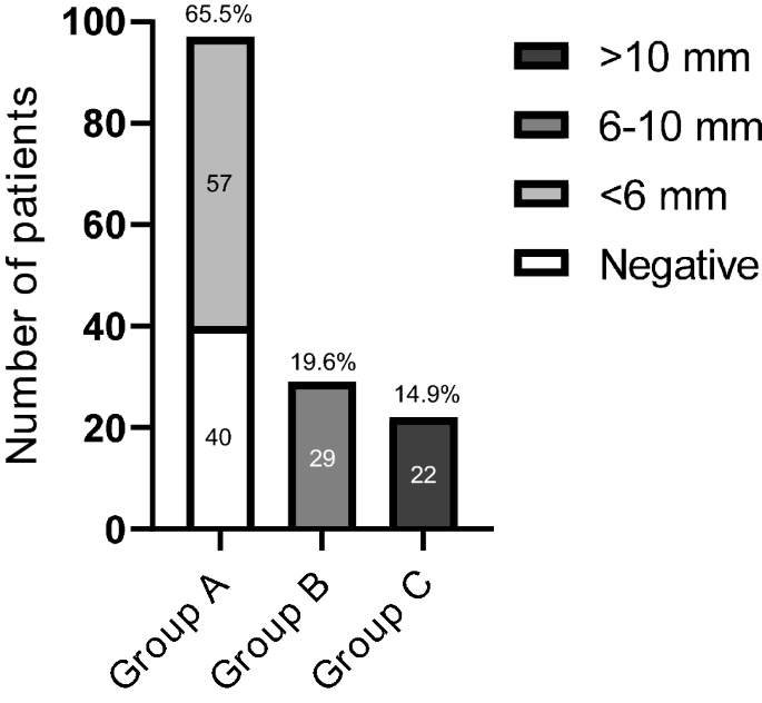
    This Reading Room is a collaboration between MedPage Today® and:

     Medpage Today

    For adrenal insufficiency (AI), reducing false positives means more than reducing resource utilization. Treatments like glucocorticoid replacement therapy can cause serious harm in people who do not actually have AI.

    Research published in the Journal of the Endocrine Society makes multiple findings that report authors say could help bring down false positive rates for AI. This retrospective study ultimately analyzed 6,531 medical records from the Imperial College Healthcare NHS Trust in the United Kingdom.

    Sirazum Choudhury, MBBS, an endocrinologist-researcher with the trust, served as a co-author of the report. He discussed the study with MedPage Today. The exchange has been edited for length and clarity.

    This study ultimately addressed two related but distinct questions. What was the first?

    Choudhury: Initially the path we were following had to do with when cortisol levels are tested.

    Cortisol levels follow a diurnal pattern; levels are highest in the morning and then decline to almost nothing overnight. This means we ought to be measuring the level in the morning. But there are logistical issues to doing so. In many hospitals, we end up taking measurements of cortisol in the afternoon. That creates a dilemma, because if it comes back low, there's an issue as to what we ought to do with the result.

    Here at Imperial, we call out results of <100 nmol/L among those taken in the afternoon. Patients and doctors then have to deal with these abnormal results, when in fact they may not actually be abnormal. We may be investigating individuals who should really not be investigated.

    So the first aim of our study was to try and ascertain whether we could bring that down to a lower level and in doing so stop erroneously capturing people who are actually fine.

    What was the second aim of the study?

    Choudhury: As we went through tens of thousands of data sets, we realized we could answer more than that one simple question. So the next part of the study became: if an individual is identified as suspicious for AI, what's the best way to prove this diagnosis?


    We do this with different tests like short Synacthen Tests (SST), all with different cutoff points. Obviously, we want to get the testing right, because if you falsely label a person as having AI, the upshot is that treatments will interfere with their cortisol access and they will not do well. Simply put, we would be shortening their life.

    So, our second goal was to look at all the SSTs we've done at the center and track them to see whether we could do better with the benchmarks.

    What did you find?

    Choudhury: When you look at the data, you see that you can bring those benchmarks down and potentially create a more accurate test.

    First, we can be quite sure that a patient who is tested in the afternoon and whose cortisol level is >234 does not have AI. If their level is <53.5 then further investigation is needed

    There were similar findings for SSTs, which in our case were processed using a platform made by Abbott. For this platform, we concluded that the existing cut-offs should be dropped down to 367 at 30 minutes or 419 at about 60 minutes.

    Did anything surprise you about the study or its findings?

    Choudhury: If you look at the literature, the number of individuals who fail at 30 minutes but pass at 60 minutes is around 5%. But I was very surprised to see that our number at Imperial was about 20%.

    This is a key issue because, as I mentioned, if individuals are wrongly labelled adrenally insufficient, you're shortening their life. It's scary to think about the number of people who might have been given steroids and treated for AI when they didn't have the condition.

    What do you see as the next steps?

    Choudhury: I see centers unifying their cutoffs for SST results and making sure we're all consistent in the way we treat these results.

    From a research perspective, on the testing we're obviously talking about one specific platform with Abbott, so research needs to be done on SST analyzers from other manufacturers to work out what their specific cutoffs should be.

    Read the study here and expert commentary on the clinical implications here.

    The study authors did not disclose any relevant relationship with industry.

     
  13. The endocrine system is a complex network of glands and organs. It uses hormones to control and coordinate your body's metabolism, energy level, reproduction, growth and development, and response to injury, stress, and mood. The following are integral parts of the endocrine system:


    • Hypothalamus. The hypothalamus is located at the base of the brain, near the optic chiasm where the optic nerves behind each eye cross and meet. The hypothalamus secretes hormones that stimulate or suppress the release of hormones in the pituitary gland, in addition to controlling water balance, sleep, temperature, appetite, and blood pressure.

    • Pineal body. The pineal body is located below the corpus callosum, in the middle of the brain. It produces the hormone melatonin, which helps the body know when it's time to sleep.

    • Pituitary. The pituitary gland is located below the brain. Usually no larger than a pea, the gland controls many functions of the other endocrine glands.

    • Thyroid and parathyroid. The thyroid gland and parathyroid glands are located in front of the neck, below the larynx (voice box). The thyroid plays an important role in the body's metabolism. The parathyroid glands play an important role in the regulation of the body's calcium balance.

    • Thymus. The thymus is located in the upper part of the chest and produces white blood cells that fight infections and destroy abnormal cells.

    • Adrenal gland. An adrenal gland is located on top of each kidney. Like many glands, the adrenal glands work hand-in-hand with the hypothalamus and pituitary gland. The adrenal glands make and release corticosteroid hormones and epinephrine that maintain blood pressure and regulate metabolism.

    • Pancreas. The pancreas is located across the back of the abdomen, behind the stomach. The pancreas plays a role in digestion, as well as hormone production. Hormones produced by the pancreas include insulin and glucagon, which regulate levels of blood sugar.

    • Ovary. A woman's ovaries are located on both sides of the uterus, below the opening of the fallopian tubes (tubes that extend from the uterus to the ovaries). In addition to containing the egg cells necessary for reproduction, the ovaries also produce estrogen and progesterone.

    • Testis. A man's testes are located in a pouch that hangs suspended outside the male body. The testes produce testosterone and sperm.

    endocrine_system.png

  14. Mineur L. · Boustany R. · Vazquez L.

    Author affiliations

    Corresponding Author

    Keywords: Ectopic Cushing syndromeNeuroendocrine tumoursWHO grade 1Paraneoplastic syndromesChromogranin A

     
    Case Rep Oncol 2021;14:1407–1413
     

    Abstract

    Ectopic production of adrenocorticotropic hormone (ACTH) by gastrointestinal neuroendocrine tumours (NETs) is relatively uncommon. We report a rare case of a liver metastatic G1 low-grade NET of the intestine that induced hypercortisolism after surgical resection. A 50-year-old man was admitted for an intestinal obstruction caused by a tumour of the intestine. Paraneoplastic Cushing syndrome was diagnosed more than a year later following the appearance of cushingoid symptoms, despite stable disease according to RECIST criteria but chromogranin A increase. Ketoconazole and sandostatin medical treatment and liver chemoembolization never managed to control the hypercortisolism unlike the bilateral adrenalectomy. The identification and effective management of this uncommon statement of ectopic ACTH secretion is important to improve the patient’s prognosis and quality of life.

    © 2021 The Author(s). Published by S. Karger AG, Basel


    Introduction

    Neuroendocrine tumours (NETs) are a relatively rare and heterogeneous tumour type, comprising about 2% of all malignancies [1]. The gastrointestinal (GI) and pancreatic tract and lungs are the most common primary tumour sites, with 62%–67% and 22%–27%, respectively, and within the GI tract, most of them occurs in the small bowel or the appendix [2, 3]. Since 2010 and the latest version of the WHO classification, GI and pancreatic NETs are subdivided according to their mitotic count or Ki67 index, associated with cellular proliferation. Well-differentiated NETs are relatively low-aggressive tumours, with a rather indolent disease course and a good prognosis in most patients. Nevertheless, some NETs with a low-grade histologic appearance may behave aggressively with rapid growth and metastasis proliferation [4, 5]. Because of this low incidence, tumour heterogeneity, lack of awareness, and non-uniform classifications, GI and pancreatic NETs remain a poorly understood disease, and delayed diagnosis is common among these [6, 7].

    Paraneoplastic Cushing syndrome (PCS) represents approximately 10% of all Cushing syndrome and is frequently caused by NETs [8, 9]. While PCS is common with lung NETs (>50% of PCS), this paraneoplastic syndrome is relatively uncommon associated with GI NETs and only described in isolated case reports. Nevertheless, knowing the indolent course of low-grade NETs and the clinical symptoms of cushingoid appearance resulting from prolonged exposure to excessive glucocorticoids, PCS is typically present before cancer detection [8], and surgery is curative in >80% of patients [10, 11]. For the remaining 20%, effective management is necessary, given the risk of infections and thromboembolic events due to the immunosuppressive effect and the hypercoagulable state [11]. For patients with medically unmanageable hypercortisolism, synchronous bilateral adrenalectomy is an effective and safe treatment [12]. We describe a case of typical metastatic intestinal NETs associated with a late ectopic Cushing syndrome, which was managed with synchronous bilateral adrenalectomy.

    Case Presentation

    We describe the case of a 50-year-old man admitted to the emergency department for an intestinal obstruction caused by an intestinal tumour. Anatomopathological analysis of the resected specimen and lymph nodes revealed an NET. Three nodes out of 12 removed were positive for cancer localization. The tumour presented serosa infiltration and perineural, vascular and lymphatics vessel invasion. The primary location could not be confirmed histologically between the ileum and appendix. Our diagnosis was pT3N1 according to the American Joint Committee on Cancer (AJCC) classification. An immunohistochemistry analysis revealed a Ki-67 expression <2%. Mitotic count/10 was 2 × 10 high-power fields, and cells showed well differentiation. So, according to the WHO classification, this tumour was classified as G1 NET. 111In-Octreoscan (Octreoscan) revealed lymph node and multifocal liver metastases.

    After discussion with a multidisciplinary team, the patient was started treatment with somatostatin analogue. Twelve months later, although computerized tomography (CT) scan showed stable disease, patient physical examination revealed facial puffiness with fatty tissue deposits in the face, generalized oedema, muscle weakness, and wasting. He also reports polydipsia, insomnia, and balance disorders. We noted however a discreet increase in the chromogranin A (CgA) value, from 55 ng/mL to 199 ng/mL (with a diagnostic value of 1,700 ng/mL) without an increase in the urinary 5-HIAA level.

    Laboratory tests revealed an 8.00 a.m. cortisol level of 888 nmol/L, an adrenocorticotropic hormone (ACTH) level of 96.5 pg/mL, and 24-h urine free cortisol of 1,494 μg. A high-dose dexamethasone suppression test showed no cortisol suppression. The patient was diagnosed with ACTH-dependent Cushing syndrome. Magnetic resonance imaging (MRI) of the brain showed a normal pituitary gland, confirming the PCS diagnosis. Ketoconazole treatment associated with sandostatin alleviated hypercortisolism within a month, with a cortisol level within normal laboratory ranges. Two months later, secondary diabetes mellitus was discovered and managed effectively with insulin glargine.

    Four months later and despite stable disease according to RECIST criteria, cortisol levels increased considerably, with cortisol values similar to diagnosis without ketoconazole increased response. Moreover, diabetes became complicated to manage. Also we noted an increase in CgA value, from 165 ng/mL to 393 ng/mL. Chemoembolization was performed on liver metastases without any effectiveness on hypercortisolism. Adding targeted therapy with mTOR inhibitor (everolimus) was considered. Nevertheless, given the magnitude of drug interaction, the use of everolimus should be avoided in ketoconazole-treated patients, or vice versa.

    Considering the risks for the patient and expected benefits, synchronous bilateral adrenalectomy was performed. It resolved hypercortisolism and permitted to stabilize diabetes (shown in Fig. 1). Everolimus treatment has been started 1 month after the surgery. Twelve months after everolimus initiation, the patient CT scan still showed stable disease, according to RECIST criteria and a stable CgA value.

    Fig. 1.

    Histogram of 8:00 a.m. plasmatic cortisol, ACTH, 24-h urinary cortisol, and CgA levels from Cushing syndrome diagnosis to bilateral adrenalectomy. ACTH, adrenocorticotropic-hormone; ULN, upper limit of normal; 8:00 a.m. cortisol normal ranges (172–497) nmol/L; ACTH normal ranges (7–63) ng/L; 24-h urinary cortisol normal ranges (20–50) µg/24 h; CgA normal ranges (27–94) ng/mL. ACTH, adrenocorticotropic hormone; CgA, chromogranin A.

    /WebMaterial/ShowPic/1364794

    Discussion

    Approximately 10% of Cushing syndrome is paraneoplastic and may result in many tumours, preferentially lung cancer (50–60% of time), with 1–2% of lung NET and about 5% of small-cell lung cancer associated with Cushing syndrome [8, 11, 13]. Others reported sites of malignancy include the thymus, thyroid, pancreas, and adrenals. Except for the pancreas, PCS secondary to GI NET (appendix, duodenum, ileum, colon, and anal canal) is extremely rare, and only isolated case reports have described this syndrome.

    In paraneoplastic endocrine syndrome cases, symptoms are due to secretion of hormones by malignant cells or secondary to the impact of neoplastic cell antibodies on normal cells. PCS arises from tumour secretion of ACTH or CRH, resulting in production and release of cortisol from the adrenal glands. Unlike paraneoplastic endocrine syndromes that present most of the time after cancer diagnosis, PCS typically appears before cancer detection and similarly relapse may herald tumour recurrence [11, 14]. In our case, no symptoms related to hypercortisolaemia led the patient to consult before obstructive syndrome. The occurrence of hypercortisolaemia 12 months after diagnosis was not linked to imaging progression according to RECIST criteria. However, concurrent CgA increase should be noted.

    Commonly measured tumour markers in NETs include serum CgA and 5-HIAA, the final secreted product of serotonin, levels in a 24-h urine sample. Elevated levels of circulating of CgA have been associated with almost all types of NETs, including those arising from GI tract but also pheochromocytomas [15]. The clinical sensitivity of CgA has been demonstrated to depend on the threshold cut-off, on NET primary location, and on the spread of the disease, especially the existence of liver metastases [16]. Indeed, a higher sensitivity was found in patients with midgut NETs and liver metastases, as in our patient. Moreover, with our cut-off level (94 ng/mL) approximately the same as used in 2 studies [16, 17], sensitivity was 62%–67% and specificity was 96%. Furthermore, Korse et al. [18]. postulated that serum CgA was superior to urinary 5-HIAA concerning the prognostic relevance in the follow-up of metastatic midgut NETs. These data are consistent with our patient outcomes for which 5-HIAA was not increased unlike CgA. However, although CgA is currently the best available tumour marker indicating tumour recurrence [19], there are many comorbidities and drugs that may increase CgA levels and lead to false-positive results. As a result, it is questionable whether the CgA increase in our patient was not rather secondary to cardiovascular or GI disorders, inflammatory diseases, diabetes, or even food intake before CgA measurement [16, 20, 21]. Similarly, many drugs, foods, natural stimulants, and comorbidities may alter the level of 24-h urinary 5-HIAA, positively or negatively.

    Cushing syndrome is due to hypercortisolism. Two-thirds of endogenous elevated cortisol is caused by ACTH-secreting pituitary tumours, 15% by primary adrenal glands and 15% by ectopic PCS [22]. The first step is laboratory tests with cortisol and ACTH levels to differentiate ACTH-dependent or ACTH-independent Cushing syndrome. When ACTH-dependent Cushing syndrome is confirmed, differentiation between PCS and Cushing disease can be difficult. The high-dose dexamethasone suppression tests help distinguish Cushing disease from PCS, as in our presented case. Indeed, no decrease in blood cortisol during the high-dose test and high ACTH levels are consistent with PCS. Nevertheless, 21–26% of ectopic ACTH secretions have a positive suppression, about one-third of MRI scans for pituitary adenoma exclusion are false-negative, and occult ectopic ACTH-secreting tumours have been described in about 15% of adult patients [23-26]. In our patient, both MRI and high-dose dexamethasone suppression test are consistent with PCS. The gold standard diagnosis – inferior petrosal sinus sampling – that demonstrates gradient in ACTH concentration between the affected side sinus and the periphery in pituitary lesions, whereas the absence of this gradient in PCS was not performed because of its invasiveness and its neurological accident risks [27]. Note however although the ACTH level at diagnosis suggests ACTH-dependent Cushing syndrome, the occurrence of adrenal metastasis few months after the diagnosis and explaining the sudden deregulation could be possible and consistent with the CgA increase but refuted by adrenal gland histology.

    Clinical features of PCS depend on the source of production and rate of ACTH synthesis. Characteristically, these patients have severe hypercortisolaemia, leading to low serum potassium levels, diabetes, generalized infections, hypertension, and psychosis. To confirm whether rapidly growing tumours produce sudden onset of symptoms, gradual physical signs are noticed in slower growing tumours [28], as for our patient for whom we suppose that liver metastases started to produce ACTH ectopically. An option for non-resectable neuroendocrine liver lesions, given that the majority of them are hypervascular, is hepatic directed procedures, which include ablative therapy, transarterial embolization, transarterial chemoembolization, and selective internal radiation therapy with yttrium-90 microspheres [29]. Hepatic artery chemoembolization for the treatment of liver metastases from NETs is useful for tumour size reduction and symptom palliation and can be associated with prolonged survival [30]. Nevertheless, chemoembolization on NET liver metastasis-producing ACTH is not well documented. Given the fact that hepatic metastasis chemoembolization was ineffective on hypercortisolism and despite Octreoscan results, there is still a small chance that he harbours somewhere else metastasis-producing ACTH. Indeed, PET-CT imaging with 68Gallium-DOTATATE has recently replaced Octreoscan as the new gold standard with a higher detection rate in GI NETs [31].

    Hypercortisolism requires a prompt therapeutic management to reduce the risk of development of a potentially fatal emergency. Synchronous bilateral adrenalectomy is an effective and safe treatment for patient with unmanageable ACTH-dependent hypercortisolism [12]. Taking account of the risks to the patient and the lack of effective medical therapeutic possibilities, we have chosen to perform this surgery.

    According to the recent consensus guidelines for digestive NETs of the jejunum and ileum, the 5-year survival rate is 36% in patients with distant metastases [32]. Several analyses suggest a significant survival benefit in patients who received surgery for the primary tumour even in the presence of metastasis [33]. Moreover, the impact of liver resection or liver-directed therapies on the survival of patients with liver metastasis is unclear with conflicting results [33]. PCS can cause a poor clinical outcome due to various complications with an increase in susceptibility to infection and GI ulceration. Indeed, for small-cell lung cancer and gynaecological malignancies, PCS is associated with accelerated decompensation and poorer response to chemotherapy (Mitchell et al. [14]). Whether these findings can be extrapolated to other malignancies is unknown. However, an early diagnosis and a prompt management can improve patient outcomes through earlier cancer diagnosis or relapse and thus earlier administration of treatment, as was the case with our patient.

    Conclusion

    We report an uncommon case with PCS due to a GI NET. The identification of this rare cause of ectopic ACTH secretion can be challenging, but aggressive management is critical to prevent or decelerate the acute decompensation of cancer patients and prolong overall survival. In this context, synchronous bilateral adrenalectomy may be the unique answer.

    Statement of Ethics

    Written informed consent was obtained from the patient for publication of this case report and any accompanying images. A copy of the written consent is available for review by the editor-in-chief of this journal.

    Conflict of interest Statement

    The authors have no conflict of interest to declare.

    Funding Sources

    No funding was received for this study.

    Author Contributions

    L.M. conceived the study and participated in data collection. L.V. performed the literature search and wrote the manuscript. L.M. and R.B. critically revised the manuscript for important intellectual content. All authors read and approved the final manuscript version.

    Data Availability Statement

    The datasets used and analysed during the current study are available from the corresponding author on reasonable request.

    From https://www.karger.com/Article/FullText/518316

    • Like 1
  15. Purpose: This study aimed to identify predictive factors and to develop a predictive model for adrenal insufficiency (AI) related to topical corticosteroids use.


    Methods: The research was conducted using a cross-sectional design. Adult patients with dermatological conditions who had been prescribed topical steroids for at least 12 months by the dermatology outpatient departments of the Faculty of Medicine, Chiang Mai University from June through October 2020 were included. Data on potential predictors, including baseline characteristics and laboratory investigations, were collected. The diagnoses of AI were based on serum 8AM cortisol and low-dose ACTH stimulation tests. Multivariable logistic regression was used for the derivation of the diagnostic score.


    Results: Of the 42 patients, 17 (40.5%) had AI. The statistically significant predictive factors for AI were greater body surface area of corticosteroids use, age < 60 years, and basal serum cortisol < 7 μg/dL. In the final predictive model, duration of treatment was added as a factor based on its clinical significance for AI. The four predictive factors with their assigned scores were: body surface area involvement 10– 30% (20), > 30% (25); age < 60 years old (15); basal serum cortisol of < 7 μg/dL (30); and duration of treatment in years. Risk of AI was categorized into three groups, low, intermediate and high risk, with total scores of < 25, 25– 49 and ≥ 50, respectively. The predictive performance for the model was 0.92 based on area under the curve.


    Conclusion: The predictive model for AI in patients using topical corticosteroids provides guidance on the risk of AI to determine which patients should have dynamic ACTH stimulation tests (high risk) and which need only close follow-up (intermediate and low risk). Future validation of the model is warranted.

    Keywords: adrenal insufficiency, topical corticosteroids, predictive model, skin diseases

    Introduction

    Topical corticosteroids are frequently used for inflammatory skin diseases owing to their anti-inflammatory and immunosuppressive effects. Common indications for use include diseases such as psoriasis, eczema, atopic dermatitis, and vitiligo.1 In clinical practice, a variety of delivery vehicles and potencies of topical corticosteroids are used.1 Prolonged and/or inappropriate use of topical corticosteroids can lead to adverse side effects.2 These adverse side effects can be categorized as cutaneous and systemic side effects. The most common cutaneous side effect is skin atrophy. Systemic side effects include hypothalamic-pituitary-adrenal (HPA) axis suppression, glaucoma, hyperglycemia and hypertension.3

    One of the most worrisome adverse side effects from the use of topical corticosteroids is adrenal insufficiency (AI) resulting from HPA axis suppression. Topically applied corticosteroids can be absorbed systemically through the skin and can suppress the HPA axis.4–8 This adverse outcome, the inability to increase cortisol production after stress, can lead to adrenal crisis, which is potentially life-threatening. Tests that are normally used to diagnose or exclude AI include serum morning cortisol and the dynamic ACTH stimulation test.9

    Secondary AI from percutaneous absorption of topical corticosteroids is less common than with parenteral or oral administration. The cumulative doses and the durations of oral corticosteroid therapy associated with HPA axis suppression have been well documented.10 Data regarding the dose and duration of oral corticosteroids and HPA axis suppression have similarly been well established. A study by Curtis et al reported that the use of oral prednisolone >7.5 mg/day for an extended period (>3 weeks) was linked to this adverse event, and that the incidence increased with duration.10 However, corresponding data for topical corticosteroids has been limited. The degree of risk of HPA axis suppression from topical corticosteroids use is associated with the level of percutaneous absorption which, in turn, depends on numerous factors including the age of the patient (younger patients are more susceptible), body surface area treated, quantity of topical corticosteroids used, potency of the drug, duration of therapy, body region of application, the associated compounds used, eg, urea or salicylic acid, the characteristics of the diseased skin, the degree of impairment of skin integrity, and the coexistence of hepatic and/or renal disease.11–13 One study reported that HPA axis suppression occurs when high potency steroids are administered at a cumulative dose per week of >50 g.2

    Presently, there is a lack of data on predictive factors for AI and no predicative model of the relationship between secondary AI resulting from HPA axis suppression and topical corticosteroids use. A simple predictive model which could help preclude and predict the risk of AI which incorporates both demographic and biochemical data could potentially reduce the number of dynamic ACTH stimulation tests performed. This study aimed to identify potential predictive factors and to design an easy-to-use model for predicting the risk of AI following topical corticosteroids use in dermatological patients.

    Materials and Methods

    This cross-sectional study was conducted with 42 patients who were seen at the dermatology outpatient departments at the Faculty of Medicine, Chiang Mai University Hospital over a 5-month period (June – October 2020). The study protocol was approved by the Faculty of Medicine, Chiang Mai University, Ethical Committee (Ethical number: MED-2563-07037). Recruited participants were adult dermatological patients (≥18 years) who had used topical corticosteroids for at least 12 months. Patients with pituitary or adrenal diseases, pregnant women and patients who had been treated with either systemic corticosteroids or other local corticosteroids were excluded. Those who meet all the inclusion criteria gave their informed consent prior to the study. This study was conducted in accordance with the Declaration of Helsinki.

    Adrenal Function Evaluation

    Adrenal function was evaluated by serum morning (8 AM) cortisol and the low-dose ACTH stimulation test. Patients were instructed to suspend use of topical corticosteroids for at least 24 hours before serum morning cortisol measurement and ACTH stimulation tests. In those with serum morning cortisol between 3 and 17.9 µg/dL, ACTH stimulation tests were performed on the same day between 9–11AM to either exclude or diagnose AI. Serum cortisol concentrations were measured at 8 AM 0 (basal cortisol) as well as 20 and 40 minutes after 5 µg ACTH was administered intravenously.

    Data Collection

    Epidemiological data collected included gender, age, blood pressure, underlying dermatologic diseases, other underlying diseases, body surface area involvement, sensitive area involvement, topical corticosteroid potency, amount and duration of topical corticosteroids use, symptoms of AI and the presence of Cushingoid features. Biochemical data included serum cortisol at 8 AM, 0 (basal cortisol) and at 20 and 40 minutes after ACTH intravenous injection, serum creatinine, electrolytes and albumin. Serum cortisol levels were measured by electrochemiluminescence assay (ECLIA) (Elecsys® Cortisol II assay, Roche Diagnostics GmbH, Mannheim, Germany).

    Definitions

    An 8AM cortisol level of ❤️ µg/dL or a peak serum cortisol level of <18 µg/dL at 20 or 40 minutes after an ACTH stimulation test was defined as having AI.14 Sensitive area involvement included the axilla, groin, face and genitalia. Topical corticosteroids are classified by potency based on a skin vasoconstriction assay, and range from ultra-high potency (class I) to low potency (class VII).15 Since some patients had concurrently used more than one class of corticosteroids in one treatment period, the new variable potency·dose·time (summary of corticosteroids potency (I–VII)16 multiplied by total doses (mg) of corticosteroids use and multiplied by duration (months) of corticosteroids use) was created. Symptoms of AI included lethargy, nausea and vomiting, orthostatic hypotension and significant weight loss. Significant weight loss was defined as a loss of 5% of body weight in one month or a loss of 10% over a period of six months.17 Having Cushingoid features was defined as at least one of the excess glucocorticoid features, eg, easy bruising, facial plethora, proximal myopathy, striae, dorsocervical fat pad, facial fullness, obesity, supraclavicular fullness, hirsutism, decreased libido and menstrual abnormalities.

    Statistical Analysis

    All statistical analyses were performed using Stata 16 (StataCorp, College Station, Texas, USA). Categorical variables are reported as frequency and percentage, while continuous variables are reported as mean ± standard deviation or median and interquartile range (IQR), according to their distribution. For univariable comparison, Fisher’s exact probability test was used for categorical variables, and the independent t-test or the Mann–Whitney U-test was used for continuous variables. p-values less than 0.05 were considered statistically significant.

    Multivariable logistic regression was used in the derivation of the prediction model for AI. Predictors with significant p-values in the univariable analysis were included in the multivariable model. We also included age and treatment duration in the model due to the clinical significance of those factors.4,18 The clinical collinearity among the predictors was also evaluated before the selection of the predictors. We generated a weighted score for each predictor by dividing the logit coefficient of the predictor by the lowest coefficient in the model. The discriminative ability of the final multivariable model was assessed using the area under the receiver operating characteristics (ROC) curve. The calibration of the scores was evaluated using the Hosmer-Lemeshow goodness-of-fit test, where a p-value >0.01 was considered a good fit. For clinical applicability, the appropriate cut-off points for the scores were identified based on sensitivity and specificity. We identified one cut-off point with high sensitivity for ruling out AI and another cut-off point with high specificity for ruling in AI. The positive predictive value for each score category with its corresponding confidence interval were presented. A sample size of at least 25 patients with at least 5 patients with AI was estimated to give 80% power at the 5% significance level.4 There was no missing data in this study.

    Results

    Baseline characteristics and biochemical investigations are shown in Table 1. Forty-two patients with dermatological diseases were included in this study. Of these, 17 patients (40.5%) had AI of whom 5 (29.4%) were female. The mean age of the group was 56.5 ±15.4 years, the mean duration of treatment was 10.1 ± 6 years, and the majority of patients had psoriasis (n = 14, 82.4%). There was no significant difference in sex, age, duration of treatment, potency dose-time, comorbidities, or underlying skin disease between the AI and non-AI groups. The average body surface area of corticosteroids use was significantly higher in patients with AI than in the non-AI group (27.5 ±18.7 m2 and 10.7 ±11.7 m2, p < 0.001, respectively). Basal serum cortisol levels were significantly lower in the AI group (6.52 ± 4.04 µg/dL) than in the non-AI group (10.48 ± 3.45 µg/dL, p 0.003). Although lower serum morning cortisol levels were observed in the AI group, the difference was not statistically significant (5.24 ± 4.65 µg/dL vs 13.39 ± 15.68 µg/dL, p = 0.069). Three patients were identified as having Cushingoid features. All patients with Cushingoid features had AI.

     
    IJGM_A_342841_t0001_Thumb.jpg

    Table 1 Comparison of Clinical Characteristics Between Patients with a History of Topical Corticosteroids Use for at Least 12 Months Who Were Diagnosed with Adrenal Insufficiency and Those without Adrenal Insufficiency (n = 42)


    Based on the multivariate logistic regression analysis (shown in Table 2), the significant predictive factors for AI in patients who used topical corticosteroids for more than 12 months were body surface area of corticosteroids use of 10–30% and >30% (POR 18.9, p =0.042, and POR 59.2, p = 0.035, respectively), age less than 60 years (POR 13.8, p = 0.04), and basal serum cortisol of <7 µg/dL (POR 131.5, p = 0.003). Only serum basal cortisol was included in the final multivariable model as there was clinical collinearity among serum morning cortisol and basal cortisol as well as 20- and 40-minute cortisol measurements.

     
    IJGM_A_342841_t0002_Thumb.jpg

    Table 2 Multivariable Model for Prediction of Adrenal Insufficiency in Patients with a History of Topical Corticosteroids Use for at Least 12 Months (n = 38)


    Predictive risk score was created to determine the probability of patients having AI using the aforementioned three significant predictive factors from the multivariable analysis (Table 2). As previous studies have demonstrated that duration of treatment is a strong predictive factor for AI in corticosteroid users,4,18 this factor was also incorporated in the model. The transformed score for body surface area, age and basal serum cortisol had a range of 0 to 30. For treatment duration, the transformed score was based on cumulative years of treatment. The total score was categorized into three groups: low, intermediate, and high risk (Table 3).

     
    IJGM_A_342841_t0003_Thumb.jpg

    Table 3 Accuracy of the Score to Rule in and Rule Out Adrenal Insufficiency in Patients with a History of Topical Corticosteroids Use for at Least 12 Months (n = 38)


    The cut-off point of ≥50 suggests high risk for developing AI with a sensitivity of 46.2% and a specificity of 100%, a score of <25 suggests a low risk with a sensitivity of 100% and a specificity of 52%, and a score between 25 and 49 indicates an intermediate risk of having AI. The ROC curve for the model assessing predictive performance which included all significant factors had an AuROC of 0.92 (Figure 1). The Hosmer-Lemeshow goodness-of-fit test revealed non-statistically significant results (p = 0.599), indicating that our newly derived scoring system fits the data well.

     
    IJGM_A_342841_O_F0001g_Thumb.jpg

    Figure 1 Model discrimination via receiver operating characteristic curve in patients with a history of topical corticosteroids use for at least 12 months (n = 42).


    Discussion

    The present study proposes an easy-to-use predictive model for AI following topical corticosteroids use in dermatological patients based on demographic and biochemical factors. The accuracy of the model shows an excellent diagnostic accuracy of 92% based on AuROC. Currently, the diagnosis of AI in dermatological patients with topical corticosteroids use involves multiple steps including screening for serum morning cortisol followed by dynamic ACTH stimulation testing. The proposed simple predictive model, which requires only three demographic data items (age, body surface area of corticosteroids use, duration of use) and one biochemical test (serum basal cortisol), could potentially reduce the number of dynamic ACTH stimulation tests performed, resulting in cost- and time-saving for both patients and health-care facilities.

    Based on the proposed cut-off points, we suggest screening of individuals at high risk for having AI, including serum morning cortisol and the ACTH stimulation tests to confirm a diagnosis of AI. If there is evidence of AI, the patient should begin to receive treatment for AI to reduce future complications. For those in the low-risk group, only clinical follow-up should be carried out. In the intermediate-risk group, we recommend regular and close biochemical follow-up including serum morning cortisol and clinical follow-up for signs and symptoms of AI. Signs and symptoms that should raise a high index of suspicion for AI include significant weight loss, nausea and/or vomiting, orthostatic hypotension and lethargy. However, this proposed predictive model was studied in adults and cannot simply be generalized and extrapolated to children or infants.

    In our study, 40.5% of the patients were determined to have AI. A previous meta-analysis by Broersen et al reported the percentage of patients with AI secondary to all potencies of topical corticosteroids based on a review of 15 studies was 4.7%, 95% CI (1.1–18.5%).19 The higher prevalence of AI in our study could be a result of differences in patients’ baseline characteristics, eg, duration of treatment, corticosteroids potency and body surface area involvement.

    In the predictive model, we incorporated both clinical and biochemical factors which are easy to obtain in actual clinical practice. Some of those predictive factors have been previously reported to be linked to AI. Body surface area of corticosteroids use larger than 10% found to be significantly related to AI, especially in patients with a lesion area of over 30%. This finding is consistent with a study by Kerner et al which suggests the extent of surface area to which the corticosteroids are applied may influence absorption of the drug.20 Regarding the age of the patients, our study found that individuals over 60 years old tended to be at high risk of AI following topical corticosteroids therapy. The underlying explanation is that the stratum corneum acts as a rate-limiting barrier to percutaneous absorption as the stratum corneum in younger individuals is thinner than in older people. Diminished effectiveness of topical corticosteroid treatment in older people was demonstrated in a study by Malzfeldt et al.21 Even though serum basal cortisol is not recommended as a standard test to diagnose AI, a prior study reported that it can be considered as an alternative choice to diagnose AI when serum morning cortisol results are not available. In fact, it has been reported that there is no difference in diagnostic accuracy between serum morning cortisol and basal cortisol22 which supports our finding that serum basal cortisol <7 µg/dL is one of the significant factors related to AI.

    The final model found no statistically significant relationship between the incidence of AI and the duration of corticosteroids treatment. However, we decided to include this factor in the final model since previous publications have reported that the duration of treatment is a relevant risk factor for developing AI following continuous topical corticosteroids use. The duration of AI events has been reported to vary between 2 weeks to 18 months.4,18 Additionally, a case report of AI demonstrated that 5 years of topical corticosteroids use can cause AI.6 Together, this suggests that patients with a longer duration of topical corticosteroids use are at increased risk of AI, especially those who also have other risk factors. Although both potency and dosage of topical corticosteroids have been reported to be significantly linked to HPA axis suppression, the present study found only a non-significance link. This could be the result of the small sample size as well as of other factors, eg, body surface area involvement and serum cortisol levels, which could have masked the association between potency and dosage of topical corticosteroids with HPA suppression.

    To the best of our knowledge, this study is the first to use these novel predictive factors to develop a predictive model for AI in patients using topical corticosteroids. This model has multiple potential implications. First, the model uses clinical and biochemical factors which are obtainable in many institutes. Second, the model’s risk score provides good diagnostic accuracy in terms of both sensitivity and specificity. Finally, each of the predictive factors in the model has an underlying pathophysiological explanation and is not due simply to chance.

    There are some limitations in this study. First, the sample size is relatively small, although it does offer sufficient statistical power for each of the predictive factors. Second, further external validation is needed to validate the predictive performance of the model. Third, the cut-off level of serum cortisol after ACTH stimulation test was based on the older generation of ECLIA assay. There was a study proposed that the cut-off for serum cortisol in the newer generation of cortisol assay should be lower (~14–15 µg/dL) than the previous one (18 µg/dL).23 However, this proposed cut-off has not yet been established in the current guideline for AI. In the future, if the newer cut-off for serum cortisol will have been employed in the standard guideline, our predictive model may lead to overdiagnosis of AI.

    Conclusions

    The proposed predictive model uses both demographic and biochemical factors to determine the risk of AI in dermatological patients following topical corticosteroids use with a high level of diagnostic accuracy. This model has advantages in terms of a reduction in the number of dynamic ACTH stimulation tests needed, thus saving time and resources. Additionally, it can provide guidance to clinical practitioners regarding which patients should be closely followed up for development of AI. Future external validation of this predictive model is warranted.

    Acknowledgments

    The authors are grateful to Lamar G. Robert, PhD and Chongchit S. Robert, PhD for editing the manuscript.

    Disclosure

    The authors report no conflict of interest in this work.

    References

    1. Ference JD, Last AR. Choosing topical corticosteroids. Am Fam Physician. 2009;79(2):135–140.

    2. Hengge UR, Ruzicka T, Schwartz RA, Cork MJ. Adverse effects of topical glucocorticosteroids. J Am Acad Dermatol. 2006;54(1):1–15;quiz 16–8. doi:10.1016/j.jaad.2005.01.010

    3. Rathi SK, D’Souza P. Rational and ethical use of topical corticosteroids based on safety and efficacy. Indian J Dermatol. 2012;57(4):251–259. doi:10.4103/0019-5154.97655

    4. Carruthers JA, August PJ, Staughton RC. Observations on the systemic effect of topical clobetasol propionate (Dermovate). Br Med J. 1975;4(5990):203–204. doi:10.1136/bmj.4.5990.203

    5. Staughton RC, August PJ. Cushing’s syndrome and pituitary-adrenal suppression due to clobetasol propionate. Br Med J. 1975;2(5968):419–421. doi:10.1136/bmj.2.5968.419

    6. Young CA, Williams IR, MacFarlane IA. Unrecognised Cushing’s syndrome and adrenal suppression due to topical clobetasol propionate. Br J Clin Pract. 1991;45(1):61–62.

    7. Abma EM, Blanken R, De Heide LJ. Cushing’s syndrome caused by topical steroid therapy for psoriasis. Neth J Med. 2002;60(3):148–150.

    8. Böckle BC, Jara D, Nindl W, Aberer W, Sepp NT. Adrenal insufficiency as a result of long-term misuse of topical corticosteroids. Dermatology. 2014;228(4):289–293. doi:10.1159/000358427

    9. Ospina NS, Al Nofal A, Bancos I, et al. ACTH stimulation tests for the diagnosis of adrenal insufficiency: systematic review and meta-analysis. J Clin Endocrinol Metab. 2016;101(2):427–434. doi:10.1210/jc.2015-1700

    10. Curtis JR, Westfall AO, Allison J, et al. Population-based assessment of adverse events associated with long-term glucocorticoid use. Arthritis Rheum. 2006;55(3):420–426. doi:10.1002/art.21984

    11. Brazzini B, Pimpinelli N. New and established topical corticosteroids in dermatology: clinical pharmacology and therapeutic use. Am J Clin Dermatol. 2002;3(1):47–58. doi:10.2165/00128071-200203010-00005

    12. Dhar S, Seth J, Parikh D. Systemic side-effects of topical corticosteroids. Indian J Dermatol. 2014;59(5):460–464. doi:10.4103/0019-5154.139874

    13. Levin C, Maibach HI. Topical corticosteroid-induced adrenocortical insufficiency: clinical implications. Am J Clin Dermatol. 2002;3(3):141–147. doi:10.2165/00128071-200203030-00001

    14. Bornstein SR, Allolio B, Arlt W, et al. Diagnosis and treatment of primary adrenal insufficiency: an endocrine society clinical practice guideline. J Clin Endocrinol Metab. 2016;101(2):364–389. doi:10.1210/jc.2015-1710

    15. Goa KL. Clinical pharmacology and pharmacokinetic properties of topically applied corticosteroids. A review. Drugs. 1988;36(Suppl 5):51–61. doi:10.2165/00003495-198800365-00011

    16. Davallow Ghajar L, Wood Heickman LK, Conaway M, Rogol AD. Low risk of adrenal insufficiency after use of low- to moderate-potency topical corticosteroids for children with atopic dermatitis. Clin Pediatr. 2019;58(4):406–412. doi:10.1177/0009922818825154

    17. Gaddey HL, Holder K. Unintentional weight loss in older adults. Am Fam Physician. 2014;89(9):718–722.

    18. Melian EB, Spencer CM, Jarvis B. Clobetasol propionate foam, 0.05%. Am J Clin Dermatol. 2001;2(2):89–92;discussion 93. doi:10.2165/00128071-200102020-00005

    19. Broersen LH, Pereira AM, Jørgensen JO, Dekkers OM. Adrenal insufficiency in corticosteroids use: systematic review and meta-analysis. J Clin Endocrinol Metab. 2015;100(6):2171–2180. doi:10.1210/jc.2015-1218

    20. Kerner M, Ishay A, Ziv M, Rozenman D, Luboshitzky R. Evaluation of the pituitary-adrenal axis function in patients on topical steroid therapy. J Am Acad Dermatol. 2011;65(1):215–216. doi:10.1016/j.jaad.2010.12.033

    21. Malzfeldt E, Lehmann P, Goerz G, Lippold BC. Influence of drug solubility in the vehicle on clinical efficacy of ointments. Arch Dermatol Res. 1989;281(3):193–197. doi:10.1007/bf00456392

    22. Manosroi W, Phimphilai M, Khorana J, Atthakomol P. Diagnostic performance of basal cortisol level at 0900-1300h in adrenal insufficiency. PLoS One. 2019;14(11):e0225255. doi:10.1371/journal.pone.0225255

    23. Vogeser M, Kratzsch J, Ju Bae Y, et al. Multicenter performance evaluation of a second generation cortisol assay. Clin Chem Lab Med. 2017;55(6):826–835. doi:10.1515/cclm-2016-0400

    Creative Commons License This work is published and licensed by Dove Medical Press Limited. The full terms of this license are available at https://www.dovepress.com/terms.php and incorporate the Creative Commons Attribution - Non Commercial (unported, v3.0) License. By accessing the work you hereby accept the Terms. Non-commercial uses of the work are permitted without any further permission from Dove Medical Press Limited, provided the work is properly attributed. For permission for commercial use of this work, please see paragraphs 4.2 and 5 of our Terms.

    Download Article [PDF] 


    From https://www.dovepress.com/novel-predictive-model-for-adrenal-insufficiency-in-dermatological-pat-peer-reviewed-fulltext-article-IJGM

  16. An updated guideline for the treatment of Cushing’s disease focuses on new therapeutic options and an algorithm for screening and diagnosis, along with best practices for managing disease recurrence.

    Despite the recent approval of novel therapies, management of Cushing’s disease remains challenging. The disorder is associated with significant comorbidities and has high mortality if left uncontrolled.

    Adrenal transparent _Adobe
    Source: Adobe Stock

    “As the disease is inexorable and chronic, patients often experience recurrence after surgery or are not responsive to medications,” Shlomo Melmed, MB, ChB, MACP, dean, executive vice president and professor of medicine at Cedars-Sinai Medical Center in Los Angeles, and an Endocrine Today Editorial Board Member, told Healio. “These guidelines enable navigation of optimal therapeutic options now available for physicians and patients. Especially helpful are the evidence-based patient flow charts [that] guide the physician along a complex management path, which usually entails years or decades of follow-up.”

    melmed_shlomo.jpg
    Shlomo Melmed

    The Pituitary Society convened a consensus workshop with more than 50 academic researchers and clinical experts across five continents to discuss the application of recent evidence to clinical practice. In advance of the virtual meeting, participants reviewed data from January 2015 to April 2021 on screening and diagnosis; surgery, medical and radiation therapy; and disease-related and treatment-related complications of Cushing’s disease, all summarized in recorded lectures. The guideline includes recommendations regarding use of laboratory tests, imaging and treatment options, along with algorithms for diagnosis of Cushing’s syndrome and management of Cushing’s disease.

    Updates in laboratory, testing guidance

    If Cushing’s syndrome is suspected, any of the available diagnostic tests could be useful, according to the guideline. The authors recommend starting with urinary free cortisol, late-night salivary cortisol, overnight 1 mg dexamethasone suppression, or a combination, depending on local availability.

    If an adrenal tumor is suspected, the guideline recommends overnight dexamethasone suppression and using late-night salivary cortisol only if cortisone concentrations can also be reported.

    The guideline includes several new recommendations in the diagnosis arena, particularly on the role of salivary cortisol assays, according to Maria Fleseriu, MD, FACE, a Healio | Endocrine Today Co-editor, professor of medicine and neurological surgery and director of the Pituitary Center at Oregon Health & Science University in Portland.

    fleseriu_maria_2020_new_web.jpg
    Maria Fleseriu

    “Salivary cortisol assays are not available in all countries, thus other screening tests can also be used,” Fleseriu told Healio. “We also highlighted the sequence of testing for recurrence, as many patients’ urinary free cortisol becomes abnormal later in the course, sometimes up to 1 year later.”

    The guideline states combined biochemical and imaging for select patients could potentially replace petrosal sinus sampling, a very specialized procedure that cannot be performed in all hospitals, but more data are needed.

    “With the corticotropin-releasing hormone stimulation test becoming unavailable in the U.S. and other countries, the focus is now on desmopressin to replace corticotropin-releasing hormone in some of the dynamic testing, both for diagnosis of pseudo-Cushing’s as well as localization of adrenocorticotropic hormone excess,” Fleseriu said.

    The guideline also has a new recommendation for anticoagulation for high-risk patients; however, the exact duration and which patients are at higher risk remains unknown.

    “We always have to balance risk for clotting with risk for bleeding postop,” Fleseriu said. “Similarly, recommended workups for bone disease and growth hormone deficiency have been further structured based on pitfalls specifically related to hypercortisolemia influencing these complications, as well as improvement after Cushing’s remission in some patients, but not all.”

    New treatment options

    The guideline authors recommended individualizing medical therapy for all patients with Cushing’s disease based on the clinical scenario, including severity of hypercortisolism. “Regulatory approvals, treatment availability and drug costs vary between countries and often influence treatment selection,” the authors wrote. “However, where possible, it is important to consider balancing cost of treatment with the cost and the adverse consequences of ineffective or insufficient treatment. In patients with severe disease, the primary goal is to treat aggressively to normalize cortisol concentrations.”

    Fleseriu said the authors reviewed outcomes data as well as pros and cons of surgery, repeat surgery, medical treatments, radiation and bilateral adrenalectomy, highlighting the importance of individualized treatment in Cushing’s disease.

    “As shown over the last few years, recurrence rates are much higher than previously thought and patients need to be followed lifelong,” Fleseriu said. “The role of adjuvant therapy after either failed pituitary surgery or recurrence is becoming more important, but preoperative or even primary medical treatment has been also used more, too, especially in the COVID-19 era.”

    The guideline summarized data on all medical treatments available, either approved by regulatory agencies or used off-label, as well as drugs studied in phase 3 clinical trials.

    “Based on great discussions at the meeting and subsequent emails to reach consensus, we highlighted and graded recommendations on several practical points,” Fleseriu said. “These include which factors are helpful in selection of a medical therapy, which factors are used in selecting an adrenal steroidogenesis inhibitor, how is tumor growth monitored when using an adrenal steroidogenesis inhibitor or glucocorticoid receptor blocker, and how treatment response is monitored for each therapy. We also outline which factors are considered in deciding whether to use combination therapy or to switch to another therapy and which agents are used for optimal combination therapy.”

    Future research needed

    The guideline authors noted more research is needed regarding screening and diagnosis of Cushing’s syndrome; researchers must optimize pituitary MRI and PET imaging using improved data acquisition and processing to improve microadenoma detection. New diagnostic algorithms are also needed for the differential diagnosis using invasive vs. noninvasive strategies. Additionally, the researchers said the use of anticoagulant prophylaxis and therapy in different populations and settings must be further studied, as well as determining the clinical benefit of restoring the circadian rhythm, potentially with a higher nighttime medication dose, as well as identifying better markers of disease activity and control.

    “Hopefully, our patients will now experience a higher quality of life and fewer comorbidities if their endocrinologist and care teams are equipped with this informative roadmap for integrated management, employing a consolidation of surgery, radiation and medical treatments,” Melmed told Healio.

  17. Jessica Rotham, National Center for Health Research

    What is it?

    Cushing’s syndrome is a condition you probably have never heard of, but for those who have it, the symptoms can be quite scary.  Worse still, getting it diagnosed can take a while.  Cushing’s syndrome occurs when the tissues of the body are exposed to high levels of cortisol for an extended amount of time. Cortisol is the hormone the body produces to help you in times of stress. It is good to have cortisol at normal levels, but when those levels get too high it causes health problems.  Although cortisol is related to stress, there is no evidence that Cushing’s syndrome is directly or indirectly caused by stress.

    Cushing’s syndrome is considered rare, but that may be because it is under-reported. As a result, we don’t have good estimates for how many people have it, which is why the estimates for the actual number of cases vary so much–from 5 to 28 million people.[1] The most common age group that Cushing’s affects are those 20 to 50 years old.  It is thought that obesity, type 2 diabetes, and high blood pressure may increase your risk of developing this syndrome.[2]

    What causes Cushing’s Syndrome?

    Cushing’s syndrome is caused by high cortisol levels. Cushing’s disease is a specific form of Cushing’s syndrome. People with Cushing’s disease have high levels of cortisol because they have a non-cancerous (benign) tumor in the pituitary gland.  The tumor releases adrenocorticotropin hormone (ACTH), which causes the adrenal glands to produce excessive cortisol.

    Cushing’s syndrome that is not Cushing’s disease can be also caused by high cortisol levels that result from tumors in other parts of the body.  One of the causes is “ectopic ACTH syndrome.” This means that the hormone-releasing tumor is growing in an abnormal place, such as the lungs or elsewhere.  The tumors can be benign, but most frequently they are cancerous. Other causes of Cushing’s syndrome are benign tumors on the adrenal gland (adrenal adenomas) and less commonly, cancerous adrenal tumors (adrenocortical carcinomas). Both secrete cortisol, causing cortisol levels to get too high.

    In some cases, a person can develop Cushing’s syndrome from taking steroid medications, such as prednisone. These drugs, known as corticosteroids, mimic the cortisol produced by the body. People who have Cushing’s syndrome from steroid medications do not develop a tumor.[3]

    What are the signs and symptoms of Cushing’s Syndrome?

    The appearance of people with Cushing’s syndrome starts to change as cortisol levels build up. Regardless of what kind of tumor they have or where the tumor is located, people tend to put on weight in the upper body and abdomen, with their arms and legs remaining thin; their face grows rounder (“moon face”); they develop fat around the neck; and purple or pink stretch marks appear on the abdomen, thighs, buttocks or arms. Individuals with the syndrome usually experience one or more of the following symptoms: fatigue, muscle weakness, high glucose levels, anxiety, depression, and high blood pressure. Women are more likely than men to develop Cushing’s syndrome, and when they do they may have excess hair growth, irregular or absent periods, and decreased fertility.[4]

    Why is Cushing’s Syndrome so frequently misdiagnosed?

    These symptoms seem distinctive, yet it is often difficult for those with Cushing’s syndrome to get an accurate diagnosis.  Why?  While Cushing’s is relatively rare, the signs and symptoms are common to many other diseases. For instance, females with excess hair growth, irregular or absent periods, decreased fertility, and high glucose levels could have polycystic ovarian syndrome, a disease that affects many more women than Cushing’s.   Also, people with metabolism problems (metabolic syndrome), who are at higher than average risk for diabetes and heart disease, also tend to have abdominal fat, high glucose levels and high blood pressure.[5]

    Problems in testing for Cushing’s

    When Cushing’s syndrome is suspected, a test is given to measure cortisol in the urine. This test measures the amount of free or unbound cortisol filtered by the kidneys and then released over a 24 hour period through the urine. Since the amount of urinary free cortisol (UFC) can vary a lot from one test to another—even in people who don’t have Cushing’s—experts recommend that the test be repeated 3 times. A diagnosis of Cushing’s is given when a person’s UFC level is 4 times the upper limit of normal.  One study found this test to be highly accurate, with a sensitivity of 95% (meaning that 95% of people who have the disease will be correctly diagnosed by this test) and a specificity of 98% (meaning that 98% of  people who do not have the disease will have a test score confirming that).[6] However, a more 2010 study estimated the sensitivity as only between 45%-71%, but with 100% specificity.[7]  This means that the test is very accurate at telling people who don’t have Cushing’s that they don’t have it, but not so good at identifying the people who really do have Cushing’s.  The authors that have analyzed these studies advise that patients use the UFC test together with other tests to confirm the diagnosis, but not as the initial screening test.[8]  

    Other common tests that may be used to diagnose Cushing’s syndrome are: 1) the midnight plasma cortisol and late-night salivary cortisol measurements, and 2) the low-dose dexamethasone suppression test (LDDST).  The first test measures the amount of cortisol levels in the blood and saliva at night.  For most people, their cortisol levels drop at night, but people with Cushing’s syndrome have cortisol levels that remain high all night. In the LDDST, dexamethasone is given to stop the production of ACTH.  Since ACTH produces cortisol, people who don’t have Cushing’s syndrome will get lower cortisol levels in the blood and urine. If after giving dexamethasone, the person’s cortisol levels remain high, then they are diagnosed with Cushing’s.[9]

    Even when these tests, alone or in combination, are used to diagnose Cushing’s, they don’t explain the cause. They also don’t distinguish between Cushing’s syndrome, and something called pseudo-Cushing state.

    Pseudo-Cushing state

    Some people have an abnormal amount of cortisol that is caused by something unrelated to Cushing’s syndrome such as polycystic ovarian syndrome, depression, pregnancy, and obesity. This is called pseudo-Cushing state.  Their high levels of cortisol and resulting Cushing-like symptoms can be reversed by treating whatever disease is causing the abnormal cortisol levels. In their study, Dr. Giacomo Tirabassi and colleagues recommend using the desmopressin (DDAVP) test to differentiate between pseudo-Cushing state and Cushing’s.  The DDAVP test is especially helpful in people who, after being given dexamethasone to stop cortisol production, continue to have moderate levels of urinary free cortisol (UFC) and midnight serum cortisol.[10]

    An additional test that is often used to determine if one has pseudo-Cushing state or Cushing’s syndrome is the dexamethasone-corticotropin-releasing hormone (CRH) test. Patients are injected with a hormone that causes cortisol to be produced while also being given another hormone to stop cortisol from being produced. This combination of hormones should make the patient have low cortisol levels, and this is what happens in people with pseudo-Cushing state.  People with Cushing’s syndrome, however, will still have high levels of cortisol after being given this combination of hormones.[11]

    How can Cushing’s be treated?

    Perhaps because Cushing’s is rare or under-diagnosed, few treatments are available. There are several medications that are typically the first line of treatment.  None of the medications can cure  Cushing’s, so they are usually taken until other treatments are given to cure Cushing’s, and only after that if the other treatment fails.

    The most common treatment for Cushing’s disease is transsphenoidal surgery, which requires the surgeon to reach the pituitary gland through the nostril or upper lip and remove the tumor.  Radiation may also be used instead of surgery to shrink the tumor.  In patients whose Cushing’s is caused by ectopic ACTH syndrome, all cancerous cells need to be wiped out through surgery, chemotherapy, radiation or a variety of other methods, depending on the location of the tumor. Surgery is also recommended for adrenal tumors.  If Cushing’s syndrome is being caused by corticosteroid (steroid medications) usage, the treatment is to stop or lower your dosage.[12]

    Medications to control Cushing’s (before treatment or if treatment fails)

    According to a 2014 study in the Journal of Clinical Endocrinology and Metabolism, almost no new treatment options have been introduced in the last decade. Researchers and doctors have focused most of their efforts on improving existing treatments aimed at curing Cushing’s. Unfortunately, medications used to control Cushing’s prior to treatment and when treatment fails are not very effective.

    Many of the medications approved by the FDA for Cushing’s syndrome and Cushing’s disease, such as pasireotide, metyrapone, and mitotane, have not been extensively studied.  The research presented to the FDA by the makers of these three drugs did not even make clear what an optimal dose was.[13] In another 2014 study, published in Clinical Epidemiology, researchers examined these three same drugs, along with ten others, and found that only pasireotide had moderate evidence to support its approval.  The other drugs, many of which are not FDA approved for Cushing’s patients, had little or no available evidence to show that they work.[14] They can be sold, however, because the FDA has approved them for other diseases.  Unfortunately, that means that neither the FDA nor anyone else has proven the drugs are safe or effective for Cushing patients.

    Pasireotide, the one medication with moderate evidence supporting its approval, caused hyperglycemia (high blood sugar) in 75% of patients who participated in the main study for the medication’s approval for Cushing’s.  As a result of developing hyperglycemia, almost half (46%) of the participants had to go on blood-sugar lowering medications. The drug was approved by the FDA for Cushing’s anyway because of the lack of other effective treatments.

    Other treatments used for Cushing’s have other risks.  Ketoconazole, believed to be the most commonly prescribed medications for Cushing’s syndrome, has a black box warning due to its effect on the liver that can lead to a liver transplant or death.  Other side effects include: headache, nausea, irregular periods, impotence, and decreased libido. Metyrapone can cause acne, hirsutism, and hypertension. Mitotane can cause neurological and gastrointestinal symptoms such as dizziness, nausea, and diarrhea and can cause an abortion in pregnant women.[15]

    So, what should you do if you suspect you have Cushing’s Syndrome?

    Cushing’s syndrome is a serious disease that needs to be treated, but there are treatment options available for you if you are diagnosed with the disease. If the symptoms in this article sound familiar, it’s time for you to go see your doctor. Make an appointment with your general practitioner, and explain your symptoms to him or her.  You will most likely be referred to an endocrinologist, who will be able to better understand your symptoms and recommend an appropriate course of action.


    All articles are reviewed and approved by Dr. Diana Zuckerman and other senior staff.

    1. Nieman, Lynette K. Epidemiology and clinical manifestations of Cushing’s syndrome, 2014. UpToDate: Wolters Kluwer Health
    2. Cushing’s syndrome/ disease, 2013. American Association of Neurological Surgeons. http://www.aans.org/Patient Information/Conditions and Treatments/Cushings Disease.aspx
    3. Cushing’s syndrome, 2012. National Endocrine and Metabolic Diseases: National Institutes of Health. http://endocrine.niddk.nih.gov/pubs/cushings/cushings.aspx#treatment
    4. Cushing’s syndrome, 2012. National Endocrine and Metabolic Diseases: National Institutes of Health. http://endocrine.niddk.nih.gov/pubs/cushings/cushings.aspx#treatment
    5. Cushing’s syndrome, 2012. National Endocrine and Metabolic Diseases: National Institutes of Health. http://endocrine.niddk.nih.gov/pubs/cushings/cushings.aspx#treatment
    6. Newell-Price, John, Peter Trainer, Michael Besser and Ashley Grossman. The diagnosis and differential diagnosis of Cushing’s syndrome and pseudo-Cushing’s states, 1998. Endocrine Reviews: Endocrine Society
    7. Carroll, TB and JW Findling. The diagnosis of Cushing’s syndrome, 2010. Reviews in Endocrinology and Metabolic Disorders: Springer
    8. Ifedayo, AO and AF Olufemi. Urinary free cortisol in the diagnosis of Cushing’s syndrome: How useful?, 2013. Nigerian Journal of Clinical Practice: Medknow.
    9. Cushing’s syndrome, 2012. National Endocrine and Metabolic Diseases: National Institutes of Health. http://endocrine.niddk.nih.gov/pubs/cushings/cushings.aspx#treatment
    10. Tirabassi, Giacomo, Emanuela Faloia, Roberta Papa, Giorgio Furlani, Marco Boscaro, and Giorgio Arnaldi. Use of the Desmopressin test in the differential diagnosis of pseudo-Cushing state from Cushing’s disease, 2013. The Journal of Clinical Endocrinology & Metabolism: Endocrine Society.
    11. Cushing’s syndrome, 2012. National Endocrine and Metabolic Diseases: National Institutes of Health. http://endocrine.niddk.nih.gov/pubs/cushings/cushings.aspx#treatment
    12. Cushing’s syndrome, 2012. National Endocrine and Metabolic Diseases: National Institutes of Health. http://endocrine.niddk.nih.gov/pubs/cushings/cushings.aspx#treatment
    13. Tirabassi, Giacomo, Emanuela Faloia, Roberta Papa, Giorgio Furlani, Marco Boscaro, and Giorgio Arnaldi. Use of the Desmopressin test in the differential diagnosis of pseudo-Cushing state from Cushing’s disease, 2013. The Journal of Clinical Endocrinology & Metabolism: Endocrine Society.
    14. Galdelha, Monica R. and Leonardo Vieira Neto. Efficacy of medical treatment in Cushing’s disease: a systematic review, 2014. Clinical Endocrinology: John Wiley & Sons.
    15. Adler, Gail. Cushing syndrome treatment & management, 2014. MedScape: WebMD.


    Adapted from https://www.center4research.org/cushings-syndrome-frequent-misdiagnosis/?fbclid=IwAR1lfJPilmaTl1BhR-Esi69eU7Xjm3RlO4f8lmFBIviCtHHXmVoyRxOlJqE

    • Like 1
  18. TAMPA, Fla.Nov. 3, 2021 /PRNewswire/ -- The Carling Adrenal Center, a worldwide destination for the surgical treatment of adrenal tumors, becomes the first center to offer adrenal vein sampling and curative surgery in one visit.

    The novel protocol and diagnostic method for adrenal tumors will condense a 2–4-week process of localization of hyper-secreting adrenal tumors and subsequent curative surgery down to just one day. The innovative approach combines highly specialized adrenal vein sampling with rapid adrenal hormone lab testing and then consultation with the world's highest volume adrenal surgeon. If appropriate, a patient may even complete their mini-surgery during that same visit.

    Established by Dr. Tobias Carling in 2020, the Carling Adrenal Center located at the Hospital for Endocrine Surgery in Tampa FL, is the highest volume adrenal surgical center in the world. The Center now averages nearly 20 adrenal tumor patients every week that could benefit from this novel diagnostic and treatment approach to address a decades-long problem for patients with adrenal tumors.  

    The Endocrine Society Clinical Practice Guideline recommends adrenal vein sampling (AVS) as the preferred method to select patients with primary hyperaldosteronism for an adrenalectomy. 

    "The difficulty and complexity of testing and diagnosing adrenal tumors secreting excess aldosterone is the primary reason why less than 5% of these adrenal tumors are diagnosed and treated," says Dr. Carling. "By combining expertise in interventional radiology for adrenal vein sampling and rapid laboratory measurements of adrenal hormones with our unique international consulting capability, we can determine which adrenal gland is bad and whether or not the patient needs that adrenal gland removed."

    Adrenal vein sampling is performed through small catheters placed in very specific veins where blood samples are obtained from both adrenal veins and the inferior vena cava. In experienced centers, the bilateral adrenal veins are catheterized and sampled with a success rate exceeding 90%. Technical success is directly associated with operator experience, leading to the recommendation that the procedure only be performed by true experts or the test will very likely be of no help.

    Dr. Carling's very high volume of adrenal surgery for many years has allowed him to publish scientific studies demonstrating that in aldosterone-producing adenomas, there is a strong correlation between the imaging phenotype (i.e., what the tumor looks like on a CT scan), histology (what the tumor looks like under the microscope) and genotype (what gene is mutated in the tumor).

    This knowledge allows Dr. Carling and his team at the Hospital for Endocrine Surgery to predict who can go straight to surgery with an excellent outcome, and who may first need adrenal vein sampling to determine which adrenal gland is over-producing the hormone causing significant morbidity and mortality.

    With adrenal vein sampling proving lateralization, the next step is surgical removal of the adrenal tumor. Dr. Carling has more experience with all types of adrenal surgery than any surgeon in the United States, but especially with advanced, minimally invasive adrenal operations which are the best options for aldosterone-secreting adrenal tumors. A fellow of the American College of Surgeons, Dr. Carling is a member of both the American Association of Endocrine Surgeons (AAES) and the International Association of Endocrine Surgeons (IAES).

    Dr. Carling moved his world-renowned adrenal surgery program from Yale University to Tampa, Florida in early 2020 to start the Carling Adrenal Center. Here, patients needing adrenal surgery have access to the best practices and best techniques the world has to offer.

    In January 2022, the Carling Adrenal Center will unite with the Norman Parathyroid Center, the Clayman Thyroid Center and the Scarless Thyroid Surgery Center at the brand-new Hospital for Endocrine Surgery located in Tampa, Florida.

    About the Carling Adrenal Center: Founded by Dr. Tobias Carling, one of the world's leading experts in adrenal gland surgery, the Carling Adrenal Center is a worldwide destination for the surgical treatment of adrenal tumors. Dr. Carling spent nearly 20 years at Yale University, including 7 as the Chief of Endocrine Surgery before leaving in 2020 to open to Carling Adrenal Center, which performs more adrenal operations than any other hospital in the world. More about adrenal vein sampling for adrenal tumors can be found at the Center's website www.adrenal.com and here. (813) 972-0000.  

    Contact:
    Julie Canan, Director of Marketing
    Carling Adrenal Center
    juliec@parathyroid.com

    SOURCE Carling Adrenal Center

    From https://www.prnewswire.com/news-releases/innovative-one-visit-adrenal-tumor-diagnosis-and-treatment-program-begins-in-tampa-301414465.html

    • Like 2
  19. Cortisol is a hormone which produced by the adrenal gland (cortex) to control blood sugar. The production of cortisol is triggered by the pituitary hormone ACTH. Cortisol is a glucocorticoid which stimulates an increase in blood glucose. Cortisol will also stimulate the release of amino acids from muscle tissue and fatty acids from adipose tissue. The amino acids are then converted in the liver to glucose (for use by the brain). The fatty acids can be used by skeletal muscles for energy (rather than glucose) thereby freeing up glucose for selective utilization by the brain. Cortisol levels are often measured to evaluate the function of the pituitary or adrenal glands. Some of the cortisol is metabolized by the liver to produce 17 hydroxycorticosteroids, which is then excreted in the urine.

    The primary stress hormone. Cortisol is the major natural GLUCOCORTICOID (GC) in humans.

    Synthetic cortisol, also known as hydrocortisone, is used as a drug mainly to fight allergies and inflammation.

    Physiology
    The amount of cortisol present in the serum undergoes diurnal variation, with the highest levels present in the early morning, and lower levels in the evening, several hours after the onset of sleep. Information about the light/dark cycle is transmitted from the retina to the paired suprachiasmatic nuclei in the hypothalamus. Changed patterns of the serum cortisol levels have been observed in connection with abnormal ACTH levels, clinical depression, psychological stress, and such physiological stressors as hypoglycemia, illness, fever, trauma, surgery, fear, pain, physical exertion or extremes of temperature. There is also significant individual variation, although a given person tends to have consistent rhythms.

    Cortisol also inhibits the secretion of corticotropin releasing hormone (CRH), resulting in feedback inhibition of ACTH secretion. Some researchers believe that this normal feedback system may break down when animals are exposed to chronic stress.

    In normal release, cortisol has widespread actions which help restore homeostasis after stress. It acts as a physiological antagonist to insulin by promoting gluconeogenesis, breakdown of lipids, and proteins, and mobilization of extrahepatic amino acids and ketone bodies. This leads to increased blood glucose concentrations, resulting in increased glycogen formation in the liver (Freeman, 2002). It also increases blood pressure. In addition, immune and inflammatory cells have their responses to stress attenuated by cortisol, and the hormone thus lowers the activity of the immune system. Bone formation is also lowered by cortisol.

    These normal endogenous functions are the basis for the physiological consequences of chronic stress - prolonged cortisol secretion causes muscle wastage, hyperglycemia, and suppresses immune / inflammatory responses. The same consequences arise from long-term use of glucocorticoid drugs.

    Also, long-term exposure to cortisol results in damage to cells in the hippocampus. This damage results in impaired learning. However, short-term exposure of cortisol helps to create memories; this is the proposed mechanism for storage of flash bulb memories.

    Pharmacology
    As an oral or injectable drug, cortisol is also known as hydrocortisone. It is used as an immunosuppressive drug, given by injection in the treatment of severe allergic reactions such as anaphylaxis and angioedema, in place of prednisolone in patients who need steroid treatment but cannot take oral medication, and peri-operatively in patients on long-term steroid treatment to prevent an Addisonian crisis.

    It is given by topical application for its anti-inflammatory effect in allergic rashes, eczema and certain other inflammatory conditions. It may also be injected into inflamed joints resulting from diseases such as gout.

    Compared to prednisolone, hydrocortisone is about 1/4th the strength. Dexamethasone is about 40 times stronger than hydrocortisone. For side effects, see corticosteroid and prednisolone.

    A certain amount of cortisol is necessary for life. Without cortisol even a small amount of stress will kill you. Addison's disease is a disease which causes low cortisol levels, and which is treated by cortisol replacement therapy.

    Cortisol...

    • helps maintain blood pressure and cardiovascular function;
    • helps slow the immune system's inflammatory response;
    • helps balance the effects of insulin in breaking down sugar for energy; and
    • helps regulate the metabolism of proteins, carbohydrates, and fats.


    • Like 1
  20. https://doi.org/10.1016/j.aace.2021.10.004Get rights and content
    Under a Creative Commons license
    open access


    Highlights


    Cushing’s Disease (CD) in pregnancy is rare, but poses many risks to the mother and fetus

    Although surgery is still considered first line, this CASE highlights the successful use of metyrapone throughout pregnancy to manage CD in patients where surgery is considered high risk or low likelihood of cure

    The dose of metyrapone can be titrated to a goal urinary free cortisol of < 150 ug/24 hours given the known rise in cortisol during gestation

    Though no fetal adverse events have been reported, metyrapone does cross the placenta and long-term effects are unknown.

    ABSTRACT

    Background

    Cushing Disease (CD) in pregnancy is a rare, but serious, disease that adversely impacts maternal and fetal outcomes. As the sole use of metyrapone in the management of CD has been rarely reported, we describe our experience using it to treat a pregnant patient with CD.

    Case Report

    34-year-old woman with hypertension who was diagnosed with adrenocorticotropic hormone-dependent CD based on a urinary free cortisol (UFC) of 290 μg/24hr (reference 6-42μg/dL) and abnormal dexamethasone suppression test (cortisol 12.4 μg/dL) before becoming pregnant. She conceived naturally 12 weeks post-transsphenoidal surgery, and was subsequently found to have persistent disease with UFC 768μg/dL. Surgery was deemed high risk given the proximity of the tumor to the right carotid artery and high likelihood of residual disease. Instead, she was managed with metyrapone throughout her pregnancy and titrated to goal UFC of <150μg/24hr due to the known physiologic rise in cortisol during gestation. The patient had diet-controlled gestational diabetes, and well-controlled hypertension. She gave birth at 37 weeks gestation to a healthy baby boy, without adrenal insufficiency in the baby or mother.

    Discussion

    This CASE highlights the successful use of metyrapone throughout pregnancy to manage CD in patients where surgery is considered high risk or low likelihood of cure. While metyrapone is effective, close surveillance is required for worsening hypertension, hypokalemia, and potential adrenal insufficiency. Though no fetal adverse events have been reported, this medication crosses the placenta and long-term effects are unknown.

    Conclusion

    We describe a CASE of CD during pregnancy that was successfully treated with metyrapone.

    Key words

    Cushing disease
    metyrapone
    pregnancy
    cortisol

    INTRODUCTION

    Cushing disease (CD) is caused by endogenous overproduction of glucocorticoids due to hypersecretion of adrenocorticotropic hormone (ACTH) by a pituitary adenoma. CD in pregnancy is very rare, and when it occurs, it is considered a high-risk pregnancy with many potential adverse outcomes for both the mother and fetus.1 Infertility is common in CD due to cortisol and androgen excess leading to hypogonadotropic hypogonadism.1 Due to the rarity of CD in pregnancy, there is little guidance in terms of treatment for this patient population. Similar to non-pregnant patients, the first-line treatment is transsphenoidal pituitary adenoma resection, with medical therapy as a second-line treatment option. This report presents a CASE that highlights the use of metyrapone, a steroidogenesis inhibitor, as a sole therapy in cases where surgery is deemed to be high risk and unlikely curative due to location of the tumor.

    CASE REPORT

    A 34-year-old woman with a past medical history of hypertension and infertility for six years presented to endocrinology for evaluation. Aside from difficulty conceiving, her only complaints were nausea and easy bruising. On exam she did not have clinical features of CD –abdominal violaceous striae, moon facies or a dorsocervical fat pad were absent. Her laboratory results revealed an elevated prolactin level (50-60ng/mL, reference range 1.4-24), an elevated ACTH level (61 pg/mL, reference range 0-46), and low FSH and LH levels (1.7mIU/mL and 1.76mIU/mL, respectively). Further testing demonstrated an elevated urinary free cortisol level (UFC) (290μg/24 hour, reference range 6-42) and her cortisol failed to suppress on a 1mg dexamethasone suppression test (cortisol 12.4μg/dL). Magnetic resonance imaging (MRI) of the pituitary with and without contrast showed a T2 hyperintense, hypoenhancing lesion within the right side of the sella touching the right cavernous internal carotid artery measuring 8x8x9 mm consistent with a pituitary adenoma (Figure 1).

    1-s2.0-S2376060521001164-gr1.jpg
    1. Download : Download high-res image (247KB)
    2. Download : Download full-size image

    Figure 1. Caption: T1 weighted post gadolinium coronal image of the pituitary gland with a small hypoenhancing lesion within the right side of the sella.

    After the presumed diagnosis of CD was made, she was referred to neurosurgery for transsphenoidal resection of the adenoma, which she underwent a few months later. Intra-operatively, a white friable tumor was found, and otherwise the surgery was uneventful. Three months later, however, she was found to have a persistent 8x8x9mm hypoenhancing lesion extending laterally over the right cavernous carotid artery on MRI. The mass approximated but did not contact the right intracranial optic nerve. The pathology from resected tissue was consistent with normal pituitary tissue with staining for growth hormone (80%), ACTH (30%), prolactin (40%), follicle stimulating hormone (5%), luteinizing hormone (40%) and thyroid stimulating hormone (15%), proving the surgery to have been unsuccessful.

    Twelve weeks post-operatively, the patient discovered she was pregnant. At 12 weeks gestation, her UFC was 768μg/24h and two midnight salivary cortisol levels were elevated at 0.175 and 0.625μg/dL (reference <0.010-0.090). She was experiencing easy bruising and taking labetalol 400 mg twice daily for hypertension. She had gained 10 pounds by 12 weeks gestation.

    A second transsphenoidal surgery during pregnancy was deemed high risk, with a high likelihood of residual disease due to the proximity of the tumor to the right carotid artery. The decision was made to treat the patient medically with metyrapone which was started at 250 mg twice per day at 12 weeks gestation and was eventually uptitrated based on UFC levels every 3-4 weeks (goal of <150μg /24h) to 1000 mg three times per day by the time of delivery with an eventual UFC level of 120μg/24h (Figure 2) . Morning ACTH and serum cortisol levels were monitored for potential adrenal insufficiency.

    1-s2.0-S2376060521001164-gr2.jpg
    1. Download : Download high-res image (375KB)
    2. Download : Download full-size image

    Figure 2. Caption: This figure depicts the patient’s 24 hour urinary cortisol levels over time as well as the titration of metyrapone dosage in mg/day.

    Her hypertension was well controlled throughout pregnancy on labetalol with the addition of nifedipine XL 30mg daily in the second trimester. She remained normokalemic with potassium ranging from 3.8-4.1mEq/L. She was diagnosed with gestational diabetes at 24 weeks by an abnormal two-step oral glucose tolerance test, which was diet-controlled. The patient was induced at 37 weeks gestation due to cervical insufficiency with cerclage in place, and was given stress dose steroids along with metyrapone. She delivered a healthy baby boy vaginally without complications. His Apgar scores were 9 and 9 and he weighed 6 pounds and 5 ounces. At the time of delivery and one week later, the baby’s cortisol levels were normal (6 μg/dL, normal 4-20), without evidence of adrenal insufficiency.

    The patient’s metyrapone dose was reduced to 500mg three times a day after pregnancy and her 2 month postpartum 24 hour UFC was 42μg/24hr. The patient stopped the metyrapone on her own four months later and her UFC was found to be elevated at 272ug/24hr (normal 6-42μg/24hr). An MRI one year postpartum revealed a 10x10x9 mm adenoma in the right sella with some suprasellar extension without compression of the optic chiasm, but with abutment of the right carotid artery. Due to the persistently elevated cortisol, large size of the tumor, and potential for cure, especially if followed by radiation therapy, a second transsphenoidal surgery was recommended. However, due to the COVID-19 pandemic the patient underwent a delayed surgery 1.5 years postpartum. The pathology was consistent with a pituitary adenoma that stained strongly and diffusely for ACTH and synaptophysin, only. Her postoperative day 2 cortisol was 1.1μg/dL (reference range 6.7-22.6) and hydrocortisone 20mg in the morning and 10mg in the afternoon was started. She remains on hydrocortisone replacement and went on to conceive again, one month after her second surgery.

    DISCUSSION

    We describe a patient with pre-existing CD who became pregnant and was managed successfully with metyrapone throughout her pregnancy.

    Although CD is rare in pregnancy, it can occur, and poses risks to both the mother and fetus.1,2 Potential maternal complications include hypertension, preeclampsia, diabetes, fractures and more uncommonly, cardiac failure, psychiatric disorders, infection and maternal death.1,2 There is also increased fetal morbidity including prematurity, intrauterine growth retardation and less commonly CD can lead to stillbirth, spontaneous abortion, intrauterine death and hypoadrenalism.1,2

    It is, therefore, imperative that these patients receive prompt care to control cortisol levels. The treatment of CD in pregnancy is challenging as there are no large research trials studying the efficacy and safety of medications in CD during pregnancy. Pituitary surgery is first-line recommendation and should be done late in the first trimester or in the second trimester to prevent spontaneous pregnancy loss.3 In this CASE, however, it was felt that a second surgery would be high-risk given the proximity of the tumor to the right carotid artery and possibly not curative, and thus surgery was not a feasible option. She was therefore successfully managed with medical therapy with metyrapone alone throughout her pregnancy.

    Metyrapone use in pregnancy has been previously reported in the literature and has been shown to be effective in reducing cortisol levels.4,5,6 Although not approved for use in pregnancy, this steroidogenesis inhibitor is the most commonly used medication to treat Cushing’s syndrome in pregnant women.3,5 Due to metyrapone’s inhibition of 11-beta-hydroxylase, there is a buildup of steroidogenesis precursors such as 11-deoxycorticosterone, which can worsen hypertension, increase frequency of preeclampsia, and cause hypokalemia.3 Metyrapone also leads to elevation of adrenal androgens, which in conjunction with accumulation of 11-deoxycorticosterone, can cause hirsutism and virilization. 8

    Though the use of Cabergoline has been reported in cases with Cushing disease during pregnancy, no long term safety data is available regarding it effects on pregnancy as well as the fetus. Moreover, studies assessing the effect of cabergoline in persistent or recurrent CD show a response rate of 20-30% only in cases with mild hypercortisolism. 9

    There is no consensus on how to medically treat patients with CD during pregnancy. We chose a goal UFC of <150μg/24 hours because of the physiological rise of cortisol to two to three times the upper limit of normal during pregnancy.3,7 During pregnancy, there is an increase in corticotropin-releasing hormone from the placenta, which is identical in structure to the hypothalamic form.7 This leads to increased levels of ACTH which stimulates the maternal adrenal glands to become slightly hypertrophic and accounts for the rise in serum cortisol levels in pregnancy.7 Corticosteroid-binding globulin also increases in pregnancy, along with serum free cortisol, leading to urinary free cortisol increasing to 3-fold the normal range.7 We therefore aimed to keep our patient’s urinary free cortisol approximately 3 times the upper limit of normal on our assay, to maintain normal cortisol levels for pregnancy.

    Close surveillance of patients is required for worsening hypertension, hypokalemia, and potential adrenal insufficiency.3 Although no fetal adverse events from metyrapone have been reported, the medication does cross the placenta, leading to the potential for fetal adrenal insufficiency, and long-term effects are unknown.3

    CONCLUSION

    This CASE demonstrates the successful use of metyrapone alone to treat CD throughout pregnancy resulting in the birth of a healthy baby without adrenal insufficiency. These cases are particularly challenging given the lack of FDA-approved therapies and the lack of consensus on directing titration of medications and the duration of therapy.

    Uncited reference

    4., 6..


    REFERENCES:


    Clinical Relevance: Cushing’s Disease (CD) in pregnancy is a rare, but serious, disease that has potential adverse effects on maternal and fetal health. Surgery is considered first line therapy, and there is little consensus on medical treatment of CD in pregnancy. This CASE demonstrates the successful use and titration of metyrapone throughout pregnancy.


    From https://www.sciencedirect.com/science/article/pii/S2376060521001164

    • Like 2
  21. This article was originally published here

    Proc (Bayl Univ Med Cent). 2021 Jul 29;34(6):715-717. doi: 10.1080/08998280.2021.1953950. eCollection 2021.

    ABSTRACT

    Cushing’s disease (CD) is the most common cause of endogenous cortisol excess. We discuss the case of a 60-year-old woman with recurrent venous thromboembolism, refractory hypokalemia, and lumbar vertebrae compression fractures with a rapidly progressive disease course. Ectopic hypercortisolism was suspected given the patient’s age and rapid onset of disease. Investigations revealed cortisol excess from a pituitary microadenoma. This case demonstrates that CD can present with severe findings and highlights the increased risk of venous thromboembolism in hypercortisolism, especially in CD.

    PMID:34732999 | PMC:PMC8545141 | DOI:10.1080/08998280.2021.1953950

    • Like 1
  22. This article was originally published here

    J Med Case Rep. 2021 Nov 1;15(1):544. doi: 10.1186/s13256-021-03127-3.

    ABSTRACT

    BACKGROUND: This report describes the case of a patient whose pituitary microadenoma resolved after he contracted coronavirus disease 2019. To our knowledge, this is one of the first reported cases of pituitary tumor resolution due to viral illness. We present this case to further investigate the relationship between inflammatory response and tumor remission.

    CASE PRESENTATION: A 32-year-old man in Yemen presented to the hospital with fever, low blood oxygen saturation, and shortness of breath. The patient was diagnosed with coronavirus disease 2019. Past medical history included pituitary microadenoma that was diagnosed using magnetic resonance imaging and secondary adrenal insufficiency, which was treated with steroids. Due to the severity of coronavirus disease 2019, he was treated with steroids and supportive care. Three months after his initial presentation to the hospital, brain magnetic resonance imaging was performed and compared with past scans. Magnetic resonance imaging revealed changes in the microadenoma, including the disappearance of the hypointense lesion and hyperintense enhancement observed on the previous scan.

    CONCLUSIONS: Pituitary adenomas rarely undergo spontaneous resolution. Therefore, we hypothesized that tumor resolution was secondary to an immune response to coronavirus disease 2019.

    PMID:34724974 | DOI:10.1186/s13256-021-03127-3

    • Wow! 1
  23. By Ed Miseta, Chief Editor, Clinical Leader
    Follow Me On Twitter @EdClinical

    Go ahead and continuously improvement iStock-1295289697

    Sparrow Pharmaceuticals is an emerging biopharma company on a mission to help patients suffering from an excess of corticosteroids, with a focus on Cushing’s syndrome, autonomous cortisol secretion (ACS), and polymyalgia rheumatica (PMR).

    Cushing’s and ACS are both caused by an excess of cortisol produced by tumors. Patients with Cushing’s can present physically with a fatty hump between their shoulders, a rounded face, and pink or purple stretch marks on their skin. Cushing’s syndrome and ACS can both result in high blood pressure, bone loss, type 2 diabetes, weight gain, and mood, cognition, and sleep disorders. Any of those symptoms may be side effects for patients with conditions such as PMR who rely on long-term treatment with corticosteroid medications such as prednisone.

    “Cushing’s syndrome impacts around 20,000 patients in the U.S. alone,” says David Katz, Chief Scientific Officer for Sparrow. “Approximately 50% of those patients can be cured by surgery, but some will develop another tumor years later. ACS is an under-recognized condition, but it may affect up to 3 million patients in the U.S. There are also around 2 million people in the U.S. who rely on long-term use of corticosteroid medications to control autoimmune diseases and other conditions.”

    The treatments being developed by Sparrow are based on recognition that cortisol and corticosteroid medications are activated in certain tissues such as the liver, bone, fat, and brain, where in excess they act to cause toxicity. The company’s investigational drugs inhibit HSD-1, the enzyme responsible for that activation.

    Sparrow is about to launch a Phase 2 trial for Cushing’s syndrome. In early 2022 the company will also begin two additional Phase 2 trials for ACS and PMR, a common autoimmune disease in elderly patients. PMR is an arthritic syndrome characterized by a phenomenon known as claudication, which means the more you use a limb, the more it hurts and the harder it is to use. “For example, the more a PMR patient walks, the more painful and stiff their legs will become,” says Katz. “If they're trying to do anything with their arms, the arms will get stiffer and more painful. The disease is pretty debilitating in terms of physical function. The only approved treatment for PMR is steroids, which have side effects such as diabetes, hypertension, osteoporosis, and fractures.”

    Unknown Clinical Challenges

    Katz is excited about the clinical trials for ACS and PMR because no sizable interventional trials have been reported in either of those conditions.

    “We're going into a completely new area, and we don't know what we're going to encounter in terms of patient recruitment and retention,” says Katz. “There is also no strong precedent for how to get approval for a drug in these conditions. The only treatment indicated for PMR is steroids, and that came without any efficacy clinical trials. There are no drugs approved for ACS. It’s hard to anticipate the challenges we will face when we are in an area that is very new.”

    Patient centricity is a topic that is very important to Katz, and he spends a lot of time thinking about how to make trials a more pleasant experience for patients by limiting the burden placed on them. He notes that can sometimes be a difficult trade-off because of the procedures that must be performed to meet regulatory standards.

    “In Cushing’s syndrome clinical care and clinical trials, the standard way for someone's cortisol level to be measured is a 24-hour urine collection,” states Katz. “That involves looking at the amount of cortisol in the urine over a 24-hour period. That collection is inconvenient and burdensome, and the patient must then carry it somewhere to be analyzed.”

    Sparrow hopes to shift that collection to a spot urine sample, like what patients would experience during a physical. The patient would urinate into a cup and hand it off to a clinic employee for analysis. The process would be much simpler and less burdensome for the patient. Sparrow will first need to prove that in a clinical trial the spot sample will work as well or better than the 24-hour collection. Subjects in the initial clinical trials will have to contribute the 24-hour collections so that Sparrow can demonstrate that future patients will not need to do so.

    The Future of Endocrinology

    Katz has a positive outlook on the future of endocrinology. Sparrow’s leading drug candidate, SPI-62, is an oral, small-molecule HSD-1 inhibitor. In four clinical trials, it demonstrated potent targeting of HSD-1 in both the brain and liver, and significantly lowered cortisol levels in the liver. The studies also showed a favorable safety and tolerability profile.

    “If we are successful at developing SPI-62, I believe it will change the field of endocrinology,” says Katz. “We aim to shift the focus in Cushing’s syndrome to intracellular cortisol as the main driver of symptoms. What I mean by that is if we find that SPI-62 substantially reduces symptoms and that the degree of inhibition of our target HSD-1 correlates well with clinical improvement, then we can get to a new standard of care. We can potentially get rid of the 24-hour urine collections, which will be a big relief to patients. Additionally, many of today's drugs have a side effect called adrenal insufficiency, which results when the drugs either reduce cortisol too much or completely block activity. Many of today's drugs also require frequent monitoring and dose titration to prevent adrenal insufficiency. We believe that with HSD-1 inhibition we might avoid adrenal insufficiency as well.”

    Katz is hopeful patients treated with SPI-62 will not require monitoring and dose titration. That proof will take years and lots of clinical trials. Sparrow may also produce the first targeted therapy for ACS. That could improve the recognition of ACS as a prevalent form of hypercortisolism and a substantial cause of morbidity and mortality.

    “ACS is probably the most under-recognized condition in endocrinology based on recent epidemiological studies,” adds Katz. “It's possible that as few as 3% of patients who have ACS actually have a diagnosis.  That is shocking for a condition that is associated with a lot of cardiometabolic and bone morbidity, negative effects on mood and cognition, sleep, and muscle strength, and is associated with excess mortality. We want to bring attention to this condition by bringing out a targeted therapy to treat a spectrum of symptoms by getting to the root cause of them.”

    From https://www.clinicalleader.com/doc/sparrow-pharmaceuticals-hopes-to-change-the-future-of-endocrinology-0001

    • Like 2
×
×
  • Create New...