Jump to content

MaryO

~Chief Cushie~
  • Posts

    8,082
  • Joined

  • Last visited

  • Days Won

    555

Posts posted by MaryO

  1. I used to carry a print out of this everywhere I go because I find it very soothing.  This print out was in a plastic page saver. On the other side there is a Psalm 116, part of the post from Day Nineteen of the 2015 Cushing’s Challenge.  These days, both these readings are available on my phone.

    I first read this in Chicken Soup for the Surviving Soul and is posted several places online.

    best-day.jpg?resize=200%2C172&ssl=1

    The Best Day Of My Life
    by Gregory M Lousignont

    Today, when I awoke, I suddenly realized that this is the best day of my life, ever! There were times when I wondered if I would make it to today; but I did! And because I did I’m going to celebrate!

    Today, I’m going to celebrate what an unbelievable life I have had so far: the accomplishments, the many blessings, and, yes, even the hardships because they have served to make me stronger.

    I will go through this day with my head held high, and a happy heart. I will marvel at God’s seemingly simple gifts: the morning dew, the sun, the clouds, the trees, the flowers, the birds. Today, none of these miraculous creations will escape my notice.

    Today, I will share my excitement for life with other people. I’ll make someone smile. I’ll go out of my way to perform an unexpected act of kindness for someone I don’t even know.

    Today, I’ll give a sincere compliment to someone who seems down. I’ll tell a child how special he is, and I’ll tell someone I love just how deeply I care for her and how much she means to me.

    Today is the day I quit worrying about what I don’t have and start being grateful for all the wonderful things God has already given me.

    I’ll remember that to worry is just a waste of time because my faith in God and his Divine Plan ensures everything will be just fine.

    And tonight, before I go to bed, I’ll go outside and raise my eyes to the heavens. I will stand in awe at the beauty of the stars and the moon, and I will praise God for these magnificent treasures.

    As the day ends and I lay my head down on my pillow, I will thank the Almighty for the best day of my life. And I will sleep the sleep of a contented child, excited with expectation because know tomorrow is going to be the best day of my life, ever!

    When I’m feeling down, depressed or low, reading my 2 special pages can help me more than anything else.

    big-smile.gif?resize=264%2C239&ssl=1

    • Like 1
  2. The Carling Adrenal Center, a worldwide destination for the surgical treatment of adrenal tumors, becomes the first center to offer the use of amniotic membrane during adrenal surgery which saves functional adrenal tissue in patients undergoing adrenal surgery. This novel technique enables more patients to have a partial adrenalectomy thereby preserving some normal adrenal physiology, potentially eliminating life-long adrenal hormone replacement.

    Preliminary clinical data from the Carling Adrenal Center suggest that the use of a human amniotic membrane allograph on the adrenal gland remnant following partial adrenal surgery leads to faster recovery of normal adrenal gland function. Rather than removing the entire adrenal gland—which has been standard of care for decades—a portion of the adrenal gland is able to be salvaged with amniotic membrane placed upon the remnant as a biologic covering.

    Preliminary clinical data from the Carling Adrenal Center suggest that the use of a human amniotic membrane allograph on the adrenal gland remnant following partial adrenal surgery leads to faster recovery of normal adrenal gland function. Rather than removing the entire adrenal gland—which has been standard of care for decades—a portion of the adrenal gland is able to be salvaged with amniotic membrane placed upon the remnant as a biologic covering. The preliminary data from an ongoing clinical trial shows this technique translates into fewer patients needing steroid hormone replacement following adrenal surgery, and if they do, it is for a significantly shorter period of time.

    "Sometimes it is possible, and preferable, to remove the adrenal tumor without removing the entire adrenal gland. This is called partial adrenal surgery and our study shows this technique is more successful when amniotic membrane is used," said Dr. Carling. He further stresses that "removing only part of the adrenal gland is a more advanced operation and is typically only performed by expert adrenal surgeons. The goal is to leave some normal adrenal tissue so that the patient can avoid adrenal insufficiency which requires a daily dose of several adrenal hormones and steroids. Partial adrenal surgery is especially beneficial for patients with pheochromocytoma, as well as Conn's and Cushing's syndrome. Avoiding daily steroids is life-changing for these patients so this is a major breakthrough."

    So how does it work? The increased viability of the adrenal gland remnant is presumed to be related to the release of growth factors known to be present in amniotic tissue which is in direct contact with the adrenal gland remnant as a covering. The results are improved rates of viable adrenal cortical tissues with faster regeneration and recovery to normal endocrine physiology by the adrenal cortical cells.

    These findings come during Adrenal Disease Awareness Month. Adrenal gland diseases cause many debilitating symptoms like chronic headaches, anxiety, depression, fatigue, brain fog, memory loss, dangerously high blood pressure, heart arrythmia, weight gain, tremors, and more, yet they are often misdiagnosed or improperly treated. Since many doctors are inexperienced in the workup of adrenal hormone problems and only see a handful of adrenal tumors during their careers, it is important for patients to know about the symptoms of adrenal tumor disease and request their doctor measure adrenal hormones.

    Adrenal.com is the leading resource for adrenal gland function, tumors and cancers, and an award-winning resource for adrenal gland surgery. The diagnosis and surgical treatment of all types of adrenal tumor types are discussed. Adrenal.com is edited by Dr. Tobias Carling who has performed more adrenal surgery than any other surgeon and has published some of the most important scientific studies of adrenal disease and adrenal surgery including the understanding of the pathogenesis of pheochromocytoma and adrenal tumors causing Conn's and Cushing's syndrome.

    Established by Dr. Tobias Carling in 2020, the Carling Adrenal Center located at the Hospital for Endocrine Surgery in Tampa FL, is the highest volume adrenal surgical center in the world. The Center now averages nearly 20 adrenal tumor patients every week. Dr Carling was the Director of Endocrine Surgery at Yale University prior to opening the Center in Tampa. At the new Hospital for Endocrine Surgery, Dr Carling joins the Norman Parathyroid Center, the Clayman Thyroid Center and the Scarless Thyroid Surgery Center as the highest volume endocrine surgery center in the world.

    About the Carling Adrenal Center: Founded by Dr. Tobias Carling, one of the world's leading experts in adrenal gland surgery, the Carling Adrenal Center is a worldwide destination for the surgical treatment of adrenal tumors. Dr. Carling spent nearly 20 years at Yale University, including 7 as the Chief of Endocrine Surgery before leaving in 2020 to open to Carling Adrenal Center, which performs more adrenal operations than any other hospital in the world. (813) 972-0000. More about partial adrenalectomy for adrenal tumors can be found at the Center's website www.adrenal.com.

    From https://www.streetinsider.com/PRNewswire/Novel+application+of+amniotic+membrane+saves+adrenal+tissue+in+patients+undergoing+adrenal+surgery/19915274.html

    • Like 1
  3. kidney-cancer-survivor.png?resize=700%2C

    Today’s Cushing’s Awareness Challenge post is about kidney cancer (renal cell carcinoma). You might wonder how in the world this is related to Cushing’s. I think it is, either directly or indirectly.

    I alluded to this a couple days ago when I said:

    I finally started the Growth Hormone December 7, 2004.
    Was the hassle and 3 year wait worth it?
    Stay tuned for tomorrow, April 15, 2016 when all will be revealed.

    So, as I said, I started Growth Hormone for my panhypopituitarism on December 7, 2004.  I took it for a while but never really felt any better, no more energy, no weight loss.  Sigh.

    April 14 2006 I went back to the endo and found out that the arginine test that was done in 2004 was done incorrectly. The directions were written unclearly and the test run incorrectly, not just for me but for everyone who had this test done there for a couple years. My endo discovered this when he was writing up a research paper and went to the lab to check on something.

    So, I went off GH again for 2 weeks, then was retested. The “good news” was that the arginine test is only 90 minutes now instead of 3 hours.

    Wow, what a nightmare my arginine retest started! I went back for that Thursday, April 27, 2006. Although the test was shorter, I got back to my hotel and just slept and slept. I was so glad that I hadn’t decided to go right home after the test.

    Friday I felt fine and drove back home, no problem. I picked up my husband for a biopsy he was having and took him to an outpatient surgical center. While I was there waiting for the biopsy to be completed, I started noticing blood in my urine and major abdominal cramps.

    There were signs all over that no cellphones were allowed so I sat in the restroom (I had to be in there a lot, anyway!) and I left messages for several of my doctors on what I should do. It was Friday afternoon and most of them were gone 🙁  I finally decided to see my PCP after I got my husband home.

    When Tom was done with his testing, his doctor took one look at me and asked if I wanted an ambulance. I said no, that I thought I could make it to the emergency room ok – Tom couldn’t drive because of the anaesthetic they had given him. I barely made it to the ER and left the car with Tom to park. Tom’s doctor followed us to the ER and instantly became my new doctor.

    They took me in pretty fast since I was in so much pain, and had the blood in my urine. At first, they thought it was a kidney stone. After a CT scan, my new doctor said that, yes, I had a kidney stone but it wasn’t the worst of my problems, that I had kidney cancer. Wow, what a surprise that was! I was admitted to that hospital, had more CT scans, MRIs, bone scans, they looked everywhere.

    My new “instant doctor” felt that he wasn’t up to the challenge of my surgery, so he called in someone else.  My next new “instant doctor” came to see me in the ER in the middle of the night.  He patted my hand, like a loving grandfather might and said “At least you won’t have to do chemotherapy”.  And I felt so reassured.

    It wasn’t until later, much after my surgery, that I found out that there was no chemo yet that worked for my cancer.  I was so thankful for the way he told me.  I would have really freaked out if he’d said that nothing they had was strong enough!

    My open radical nephrectomy was May 9, 2006 in another hospital from the one where the initial diagnosis was made. My surgeon felt that he needed a specialist from that hospital because he believed preop that my tumor had invaded into the vena cava because of its appearance on the various scans. Luckily, that was not the case.

    My entire left kidney and the encapsulated cancer (10 pounds worth!) were removed, along with my left adrenal gland and some lymph nodes. Although the cancer (renal cell carcinoma AKA RCC) was very close to hemorrhaging, the surgeon believed he got it all.

    He said I was so lucky. If the surgery had been delayed any longer, the outcome would have been much different. I will be repeating the CT scans every 3 months, just to be sure that there is no cancer hiding anywhere. As it turns out, I can never say I’m cured, just NED (no evidence of disease). This thing can recur at any time, anywhere in my body.

    I credit the arginine re-test with somehow aggravating my kidneys and revealing this cancer. Before the test, I had no clue that there was any problem. The arginine test showed that my IGF is still low but due to the kidney cancer I couldn’t take my growth hormone for another 5 years – so the test was useless anyway, except to hasten this newest diagnosis.

    So… either Growth Hormone helped my cancer grow or testing for it revealed a cancer I might not have learned about until later.

    My five years are up now.  When I was 10 years free of this cancer my kidney surgeon *thought* it would be ok to try the growth hormone again.  I was a little leery about this, especially where I didn’t notice that much improvement.

    What to do?

    question.png?resize=398%2C398&ssl=1

    BTW, I decided to…do-it.png?resize=398%2C398&ssl=1

    • Like 1
  4. 7-dwarves.jpg?resize=700%2C463&ssl=1


    Way back when we first got married, my husband thought we might have a big family with a lot of kids.  He was from a family of 6 siblings, so that’s what he was accustomed to.  I am an only child so I wasn’t sure about having so many.

    I needn’t have worried.

    In January 1974 I had a miscarriage.  I was devastated. My father revealed that my mother had also had a miscarriage.  I had no idea.

    At some point later I tried fertility drugs.  Clomid and another drug.  One or both drugs made me very angry/depressed/bitchy (one dwarf I left off the image)  Little did I know that these meds were a waste of time.

    Eventually,  I did get pregnant and our wonderful son, Michael was born.  It wasn’t until he was seven that I was finally, actually diagnosed with Cushing’s.

    When I had my early Cushing’s symptoms, I thought I was pregnant again but it was not to be.

    I’ll never forget the fall when he was in second grade.  He was leaving for school and I said goodbye to him.  I knew I was going into NIH that day for at least 6 weeks and my future was very iffy.  The night before, I had signed my will – just in case.  He just turned and headed off with his friends…and I felt a little betrayed.

    Michael wrote this paper on Cushing’s when he was in the 7th grade. From the quality of the pages, he typed this on typing paper – no computers yet!

    Click on each page to enlarge.

    http://cushieblog.files.wordpress.com/2012/04/michael-1.jpg?w=150&h=150&resize=150%2C150

    http://cushieblog.files.wordpress.com/2012/04/michael2.jpg?w=150&h=150&resize=150%2C150

    michael3.jpg?resize=127%2C150&ssl=1

    When Michael started having headache issues in middle school, I had him tested for Cushing’s.  I had no idea yet if it could be familial but I wasn’t taking any chances.  It turned out that my father had also had some unnamed endocrine issues.  Hmmm…

    I survived my time and surgery at NIH and Michael grew up to be a wonderful young man, if an only child.  🙂

    http://cushieblog.files.wordpress.com/2012/04/michael-belize.jpg?w=300&h=225&resize=300%2C225After I survived kidney cancer (Day Twelve, Cushing’s Awareness Challenge 2015) Michael and I went zip-lining – a goal of mine after surviving that surgery.  This photo was taken in a treetop restaurant in Belize.

    For the mathematically inclined, this is his blog.  Xor’s Hammer.  I understand none of it.  He also has a page of Math and Music, which I also don’t understand.

    I know it doesn’t fit into a Cushing’s awareness post but just because I’m a very proud mama – Michael got a PhD in math from Cornell and his thesis was Using Tree Automata to Investigate Intuitionistic Propositional Logic

    Michael explains How do we know the quintic is unsolvable?

    And, from his wedding:

    michael-lingyi1.jpg?resize=700%2C933&ssl

    proud-mom

    And, as of March 7, 2021:

    michael-leo.jpg?resize=700%2C525&ssl=1

    • Like 1
  5. http://cushieblog.files.wordpress.com/2012/04/uva20041.jpg?w=1428&resize=468%2C351

    Cushing’s Conventions have always been special times for me – we learn a lot, get to meet other Cushies, even get referrals to endos!

    As early as 2001 (or before) my pituitary function was dropping.  My former endo tested annually but did nothing to help me with the symptoms.

    In the fall of 2002 my endo refused to discuss my fatigue or anything at all with me until I lost 10 pounds. He said I wasn’t worth treating in my overweight condition and that I was setting myself up for a heart attack. He gave me 3 months to lose this weight. Those 3 months included Thanksgiving, Christmas and New Years.  Needless to say, I left his office in tears, again.

    Fast forward 2 years to 2004.  I had tried for a while to get my records from this endo. He wouldn’t send them, even at doctors’ or my requests.

    I wanted to go see Dr. Vance at UVa but I had no records so she wouldn’t see me until I could get them.

    Finally, my husband went to the former endo’s office and threatened him with a court order. The office manager managed to come up with about 13 pages of records. For going to him from 1986 to 2001 including weeks and weeks at NIH and pituitary surgery, that didn’t seem like enough records to me.

    In April of 2004, many of us from the message boards went to the UVa Pituitary Days Convention. That’s where the picture above comes in.  Other pictures from that convention are here.

    By chance, we met a wonderful woman named Barbara Craven. She sat at our table for lunch on the last day and, after we learned that she was a dietitian who had had Cushing’s, one of us jokingly asked her if she’d do a guest chat for us. I didn’t follow through on this until she emailed me later. In the email, she asked how I was doing. Usually I say “fine” or “ok” but for some reason, I told her exactly how awful I was feeling.

    Barbara emailed me back and said I should see a doctor at Johns Hopkins. I said I didn’t think I could get a recommendation to there, so SHE referred me. The doctor got right back to me, set up an appointment. Between his vacation and mine, that first appointment turned out to be Tuesday, Sept 14, 2004.

    Just getting through the maze at Johns Hopkins was amazing. They have the whole system down to a science, moving from one place to another to sign in, then go here, then window 6, then… But it was very efficient.

    My new doctor was wonderful. Understanding, knowledgeable. He never once said that I was “too fat” or “depressed” or that all this was my own fault. I feel so validated, finally.

    He looked through my records, especially at my 2 previous Insulin Tolerance Tests (ITT). From those, he determined that my growth hormone has been low since at least August 2001 and I’ve been adrenal insufficient since at least Fall, 1999 – possibly as much as 17 years! I was amazed to hear all this, and astounded that my former endo not only didn’t tell me any of this, he did nothing. He had known both of these things – they were in the past records that I took with me. Perhaps that was why he had been so reluctant to share copies of those records. He had given me Cortef in the fall of 1999 to take just in case I had “stress” and that was it.

    The new endo took a lot of blood (no urine!) for cortisol and thyroid stuff. I went back on Sept. 28, 2004 for arginine, cortrosyn and IGF testing.

    He said that I would end up on daily cortisone – a “sprinkling” – and some form of GH, based on the testing the 28th.

    For those who are interested, my new endo is Roberto Salvatori, M.D.
    Assistant Professor of Medicine at Johns Hopkins

    Medical School: Catholic University School of Medicine, Rome, Italy
    Residency: Montefiore Medical Center
    Fellowship: Cornell University, Johns Hopkins University
    Board Certification: Endocrinology and Metabolism, Internal Medicine

    Clinical Interests: Neuroendocrinology, pituitary disorders, adrenal disorders

    Research Interests: Control of growth hormone secretion, genetic causes of growth hormone deficiency, consequences of growth hormone deficiency.

    Although I have this wonderful doctor, a specialist in growth hormone deficiency at Johns Hopkins, in November, 2004, my insurance company saw fit to over-ride his opinions and his test results based on my past pharmaceutical history! Hello??? How could I have a history of taking GH when I’ve never taken it before?

    Of course, I found out late on a Friday afternoon. By then it was too late to call my case worker at the drug company, so we had to appeal on Monday. My local insurance person also worked on an appeal, but the whole thing was just another long ordeal of finding paperwork, calling people, FedExing stuff, too much work when I just wanted to start feeling better by Thanksgiving.

    As it turned out the insurance company rejected the brand of hGH that was prescribed for me. They gave me the ok for a growth hormone was just FDA-approved for adults on 11/4/04. The day this medication was approved for adults was the day after my insurance said that’s what is preferred for me. In the past, this form of hGH was only approved for children with height issues. Was I going to be a guinea pig again?

    The new GH company assigned a rep for me, submitted info to pharmacy, and waited for insurance approval, again.

    I finally started the Growth Hormone December 7, 2004.

    Was the hassle and 3 year wait worth it?

    Stay tuned when all will be revealed.

    Read Dr. Barbara Craven’s Guest Chat, October 27, 2004

    Thanks for reading 🙂

    http://cushieblog.files.wordpress.com/2012/04/maryo-butterfly-script.gif?w=468&resize=251%2C121

    • Like 1
  6. nih.jpeg?resize=250%2C188&ssl=1

    In March of 1987, after the endo finally confirmed that I had Cushing’s, I was sent to a local hospital where they repeated all those same tests for another week and decided that it was not my adrenal gland (Cushing’s Syndrome) creating the problem. The doctors and nurses had no idea what to do with me, so they put me on the brain cancer ward.

    When I left this hospital after a week, we didn’t know any more than we had before.

    As luck would have it, NIH (National Institutes of Health, Bethesda, Maryland) was doing a clinical trial of Cushing’s. I live in the same area as NIH so it was not too inconvenient but very scary at first to think of being tested there. At that time I only had a choice of NIH, Mayo Clinic and a place in Quebec to do this then-rare pituitary surgery called a Transsphenoidal Resection.

    My husband asked my endo if it were his wife, if he would recommend this surgery.  The endo responded that he was divorcing his wife – he didn’t care what happened to her.  Oh, my!

    I chose NIH – closest and free. After I was interviewed by the doctors there, I got a letter that I had been accepted into the clinical trial.

    The night before I was admitted, I signed my will.  I was sure I was going to die there.  If not during testing, as a result of surgery.


    The first time I was there was for 6 weeks as an inpatient. More of the same tests.

    There were about 12 of us there and it was nice not to be alone with this mystery disease. Many of these Cushies (mostly women) were getting bald, couldn’t walk, having strokes, had diabetes. One was blind, one had a heart attack while I was there. Several were from Greece.

    My first roommate was a nurse.  She spent the entire first night screaming in pain.  I was very glad when they moved me to a new room!

    Towards the end of my testing period, I was looking forward to the surgery just to get this whole mess over with – either a cure or dying. While I was at NIH, I was gaining about a pound a day!

    During the time I was home the weekend  before surgery, a college classmate of mine (I didn’t know her) DID die at NIH of a Cushing’s-related problem. I’m so glad I didn’t find out until reading the alumnae magazine a couple months later!  She was the same class, same major, same home-town, same disease…

    We have a Scottish doctor named James Lind to thank for the clinical trial.  He  conducted the first ever clinical trial in 1747 and developed the theory that citrus fruits cured scurvy.  Lind  compared the effects of various different acidic substances, ranging from vinegar to cider, on groups of afflicted sailors, and found that the group who were given oranges and lemons had largely recovered from scurvy after 6 days.

    I’d like to think that I advanced the knowledge of Cushing’s at least a little bit by being a guinea  pig in 1987-1989.

    From the NIH: http://endocrine.niddk.nih.gov/pubs/cushings/cushings.aspx

    Hope through Research

    Several components of the National Institutes of Health (NIH) conduct and support research on Cushing’s syndrome and other disorders of the endocrine system, including the National Institute of Diabetes and Digestive and Kidney Diseases, the National Institute of Child Health and Human Development (NICHD), the National Institute of Neurological Disorders and Stroke, the National Cancer Institute, and the National Center for Research Resources.

    NIH-supported scientists are conducting intensive research into the normal and abnormal function of the major endocrine glands and the many hormones of the endocrine system. Researchers continue to study the effects of excess cortisol, including its effect on brain structure and function. To refine the diagnostic process, studies are under way to assess the accuracy of existing screening tests and the effectiveness of new imaging techniques to evaluate patients with ectopic ACTH syndrome. Researchers are also investigating jugular vein sampling as a less invasive alternative to petrosal sinus sampling. Research into treatment options includes study of a new drug to treat the symptoms of Cushing’s syndrome caused by ectopic ACTH secretion.

    Studies are under way to understand the causes of benign endocrine tumor formation, such as those that cause most cases of Cushing’s syndrome. In a few pituitary adenomas, specific gene defects have been identified and may provide important clues to understanding tumor formation. Endocrine factors may also play a role. Increasing evidence suggests that tumor formation is a multistep process. Understanding the basis of Cushing’s syndrome will yield new approaches to therapy.

    The NIH supports research related to Cushing’s syndrome at medical centers throughout the United States. Scientists are also treating patients with Cushing’s syndrome at the NIH Clinical Center in Bethesda, MD. Physicians who are interested in referring an adult patient may contact Lynnette Nieman, M.D., at NICHD, 10 Center Drive, Room 1-3140, Bethesda, MD 20892-1109, or by phone at 301-496-8935. Physicians interested in referring a child or adolescent may contact Constantine Stratakis, M.D., D.Sc., at NICHD, 10 Center Drive, Room 1-3330, Bethesda, MD 20892-1103, or by phone at 301-402-1998.


    maryofloralspray.gif?resize=327%2C192&ss
    • Like 1
  7. Blue and Yellow – we have those colors on ribbons, websites, T-shirts, Cushing’s Awareness Challenge logos and even cars.

    cushie-car-yellow.jpg?w=300&resize=300%2This is the yellow PT cruiser I had rented for the Columbus, OH meeting in 2007.  I didn’t ask for yellow.  That’s just what the rental company gave me.  Somehow, they knew.

    This meeting is the one when we all met at Hoggy’s for dinner although some of us travelers stayed at this hotel.

    I’m the one in yellow and blue.

    Later in 2007, I bought my own truly Cushie Car.  I even managed to get a butterfly on the tags.

    http://cushieblog.files.wordpress.com/2012/04/cushie-car1.jpg?w=1428&resize=468%2C351

    So, where did all this blue and yellow come from, anyway?  The answer is so easy and without any thought that it will amaze you!

    In July of 2000, I was talking with my dear friend Alice, who ran a wonderful menopause site, Power Surge.  We wondering why there weren’t many support groups online (OR off!) for Cushing’s and I wondered if I could start one myself and we decided that maybe I could.

    This website (http://www.cushings-help.com) first went “live” July 21, 2000.  It was a one-page bit of information about Cushing’s.  Nothing fancy.  No message boards, no blogs, no wiki, no image galleries…  Certainly no Cushing’s Awareness Challenges.

    I didn’t know much about HTML (yet!) but I knew a little from what Alice had taught me and I used on my music studio site.  I didn’t want to put as much work <COUGH!> into the Cushing’s site as I had on the music studio site so I used a now defunct  WYSIWYG (What You See Is What You Get) web editor called Microsoft FrontPage.

    One of their standard templates was – you guessed it! – blue and yellow.

    TaDa!  Instant Cushie color scheme forever.  Turns out that the HTML that this software churned out was really awful and had to be entirely redone as the site grew.  But the colors stuck.

    Now, in this day of mobile web browsers and people going online on their cellphones, the website is being redone yet again.  But the colors are still, and always, blue and yellow.


    maryoribbon3.gif?resize=380%2C143&ssl=1
    • Like 1
  8. Richard M. Plotzker, MD


    Although adrenal insufficiency is uncommon, it is not so infrequent that we never encounter patients who have it. The sometimes-tricky presentations, along with the glorious responses to therapy, provide many endocrinologists with a blend of fascination and professional gratification. We think of the condition as a possibility more often than we find the real thing, doing cosyntropin tests that yield normal results for a variety of presentations, from critical illness to hyponatremia. We inherit people with panhypopituitarism and figure it out ourselves when an adult survivor of cancer who received central nervous system radiation years ago begins to gradually underperform.

    Despite our fascination with the condition and the frequent challenge of figuring out a clinical puzzle, few of us see more than a few dozen of these individuals, each unique in glucocorticoid and mineralocorticoid replacement requirements, and often without reliable laboratory tracking of our therapeutic judgments. These people have a way of generating calls to our offices, either from their own setbacks or emergency department (ED) doctors and surgeons who are skittish about elective outpatient surgery. But despite the central role of the endocrinologist in ongoing care, we have surprisingly little data on how these patients fare over time as they navigate the increasingly baffling maze of medical care of chronic diseases. Until now.

    A group in Alberta, Canada, performed a review of medical encounters among patients with primary or secondary adrenal insufficiency over a 5-year interval. An accumulation of data from a few central locations combined with computerized sorting capabilities has enabled better exploration of relatively rare conditions like adrenal insufficiency, which, when pooled, exposes common paths this condition takes. We sense from our phone calls that these people need emergency care, but the frequency of setbacks varies from never to a lot. Thus, large clinical databases have made health planning less laborious and more reliable but, as this study reveals, not exactly glitch-free.

    In Alberta, each person is assigned a personal identification number (PIN) that remains constant for all physician and pharmacy encounters. This allowed the research group to match diagnostic codes for primary and secondary adrenal insufficiency, and to capture individuals receiving multiple refills of glucocorticoids and mineralocorticoids. Because there were three active databases, however, some encounters appeared in multiple places, which the authors corrected to avoid overcounting.


    They found 2637 unique individuals seeking outpatient or ED care who had a coding diagnosis of primary or secondary adrenal insufficiency. An estimated prevalence of 839 per million Alberta residents exceeded the frequency of other national or regional estimates.


    The limitations of teasing out the realities of medical care from administrative codes may be best illustrated by the researchers' attempts to sort out how much medical care these people received and for what purpose. On average, the claims and encounter data show 2.2 ED visits and 17.8 outpatient visits per person with adrenal insufficiency. However, the diagnostic codes show that only a small fraction of these have an adrenal or pituitary diagnostic code for most episodes of medical care. The inclusion of physical therapy and radiologic procedures as outpatient visits makes it difficult to assess how the adrenal insufficiency affected other elements of health.

    The ED visits may be more revealing. Adrenal or pituitary codes rarely appeared as the primary diagnosis; however, among the top 10 were abdominal pain, volume depletion, electrolyte disorders, chest pain, and abscesses, often the sequelae of either adrenal insufficiency or its treatment. The authors determined who the patients were by their PINs and got some sense of what happened to them by tabulating the complications that resulted in emergency care. Unfortunately, the top 10 primary ED codes comprised only a small fraction of the 2699 different diagnostic codes submitted, so it remains unclear how much ED attention was directly or indirectly a consequence of hypoadrenalism, only that these patients seek emergency care considerably more frequently than others in Alberta.

    This analysis of administrative data confirms what most clinicians providing longitudinal care to these patients already suspected: they need more than average amounts of physician attention and they sometimes need rescue from crises unique to adrenal impairment. However, as we depend more and more on large amounts of aggregated data to isolate specific conditions and assess the treatment history, this study also exposes some of the limitations of how we code, classify, sort, and retrieve these patient encounters. Even data that should be binary, such as you went to the doctor or you didn't, have a way of slipping through the cracks, as not all medical visits in Alberta needed to be centrally reported.

    As much as physicians, myself among them, dislike our secondary roles as coding clerks, the defaults permitted, including "follow-up visit," often give no medical information yet are sufficient for payment. Having 2699 unique ED visit diagnoses impedes sorting them into the manageable categories we depend on as clinicians to understand diseases and as planners to assess public needs. Even things as basic as why the adrenals or pituitary failed, whether by immune, surgical, trauma, or medication (including opiate use) causes, are not adequately captured for later assessment.

    Big Data and the ability to access it has expanded our capabilities but, as this study demonstrates, more can be done to make it reveal to clinicians and planners what we most need to know. How many people with other uncommon but threatening diseases still drift in and out of our exam rooms and EDs, controlled by the appropriate specialist but shared with many others? It looks like we can figure out from administrative data more than we knew before but less than we would seek to find out.

    From https://www.medscape.com/viewarticle/971618

    • Like 1
  9. knowledge.png?resize=398%2C398&ssl=1


    This is one of the suggestions from the Cushing’s Awareness Challenge post:

    What have you learned about the medical community since you have become sick?

    This one is so easy. I’ve said it a thousand times – you know your own body better than any doctor will. Most doctors have never seen a Cushing’s patient, few ever will in the future.

    If you believe you have Cushing’s (or any other rare disease), learn what you can about it, connect with other patients, make a timeline of symptoms and photographs. Read, take notes, save all your doctors’ notes, keep your lab findings, get second/third/ten or more opinions.  Make a calendar showing which days you had what symptoms.  Google calendars are great for this.

    This is your life, your one and only shot (no pun intended!) at it. Make it the best and healthiest that you can.

    When my friend and fellow e-patient Dave deBronkart learned he had a rare and terminal kidney cancer, he turned to a group of fellow patients online and found a medical treatment that even his own doctors didn’t know. It saved his life.

    In this video, he calls on all patients to talk with one another, know their own health data, and make health care better one e-Patient at a time.

    7a4e4-maryoonerose

     
    • Like 1
  10. More than three-quarters of adults with Cushing’s disease assigned osilodrostat had a normalized mean urinary free cortisol level at 12 weeks and maintained a normal level at 36 weeks, according to data from the LINC 4 phase 3 trial.

    In findings published in The Journal of Clinical Endocrinology & Metabolism, 77% of adults with Cushing’s disease randomly assigned to osilodrostat (Isturisa, Recordati) had mean urinary free cortisol (UFC) levels reduced to below the upper limit of normal at 12 weeks compared with 8% of adults assigned to placebo.

    Osilodrostat normalizes UFC in most people with Cushing's disease at 12 weeks
    Most adults with Cushing's disease taking 2 mg twice daily osilodrostat had normalized mean UFC levels at 12 weeks compared with placebo. Data were derived from Gadelha M, et al. J Clin Endocrinol Metab. 2022;doi:10.1210/clinem/dgac178.

    Osilodrostat is a highly effective treatment for Cushing’s disease, normalizing urinary free cortisol excretion in 77% of patients after 12 weeks’ treatment,” Mônica Gadelha, MD, professor of endocrinology at The Federal University of Rio de Janeiro, and colleagues wrote. “Cortisol reductions were maintained throughout 48 weeks of treatment and were accompanied by improvements in clinical signs of hypercortisolism and quality of life.”

    Gadelha and colleagues enrolled 73 adults aged 18 to 75 years with Cushing’s disease from 40 centers in 14 countries into the LINC 4 phase 3 trial. Participants were randomly assigned to 2 mg osilodrostat twice daily (n = 48) or placebo (n = 25) for 12 weeks. Urinary samples were collected at weeks 2, 5 and 8 to measure mean UFC, and dosage was adjusted based on efficacy and tolerability. After 12 weeks, participants from both groups received osilodrostat in a 36-week open-label treatment period. All participants restarted the open-label portion of the trial at 2 mg osilodrostat unless they were on a lower dose at week 12. Dose adjustments in the open-label phase were made using the same guidelines in the randomized, double-blind, placebo-controlled trial. The primary endpoint was the efficacy of osilodrostat at achieving a mean UFC below the upper limit of normal of 138 nmol per 24 hours at 12 weeks vs. placebo; the key secondary endpoint was the percentage of participants achieving a normal mean UFC at 36 weeks.

    At 12 weeks, the percentage of adults with a normalized mean UFC level was higher in the osilodrostat group compared with placebo (77.1% vs. 8%; P < .0001).

    At 36 weeks, 80.8% of all participants had a normal mean UFC level. The overall response rate was 79.5% at 48 weeks.

    Median time to first controlled mean UFC response was 35 days for those randomly assigned to osilodrostat as well as those randomly assigned to placebo who crossed over to osilodrostat for the open-label phase. At 48 weeks, 84% of participants were receiving 10 mg or less of osilodrostat per day, including 56% receiving 4 mg or less daily.

    At 12 weeks, the osilodrostat group had several cardiovascular and metabolic-related improvements, including systolic and diastolic blood pressure, HbA1c, HDL cholesterol, body weight and waist circumference. No changes were observed in the placebo group.

    “The improvements in cardiovascular and metabolic parameters were sustained throughout osilodrostat treatment and have the potential to alleviate the burden of comorbidities in many patients with Cushing’s disease,” the researchers wrote.

    At 12 weeks, 52.5% of those receiving osilodrostat had a reduction in supraclavicular fat pad and 50% had a reduction in dorsal fat pad. At least 25% of participants also had improvements in facial redness, striae, proximal muscle atrophy and central obesity. Improvements were sustained through week 48.

    During the placebo-controlled trial, grade 3 and 4 adverse events occurred for about 20% of participants in both groups. For the entire study, 38.4% of adults reported grade 3 and 4 adverse events, with the most common being hypertension. Eight participants discontinued the study due to adverse events.

    From https://www.healio.com/news/endocrinology/20220408/osilodrostat-normalizes-urinary-free-cortisol-in-most-adults-with-cushings-disease

  11. Highlights


    Cushing syndrome (CS) is a rare disorder with a variety of underlying etiologies.

    CS is expected to affect 0.2 to 5 people per million per year.

    Adrenal-dependent CS is an uncommon variant of CS.

    This study reports a rare occurrence of pituitary and adrenal adenoma with CS.

    Abstract

    Introduction

    Cushing syndrome is a rare disorder with a variety of underlying etiologies, that can be exogenous or endogenous (adrenocorticotropic hormone (ACTH)-dependent or ACTH-independent). The current study aims to report a case of ACTH-independent Cushing syndrome with adrenal adenoma and nonfunctioning pituitary adenoma.

    Case report

    A 37–year–old female presented with amenorrhea for the last year, associated with weight gain. She had a moon face, buffalo hump, and central obesity. A 24-hour urine collection for cortisol was performed, revealing elevated cortisol. Cortisol level was non-suppressed after administering dexamethasone. MRI of the pituitary revealed a pituitary microadenoma, and the CT scan of the abdomen with adrenal protocol revealed a left adrenal adenoma.

    Discussion

    Early diagnosis may be postponed due to the variety of clinical presentations and the referral of patients to different subspecialists based on their dominant symptoms (gynecological, dermatological, cardiovascular, psychiatric); it is, therefore, critical to consider the entire clinical presentation for correct diagnosis.

    Conclusion

    Due to the diversity in the presentation of CS, an accurate clinical, physical and endocrine examination is always recommended.

    Keywords

    Cushing syndrome
    Cushing's disease
    Adrenal adenoma
    Pituitary adenoma
    Urine free cortisol

    1. Introduction

    Cushing syndrome (CS) is a collection of clinical manifestations caused by an excess of glucocorticoids [1]. CS is a rare disorder with a variety of underlying etiologies that can be exogenous due to continuous corticosteroid therapy for any underlying inflammatory illness or endogenous due to either adrenocorticotropic hormone (ACTH)-dependent or ACTH-independent [2], [3]. Cushing syndrome is expected to affect 0.2 to 5 people per million per year. Around 10% of such cases involve children [4], [5]. ACTH-dependent glucocorticoid excess owing to pituitary adenoma accounts for the majority (60–70%) of endogenous CS, with primary adrenal causes accounting for only 20–30% and ectopic ACTH-secreting tumors accounting for the remaining 5–10% [6]. Adrenal-dependent CS is an uncommon variant of CS caused mostly by benign (90%) or malignant (8%) adrenal tumors or, less frequently, bilateral micronodular (1%) or macronodular (1%) adrenal hyperplasia [7].

    The current study aims to report a case of ACTH-independent Cushing syndrome with adrenal adenoma and nonfunctioning pituitary adenoma. The report has been arranged in line with SCARE guidelines and includes a brief literature review [8].

    2. Case report

    2.1. Patient's information

    A 37–year–old female presented with amenorrhea for the last year, associated with weight gain. She denied having polyuria, polydipsia, headaches, visual changes, dizziness, dryness of the skin, cold intolerance, or constipation. She had no history of chronic disease and denied using steroids. She visited an internist, a general surgeon, and a gynecologist and was treated for hypothyroidism. She was put on Thyroxin 100 μg daily, and oral contraceptive pills were given for her menstrual problems. Last time, the patient was referred to an endocrinology clinic, and they reviewed the clinical and physical examinations.

    2.2. Clinical examination

    She had a moon face, buffalo hump, central obesity, pink striae over her abdomen, and proximal weakness of the upper limbs. After reviewing the history and clinical examination, CS was suspected.

    2.3. Diagnostic assessment

    Because the thyroid function test revealed low thyroid-stimulating hormone (TSH), free T3, and freeT4, the patient was sent for a magnetic resonance imaging (MRI) of the pituitary, which revealed a pituitary microadenoma (7 ∗ 6 ∗ 5) mm (Fig. 1). Since the patient was taking thyroxin and oral contraceptive pills, the investigations were postponed for another six weeks due to the contraceptive pills' influence on the results of the hormonal assessment for CS. After six weeks of no medication, a 24-hour urinary free cortisol (UFC) was performed three times, revealing elevated cortisol levels (1238, 1100, and 1248) nmol (normal range, 100–400) nmol. A dexamethasone suppression test was done (after administering dexamethasone tab 1 mg at 11 p.m., serum cortisol was measured at 9 a.m.). The morning serum cortisol level was 620 nmol (non-suppressed), which normally should be less than 50 nmol. The ACTH level was below 1 pg/mL.

    Fig. 1
    1. Download : Download high-res image (103KB)
    2. Download : Download full-size image

    Fig. 1. Contrast enhanced T1W weighted MRI (coronal section) showing small 7 mm hypo-enhanced microadenoma (yellow arrow) in right side of pituitary gland with mild superior bulge.

    Based on these findings, ACTH independent CS was suspected. The computerized tomography (CT) scan of the abdomen with adrenal protocol revealed a left adrenal adenoma (33 mm × 25 mm) without features of malignancy (Fig. 2).

    Fig. 2
    1. Download : Download high-res image (168KB)
    2. Download : Download full-size image

    Fig. 2. Computed tomography scan of the abdomen with IV contrast, coronal section, showing 33 mm × 25 mm lobulated enhanced left adrenal tumor (yellow arrow), showing absolute washout on dynamic adrenal CT protocol, consistent with adrenal adenoma.

    2.4. Therapeutic intervention

    The patient was referred to the urologist clinic for left adrenalectomy after preparation for surgery and perioperative hormonal management. She underwent laparoscopic adrenalectomy and remained in the hospital for two days. The histopathology results supported the diagnosis of adrenal adenoma.

    2.5. Follow-up

    She was released home after two days on oral hydrocortisone 20 mg in the morning and 10 mg in the afternoon. After one month of follow-up, serum cortisol was 36 nmol, with the resolution of some features such as weight reduction (3 kg) and skin color (pink striae became white).

    3. Discussion

    Cushing's syndrome is a serious and well-known medical condition that results from persistent exposure of the body to excessive glucocorticoids, either from endogenous or, most frequently, exogenous sources [9]. The average age of diagnosis is 41.4 years, with a female-to-male ratio of 3:1 [10]. ACTH-dependent CS accounts for almost 80% of endogenous CS, while ACTH-independent CS accounts for nearly 20% [10]. This potentially fatal condition is accompanied by several comorbidities, including hypertension, diabetes, coagulopathy, cardiovascular disease, infections, and fractures [11]. Exogenous CS, also known as iatrogenic CS, is more prevalent than endogenous CS and is caused by the injection of supraphysiologic glucocorticoid dosages [12]. ACTH-independent CS is induced by uncontrolled cortisol release from an adrenal gland lesion, most often an adenoma, adrenocortical cancer, or, in rare cases, ACTH-independent macronodular adrenal hyperplasia or primary pigmented nodular adrenal disease [13].

    The majority of data suggests that early diagnosis is critical for reducing morbidity and mortality. Detection is based on clinical suspicion initially, followed by biochemical confirmation [14]. The clinical manifestation of CS varies depending on the severity and duration of glucocorticoid excess [14]. Some individuals may manifest varying symptoms and signs because of a rhythmic change in cortisol secretion, resulting in cyclical CS [15]. The classical symptoms of CS include weight gain, hirsutism, striae, plethora, hypertension, ecchymosis, lethargy, monthly irregularities, diminished libido, and proximal myopathy [16]. Neurobehavioral presentations include anxiety, sadness, mood swings, and memory loss [17]. Less commonly presented features include headaches, acne, edema, abdominal pain, backache, recurrent infection, female baldness, dorsal fat pad, frank diabetes, electrocardiographic abnormalities suggestive of cardiac hypertrophy, osteoporotic fractures, and cardiovascular disease from accelerated atherosclerosis [10]. The current case presented with amenorrhea, weight gain, moon face, buffalo hump, and skin discoloration of the abdomen.

    Similar to the current case, early diagnosis may be postponed due to the variety of clinical presentations and the referral of patients to different subspecialists based on their dominant symptoms (gynecological, dermatological, cardiovascular, psychiatric); it is, therefore, critical to consider the entire clinical presentation for correct diagnosis [18]. Weight gain may be less apparent in children, but there is frequently an arrest in growth with a fall in height percentile and a delay in puberty [19].

    The diagnosis and confirmation of the etiology can be difficult and time-consuming, requiring a variety of laboratory testing and imaging studies [20]. According to endocrine society guidelines, the initial assessment of CS must include one or more of the three following tests: 24-hour UFC measurement; evaluation of the diurnal variation of cortisol secretion by assessing the midnight serum or salivary cortisol level; and a low-dose dexamethasone suppression test, typically the 1 mg overnight test [21]. Although UFC has sufficient sensitivity and specificity, it does not function well in milder cases of Cushing's syndrome [22]. In CS patients, the typical circadian rhythm of cortisol secretion is disrupted, and a high late-night cortisol serum level is the earliest and most sensitive diagnostic indicator of the condition [23]. In the current case, the UFC was elevated, and cortisol was unsuppressed after administration of dexamethasone.

    All patients with CS should have a high-resolution pituitary MRI with a gadolinium-based contrast agent to prove the existence or absence of a pituitary lesion and to identify the source of ACTH between pituitary adenomas and ectopic lesions [24]. Adrenal CT scan is the imaging modality of choice for preoperatively localizing and subtyping adrenocortical lesions in ACTH-independent Cushing's syndrome [9]. MRI of the pituitary gland of the current case showed a microadenoma and a CT scan of the adrenals showed left adrenal adenoma.

    Surgical resection of the origin of the ACTH or glucocorticoid excess (pituitary adenoma, nonpituitary tumor-secreting ACTH, or adrenal tumor) is still the first-line treatment of all forms of CS because it leaves normal adjacent structures and results in prompt remission and inevitable recovery of regular adrenal function [12], [25]. Laparoscopic (retroperitoneal or transperitoneal) adrenalectomy has become the gold standard technique for adrenal adenomas since it is associated with fewer postoperative morbidity, hospitalization, and expense when compared to open adrenalectomy [17]. In refractory cases, or when a patient is not a good candidate for surgery, cortisol-lowering medication may be employed [26]. The current case underwent left adrenalectomy.

    Symptoms of CS, such as central obesity, muscular wasting or weakness, acne, hirsutism, and purple striae generally improve first and may subside gradually over a few months or even a year; nevertheless, these symptoms may remain in 10–30% of patients [27]. Glucocorticoid replacement is essential after adrenal-sparing curative surgery until the pituitary-adrenal function returns, which might take up to two years, especially if adrenal adenomas have been resected [25]. Chronic glucocorticoid excess causes lots of new co-morbidities, lowering the quality of life and increasing mortality. The most common causes of mortality in CS are cardiovascular disease and infections [28]. After one month of follow-up, serum cortisol was 36 nmol, and several features, such as weight loss (3 kg) and skin color, were resolved (pink striae became white).

    In conclusion, the coexistence of adrenal adenoma and pituitary adenoma with CS is a rare possibility. Due to the diversity in the presentation of CS, an accurate clinical, physical and endocrine examination is always recommended. Laparoscopic adrenalectomy is the gold standard for treating adrenal adenoma.

    Consent

    Written informed consent was obtained from the patient's family for publication of this case report and accompanying images. A copy of the written consent is available for review by the Editor-in-Chief of this journal on request.

    Provenance and peer review

    Not commissioned, externally peer-reviewed.

    Ethical approval

    Approval is not necessary for case report (till 3 cases in single report) in our locality.

    The family gave consent for the publication of the report.

    Funding

    None.

    Guarantor

    Fahmi Hussein Kakamad, Fahmi.hussein@univsul.edu.iq.

    Research registration number

    Not applicable.

    CRediT authorship contribution statement


    Abdulwahid M. Salh: major contribution of the idea, literature review, final approval of the manuscript.

    Rawa Bapir: Surgeon performing the operation, final approval of the manuscript.

    Fahmi H. Kakamad: Writing the manuscript, literature review, final approval of the manuscript.

    Soran H. Tahir, Fattah H. Fattah, Aras Gh. Mahmood, Rawezh Q. Salih, Shaho F. Ahmed: literature review, final approval of the manuscript.

    Declaration of competing interest

    None to be declared.


    References

    • Like 1
  12. day-late.jpeg?resize=222%2C227&ssl=1

    Uh, Oh – I’m nearly a day late (and a dollar short?)…and I’m not yet sure what today’s topic will be.  I seem swamped by everything lately, waking up tired, napping, going to bed tired, waking up in the middle of the night, starting all over again…and the coronavirus which makes everything more hectic, stressful and tiring.

    It’s been like this since I was being diagnosed with Cushing’s in the mid-1980’s.  You’d think things would be improved in the last 33 years.  But, no.

    My mind wants things to have improved, so I’ve taken on more challenges, and my DH has provided some for me (see one of my other blogs, MaryOMedical).

    Thank goodness, I have only part-time jobs (4 0f them!), that I can mostly do from home.  I don’t know how anyone post-Cushing’s could manage a full-time job!

    I can see this post morphing into the topic “My Dream Day“…

    I’d wake up refreshed and really awake at about 7:00AM and take the dog out for a brisk run.

    Get home about 8:00AM and start on my website work.

    Later in the morning, I’d get some bills paid – and there would be enough money to do so!

    After lunch, out with the dog again, then practice the piano some, read a bit, finish up the website work, teach a few piano students, work on my church job, then dinner.

    After dinner, check email, out with the dog, maybe handbell or choir practice, a bit of TV, then bed about 10PM

    Nothing fancy but NO NAPS.  Work would be getting done, time for hobbies, the dog, 3 healthy meals.

    Just a normal life that so many take for granted. Or, do they?


    me-tired

    • Like 1
  13. It’s Here!

    cushings-awareness.jpg?resize=700%2C700&

    Dr. Cushing was born in Cleveland Ohio. The fourth generation in his family to become a physician, he showed great promise at Harvard Medical School and in his residency at Johns Hopkins Hospital (1896 to 1900), where he learned cerebral surgery under William S. Halsted

    After studying a year in Europe, he introduced the blood pressure sphygmomanometer to the U.S.A. He began a surgical practice in Baltimore while teaching at Johns Hopkins Hospital (1901 to 1911), and gained a national reputation for operations such as the removal of brain tumors. From 1912 until 1932 he was a professor of surgery at Harvard Medical School and surgeon in chief at Peter Bent Brigham Hospital in Boston, with time off during World War I to perform surgery for the U.S. forces in France; out of this experience came his major paper on wartime brain injuries (1918). In addition to his pioneering work in performing and teaching brain surgery, he was the reigning expert on the pituitary gland since his 1912 publication on the subject; later he discovered the condition of the pituitary now known as “Cushing’s disease“.

    Read more about Dr. Cushing

    Today, April 8th, is Cushing’s Awareness Day. Please wear your Cushing’s ribbons, t-shirts, awareness bracelets or Cushing’s colors (blue and yellow) and hand out Robin’s wonderful Awareness Cards to get a discussion going with anyone who will listen.

    And don’t just raise awareness on April 8.  Any day is a good day to raise awareness.


    harvey-book

    I found this biography fascinating!

    I found Dr. Cushing’s life to be most interesting. I had previously known of him mainly because his name is associated with a disease I had – Cushing’s. This book doesn’t talk nearly enough about how he came to discover the causes of Cushing’s disease, but I found it to be a valuable resource, anyway.
    I was so surprised to learn of all the “firsts” Dr. Cushing brought to medicine and the improvements that came about because of him. Dr. Cushing introduced the blood pressure sphygmomanometer to America, and was a pioneer in the use of X-rays.

    He even won a Pulitzer Prize. Not for medicine, but for writing the biography of another Doctor (Sir William Osler).

    Before his day, nearly all brain tumor patients died. He was able to get the number down to only 5%, unheard of in the early 1900s.

    This is a very good book to read if you want to learn more about this most interesting, influential and innovative brain surgeon.


    What Would Harvey Say?

    http://cushieblog.files.wordpress.com/2013/08/harvey-book.jpeg?resize=183%2C276

    (BPT) – More than 80 years ago renowned neurosurgeon, Dr. Harvey Cushing, discovered a tumor on the pituitary gland as the cause of a serious, hormone disorder that leads to dramatic physical changes in the body in addition to life-threatening health concerns. The discovery was so profound it came to be known as Cushing’s disease. While much has been learned about Cushing’s disease since the 1930s, awareness of this rare pituitary condition is still low and people often struggle for years before finding the right diagnosis.

    Read on to meet the man behind the discovery and get his perspective on the present state of Cushing’s disease.

    * What would Harvey Cushing say about the time it takes for people with Cushing’s disease to receive an accurate diagnosis?

    Cushing’s disease still takes too long to diagnose!

    Despite advances in modern technology, the time to diagnosis for a person with Cushing’s disease is on average six years. This is partly due to the fact that symptoms, which may include facial rounding, thin skin and easy bruising, excess body and facial hair and central obesity, can be easily mistaken for other conditions. Further awareness of the disease is needed as early diagnosis has the potential to lead to a more favorable outcome for people with the condition.

    * What would Harvey Cushing say about the advances made in how the disease is diagnosed?

    Significant progress has been made as several options are now available for physicians to use in diagnosing Cushing’s disease.

    In addition to routine blood work and urine testing, health care professionals are now also able to test for biochemical markers – molecules that are found in certain parts of the body including blood and urine and can help to identify the presence of a disease or condition.

    * What would Harvey Cushing say about disease management for those with Cushing’s disease today?

    Patients now have choices but more research is still needed.

    There are a variety of disease management options for those living with Cushing’s disease today. The first line and most common management approach for Cushing’s disease is the surgical removal of the tumor. However, there are other management options, such as medication and radiation that may be considered for patients when surgery is not appropriate or effective.

    * What would Harvey Cushing say about the importance of ongoing monitoring in patients with Cushing’s disease?

    Routine check-ups and ongoing monitoring are key to successfully managing Cushing’s disease.

    The same tests used in diagnosing Cushing’s disease, along with imaging tests and clinical suspicion, are used to assess patients’ hormone levels and monitor for signs and symptoms of a relapse. Unfortunately, more than a third of patients experience a relapse in the condition so even patients who have been surgically treated require careful long-term follow up.

    * What would Harvey Cushing say about Cushing’s disease patient care?

    Cushing’s disease is complex and the best approach for patients is a multidisciplinary team of health care professionals working together guiding patient care.

    Whereas years ago patients may have only worked with a neurosurgeon, today patients are typically treated by a variety of health care professionals including endocrinologists, neurologists, radiologists, mental health professionals and nurses. We are much more aware of the psychosocial impact of Cushing’s disease and patients now have access to mental health professionals, literature, patient advocacy groups and support groups to help them manage the emotional aspects of the disease.

    Learn More

    Novartis is committed to helping transform the care of rare pituitary conditions and bringing meaningful solutions to people living with Cushing’s disease. Recognizing the need for increased awareness, Novartis developed the “What Would Harvey Cushing Say?” educational initiative that provides hypothetical responses from Dr. Cushing about various aspects of Cushing’s disease management based on the Endocrine Society’s Clinical Guidelines.

    For more information about Cushing’s disease, visit www.CushingsDisease.com or watch educational Cushing’s disease videos on the Novartis YouTube channel at www.youtube.com/Novartis.

    From http://www.jsonline.com/sponsoredarticles/health-wellness/what-would-harvey-cushing-say-about-cushings-disease-today8087390508-253383751.html

    maryo-webmistress.png?resize=594%2C602&s

    • Like 1
  14. cushings-diagnosis.gif?resize=515%2C356


    The above is the official Cushing’s path to a diagnosis but here’s how it seems to be in real life:


    http://cushieblog.files.wordpress.com/2012/03/cushie-diagnosis.gif?resize=500%2C500

    Egads!  I remember the naive, simple days when I thought I’d give them a tube or two of blood and they’d tell me I had Cushing’s for sure.

    Who knew that diagnosing Cushing’s would be years of testing, weeks of collecting every drop of urine, countless blood tests, many CT and MRI scans…

    Then going to NIH, repeating all the above over 6 weeks inpatient plus an IPSS test, apheresis (this was experimental at NIH) and specialty blood tests…

    The path to a Cushing’s diagnosis is a long and arduous one but you have to stick with it if you believe you have this Syndrome.


    stick-with-it.jpeg?resize=575%2C87&ssl=1


    maryo-blue.gif?resize=228%2C70&ssl=1
    • Like 1
  15. In Day 9 on April 9, 2015, I wrote about how we got the Cushing’s colors of blue and yellow.  This post is going to be about the first Cushing’s ribbons.

    http://cushieblog.files.wordpress.com/2012/04/janice-ribbon.jpg?w=500

    I was on vacation  in September, 2001 when SuziQ called me to let me know that we had had our first Cushie casualty (that we knew about).

    On the message boards, Lorrie wrote: Our dear friend, Janice died this past Tuesday, September 4, 2001. I received an IM from her best friend Janine, tonight. Janine had been reading the boards, as Janice had told her about this site, and she came upon my name and decided to IM me. I am grateful that she did. She said that she knew that Janice would want all of us to know that she didn’t just stop posting.

    For all of the newcomers to the board that did not know Janice, she was a very caring individual. She always had something positive to say. Janice was 36 years old, was married and had no children. She had a miscarriage in December and began to have symptoms of Cushing’s during that pregnancy. After the pregnancy, she continued to have symptoms. When discussing this with her doctor, she was told that her symptoms were just related to her D&C. She did not buy this and continued until she received the accurate diagnosis of Cushing’s Syndrome (adrenal) in March of 2001. Tragically, Janice’s tumor was cancerous, a very rare form of Cushing’s.

    Janice then had her tumor and adrenal gland removed by open adrenalectomy, a few months ago. She then began chemotherapy. She was very brave through this even though she experienced severe side effects, including weakness and dizziness. She continued to post on this board at times and even though she was going through so much, she continued with a positive attitude. She even gave me a referral to a doctor a few weeks ago. She was my inspiration. Whenever I thought I had it bad, I thought of what she was dealing with, and I gained more perspective.

    Janice was having difficulty with low potassium levels and difficulty breathing. She was admitted to the hospital, a CT scan was done and showed tumor metastasis to the lungs. She then was begun on a more aggressive regimen of chemo. She was discharged and apparently seemed to be doing well.

    The potassium then began to drop again, she spiked a temp and she was again admitted to the hospital. She improved and was set to be discharged and then she threw a blood clot into her lungs. She was required to be put on a ventilator. She apparently was at high risk for a heart attack. Her husband did not want her to suffer anymore and did not want her to suffer the pain of a heart attack and so chose for the doctors to discontinue the ventilator on Tuesday. She died shortly thereafter.

    Janice was our friend. She was a Cushie sister. I will always remember her. Janine asked me to let her know when we get the Cushing’s ribbons made as she and the rest of Janice’s family would like to wear them in her memory. She said that Janice would want to do anything she could to make others more aware of Cushing’s.

    The image at the top of the page shows the first blue and yellow ribbon which were worn at Janice’s funeral.  When we had our “official ribbons” made, we sent several to Janice’s family.

    Janice was the first of us to die but there have been more, way too many more, over the years.  I’ll write a bit more about that on Day 21.

    maryoribbon.gif?resize=248%2C137&ssl=1

     
  16. This is one of the suggestions from the Cushing’s Awareness Challenge post:

    “Give yourself, your condition, or your health focus a mascot. Is it a real person? Fictional? Mythical being? Describe them. Bonus points if you provide a visual!”

    Our “Official mascot” is the zebra.

    Our mascot
    Our mascot

    In med school, student doctors are told “When you hear hoofbeats, think horses, not zebras“.

    According to Wikipedia: “Zebra is a medical slang term for a surprising diagnosis. Although rare diseases are, in general, surprising when they are encountered, other diseases can be surprising in a particular person and time, and so “zebra” is the broader concept.

    The term derives from the aphorism ‘When you hear hoofbeats behind you, don’t expect to see a zebra’, which was coined in a slightly modified form in the late 1940s by Dr. Theodore Woodward, a former professor at the University of Maryland School of Medicine in Baltimore.  Since horses are the most commonly encountered hoofed animal and zebras are very rare, logically you could confidently guess that the animal making the hoofbeats is probably a horse.

    zebra-mug
    A zebra cup my DH bought me 🙂

    By 1960, the aphorism was widely known in medical circles.”

    Why? Because those of us who DO have a rare disorder know from personal experience what it feels like to be dismissed by a doctor or in many cases, multiple doctors. Many physicians have completely lost the ability to even imagine that zebras may exist!  Cushing’s is too rare – you couldn’t possible have that.  Well… rare means some people get it.  Why couldn’t it be me?

    Although one of my signature images has a zebra, many have rainbows or butterflies in them so I guess that I consider those my own personal mascots.

    I posted this in 2010 in 40 Days of Thankfulness: Days Twenty-Two through Thirty

    I have a special affinity for rainbows. To me, a rainbow is a sign that things are going to be ok.

    Years ago, our little family was in Florida. I felt guilty about going because my dad was terminally ill with his second bout of colon cancer. I was worried about him and said a little prayer for him.

    I was lying on the beach while DH and our son were in the ocean and I looked up and saw a rainbow. It was a perfectly clear, sunny afternoon. I even called the people out of the water, in case it was something I wanted to see that didn’t really exist. They saw it, too.

    Where in the world did that rainbow come from, if it wasn’t a sign that everything would be ok?

    Butterflies are something else again.  I like them because I would like to think that my life has evolved like a butterfly’s, from something ugly and unattractive to something a big easier on the eye.

    My Cushie self was the caterpillar, post-op is more butterfly-ish, if not in looks, in good deeds.

    From July, 2008

    For as long as I can remember, I’ve loved butterflies for their beauty and what they stood for. I’ve always wanted to shed my cocoon and become someone else, someone beautiful, graceful.

    One of my first memories as a kid was knocking on the back door of my house and when my mom answered, I’d pretend to somehow be an orphan, looking for some kind person to take me in. And I would try to be that different child, with new habits, in the hopes that my parents would somehow think better of me, love me more as this poor homeless kid than they did as their own.

    The butterfly was trying to emerge but it never got too far. Somehow, I would slip into my original self and be a bother to my parents.

    Hope springs eternal, though!

    maryo2butterfliesrainbow.gif?resize=181%

    • Like 1
  17. me-tired.jpeg?resize=700%2C416

    Sleep.  Naps.  Fatigue, Exhaustion.  I still have them all.  I wrote on my bio in 1987 after my pituitary surgery “I am still and always tired and need a nap most days. I do not, however, still need to take whole days off just to sleep.

    That seems to be changing back, at least on the weekends.  A recent weekend, both days, I took 7-hour naps each day and I still woke up tired. That’s awfully close to taking a whole day off to sleep again.

    In 2006, I flew to Chicago, IL for a Cushing’s weekend in Rockford.  Someone else drove us to Lake Geneva, Wisconsin for the day.  Too much travel, too Cushie, whatever, I was too tired to stay awake.  I actually had put my head down on the dining room table and fallen asleep but our hostess suggested the sofa instead.  Amazing that I traveled that whole distance – and missed the main event 🙁

    asleep-in-rockford.jpg?resize=700%2C468&

    This sleeping thing really impacts my life.  Between piano lessons, I take a nap.  I sleep as late as possible in the mornings and afternoons are pretty much taken up by naps.  I nod off at night during TV. One time I came home between church services and missed the third service because I fell asleep.

    I only TiVo old tv shows that I can watch and fall asleep to since I already know the ending.

    A few years ago I was doing physical therapy twice a week for 2 hours at a time for a knee injury (read more about that in Bees Knees).  I come home from that exhausted – and in more pain than when I went.  I knew it was working and my knee got better for a while, but it’s such a time and energy sapper.  Neither of which I can really spare.

    Maybe now that I’m nearly 15  years out from my kidney cancer (May 9, 2006) I’ve been back on Growth Hormone again.  My surgeon says he “thought” it’s ok.  I was sort of afraid to ask my endo about it, though but he gave me the go-ahead.  I want to feel better and get the benefits of the GH again but I don’t want any type of cancer again and I certainly can’t afford to lose another kidney.

    I always laugh when I see that commercial online for something called Serovital.  I saw it in Costco the other day and it mentions pituitary right on the package.  I wish I could take the people buying this, sit them down and tell them not to mess with their pituitary glands.  But I won’t.  I’ll take a nap instead because I’m feeling so old and weary today, and yesterday.

    Eventually, I did restart the GH, this time Omnitrope.

    And tomorrow…

    nap-work.png?resize=398%2C398&ssl=1

    • Like 1
  18. Sponsor:
    Information provided by (Responsible Party):
    National Institutes of Health Clinical Center (CC) ( Eunice Kennedy Shriver National Institute of Child Health and Human Development (NICHD) 
     
    Brief Summary:

    Background:

    The pituitary gland produces hormones. A tumor in this gland can cause it to produce too much of the hormone cortisol. Too much cortisol in the body causes Cushing disease. This disease causes many problems. Some of these problems might persist after the disease is cured.

    Objective:

    To find out the long-term effects of exposure to high levels of cortisol during childhood and adolescence.

    Eligibility:

    People ages 10-42years who were diagnosed with Cushing disease before age 21 and are now cured and have normal or low cortisol levels

    People related to someone with Cushing disease

    Design:

    Participants will be screened with a medical history.

    Participants will complete an online survey. This will include questions about their or their child s physical and mental health.

    All participants will be seen at 5 -year intervals after cure of Cushing disease (5yr, 10yr, 15yr, 20yr (last visit))

    Participants who have a relative with Cushing disease will have a medical history and blood tests or cheek swabs.

    Participants who have the disease will have:

    Physical exam

    Blood tests

    Cheek swab

    DXA scan: A machine will x-ray the participant s body to measure bone mineral content.

    For participants who are still growing, a hand x-ray

    Participants with the disease may also have:

    Hormone stimulation test: Participants will get a hormone or another substance that will be measured.

    Serial hormone sampling: Participants blood will be measured several times through a thin plastic tube in an arm vein.

    Urine tests: Participants urine may be collected over 24 hours.

    MRI: Participants may have a dye injected into a vein. They will lie on a table that slides into a machine. The machine will take pictures of the body.

    Read more https://clinicaltrials.gov/ct2/show/NCT03831958#eligibility

    • Like 1
  19. Background: In Cushing’s syndrome (CS), chronic glucocorticoid excess (GC) and disrupted circadian rhythm lead to insulin resistance (IR), diabetes mellitus, dyslipidaemia and cardiovascular comorbidities. As undifferentiated, self-renewing progenitors of adipocytes, mesenchymal stem cells (MSCs) may display the detrimental effects of excess GC, thus revealing a promising model to study the molecular mechanisms underlying the metabolic complications of CS.

    Methods: MSCs isolated from the abdominal skin of healthy subjects were treated thrice daily with GCs according to two different regimens: lower, circadian-decreasing (Lower, Decreasing Exposure, LDE) versus persistently higher doses (Higher, Constant Exposure, HCE), aimed at mimicking either the physiological condition or CS, respectively. Subsequently, MSCs were stimulated with insulin and glucose thrice daily, resembling food uptake and both glucose uptake/GLUT-4 translocation and the expression of LIPE, ATGL, IL-6 and TNF-α genes were analyzed at predefined timepoints over three days.

    Results: LDE to GCs did not impair glucose uptake by MSCs, whereas HCE significantly decreased glucose uptake by MSCs only when prolonged. Persistent signs of IR occurred after 30 hours of HCE to GCs. Compared to LDE, MSCs experiencing HCE to GCs showed a downregulation of lipolysis-related genes in the acute period, followed by overexpression once IR was established.

    Conclusions: Preserving circadian GC rhythmicity is crucial to prevent the occurrence of metabolic alterations. Similar to mature adipocytes, MSCs suffer from IR and impaired lipolysis due to chronic GC excess: MSCs could represent a reliable model to track the mechanisms involved in GC-induced IR throughout cellular differentiation.


    Introduction

    Glucocorticoids (GCs) regulate a variety of physiological processes, such as metabolism, immune response, cardiovascular activity and brain function (1, 2). Chronic excess and dysregulation of GCs induces Cushing’s syndrome (CS), a complex clinical condition characterized by multisystem morbidities such as central obesity, hypertension, type 2 diabetes mellitus, insulin resistance (IR), dyslipidaemia, fatty liver, hypercoagulability, myopathy and osteoporosis (35). In patients with CS, GC secretion does not follow the circadian rhythm and consistently high serum GC levels are observed throughout the day (6, 7).

    IR, defined as the reduced ability of insulin to control the breakdown of glucose in target organs, represents the common thread among obesity, metabolic syndrome and type 2 diabetes mellitus (8). GCs induce IR, but the mechanisms are complex and not completely understood. Under physiological conditions, the binding of insulin to its receptor on the cell surface induces the autophosphorylation of tyrosine in the insulin receptor substrate (IRS)-1 subunit with a consequent complex cascade of intracellular signals that leads to the inhibition of glycogen synthase kinase 3, the inhibition of apoptosis and the translocation of glucose transporter 4 (GLUT4) to the cell membrane with consequent glucose uptake (9, 10). Several studies have shown how chronic exposure to high levels of GCs reduces IRS-1 phosphorylation and protein expression, resulting in a lack of GLUT4 translocation and a reduction in glucose uptake in adipose tissue (11). In addition, the chronic excess of GCs increases lipoprotein activity and expression with subsequent release of circulating fatty acids, which, in turn, induce the phosphorylation of serine in IRS-1, thus compromising the mechanisms that lead to glucose transport into the cell (12).

    In recent years, the involvement of mesenchymal stem cells (MSCs) in the onset of different pathologies has been addressed, and for some of them, MSCs have been identified as the real target for lasting therapeutic approaches (13, 14). MSCs are undifferentiated cells inside many tissues that are able to self-renew and differentiate into adipocytes, osteocytes and chondrocytes (15).

    Adipose tissue, muscle tissue and bone are compromised in CS, so the involvement of MSCs in CS complications has been hypothesized; this was confirmed by our previous work reporting that MSCs isolated from the skin of patients affected by CS showed an altered wound healing process that is recognized as a clinical manifestation of CS (16).

    In this scenario, it is tempting to speculate that the detrimental effects of excess GC could also affect MSCs, which may represent a promising cellular model to study the mechanisms leading to IR. The choice to use MSCs as a model is particularly interesting, since MSCs are the progenitors of mature adipocytes that may inherit and spread dysregulated mechanisms already present in MSCs.

    Here, MSCs isolated from the abdominal skin of healthy subjects were treated in vitro with two different GC regimens, mimicking circadian cortisol rhythm and chronic hypercortisolism. Subsequently, cells were stimulated with insulin and glucose three times/day, resembling the normal uptake of food, and both glucose uptake and the expression of selected genes were analyzed to clarify the mechanisms underlying the development of IR and the occurrence of altered carbohydrate and lipid metabolism under chronic exposure to high levels of GCs.

    Materials and Methods

    Sample Collection

    Seven abdominal skin samples were collected from healthy subjects (four males and three females age matched 42.3 ± 3.4) undergoing abdominoplasty at the Clinic of Plastic and Reconstructive Surgery, Università Politecnica delle Marche. Patients gave their informed consent; the study was approved by the Università Politecnica delle Marche Ethical Committee and conducted in accordance with the Declaration of Helsinki. The main demographical and clinical characteristics of enrolled patients are summarized in Table 1.

     
    TABLE 1
    www.frontiersin.org

    Table 1 Demographical and functional characteristics of enrolled patients.


    Isolation and Characterization of MSCs

    Cells were isolated from abdominal skin and then cultured with a Mesenchymal Stem Cell Growth Medium bullet kit (MSCGM, Lonza Group® Ltd) as previously described (16) and characterized according to the criteria by Dominici (15). Plastic adherence, immunophenotype and multipotency were tested as already described (1719). After the Oil Red staining, a semiquantitative analysis was carried out by dissolving the staining with 100% isopropanol and the absorbance was measured at 510nm in a microplate reader (Thermo Scientific Multiskan GO Microplate Spectrophotometer, Milano, Italy). In addition, the expression of PPAR-γ (peroxisome proliferator-activated receptor gamma) and C/EBP-α (CCAAT/enhancer-binding protein alpha) was tested by Real time PCR to confirm the adipocytes differentiation. Undifferentiated MSCs were used as control (C-MSCs). Briefly, after 21 days of culture in adipocytes differentiation medium, 2.5x10⁵ cells from the 7 patients were collected; cDNA synthesis and qRT–PCR were carried out as previously described (20). The primer sequences are summarized in Table 2. mRNA expression was calculated by the 2−ΔΔCt method (21), where ΔCt=Ct (gene of interest)—Ct (control gene) and Δ (ΔCt)=ΔCt (differentiated MSCs)—ΔCt (undifferentiated MSCs). Genes were amplified in triplicate with the housekeeping genes RPLP0 (Ribosomal Protein Lateral Stalk Subunit P0) and GAPDH (Glyceraldehyde-3-Phosphate Dehydrogenase) for data normalization. Of the two, GAPDH was the most stable and was used for subsequent normalization. The values of the relative expression of the genes are mean ± SD of three independent experiments.

     
    TABLE 2
    www.frontiersin.org

    Table 2 Primer sequences.


    Experimental Design: In Vitro Reproduction of Both Circadian Rhythm and Chronic Excess GCs and Food Uptake

    Cells were treated with two different GC regimens: some were given lower, circadian-decreasing GC doses (Lower and Decreasing Exposure, LDE), some were exposed to persistently higher GC doses (Higher and Constant Exposure, HCE), to mimic in vitro either the preserved circadian rhythm or its pathologic abolishment in CS, as shown in Figure 1A and described in detail below. LDE cells were first exposed (8:00 a.m.-9:50 a.m.) to 500 nM hydrocortisone (MedChemExpress, MCE, Monmouth Junction, NJ, USA) and then to decreasing concentrations by replacing the medium with a fresh medium containing 250 nM hydrocortisone (9:50 a.m.-01:50 p.m.) and 100 nM (01:50 p.m.-05:50 p.m. and 05:50 p.m.-08:00 a.m.) of hydrocortisone (22). To mimic CS, HCE cells were exposed to 500 nM hydrocortisone for 24/24 hours. The 500 nM hydrocortisone medium was replaced with fresh medium at the same time as the physiological condition medium was changed.

     
    FIGURE 1
    www.frontiersin.org

    Figure 1 (A) In vitro reproduction of preserved versus abolished GC circadian rhythm. (B). Daily experimental design.


    Cells were starved and exposed three times/day to 10 mM glucose with or without prestimulation with 1 μM insulin (Sigma–Aldrich, Milano, Italy) to resemble daily food uptake.

    Protocol is resumed in Figure 1B.

    Cells derived from each single patient were divided into six experimental groups (Exp):

    1) Exp 1, GLU: Cells exposed to glucose;

    2) Exp 2, INS+GLU: Cells stimulated with insulin before glucose exposure;

    3) Exp 3, LDE+GLU: LDE cells treated with glucose;

    4) Exp 4, HCE+GLU: HCE cells treated with glucose;

    5) Exp 5, LDE+INS+GLU: LDE cells stimulated with insulin before glucose exposure;

    6) Exp 6, HCE+INS+GLU: HCE cells stimulated with insulin before glucose exposure.

    In detail, cells were seeded in DMEM/F-12+10% FBS (Corning, NY, USA). After 24 hours, the medium was changed, and the cells were starved overnight with Advanced DMEM/F-12 w/o glucose (Lonza) with 0.5% FBS. At 8:00 a.m., starvation medium was replaced with a new medium containing hydrocortisone 500 nM for 30 minutes in groups exposed to GCs. After washing, the cells were glucose starved with KRPH buffer (20 mM HEPES, 5 mM KH2PO4, 1 mM MgSO4, 1 mM CaCl2, 136 mM NaCl and 4.7 mM KCl, pH 7.4) containing 2% BSA (Sigma–Aldrich) and hydrocortisone for 40 minutes. Cells from Exp 2, 5 and 6 were then stimulated with 1 μM insulin (Sigma–Aldrich) for 20 minutes. Finally, 10 mM glucose was added, and the time sampling was after 20 minutes.

    The same protocol starting with starvation for 2 hours in DMEM/F-12 w/o glucose was repeated two times during the day, and the hydrocortisone concentration in the medium of LDE and HCE cells varied accordingly.

    To evaluate the long-term impact on metabolism and IR, the experiment was performed for three days with repeated sampling times after glucose administration: T1, T2 and T3 at 9:50 a.m., 1:50 p.m., 5:50 p.m. of the first day; T4, T5 and T6 at 9:50 a.m., 1:50 p.m., 5:50 p.m. of the second day; T7 at 1:50 p.m. of the third day (Figure 1A).

    The entire experiment (Exp 1-6, from T1 to T7) was repeated thrice, and data are reported as mean± standard deviation (SD) over the three independent experiments.

    XTT Assay

    To evaluate whether repeated starvation steps and treatments would affect cell viability and consequently influence the measurement of glucose uptake, an XTT assay (Sigma–Aldrich) was initially performed. A total of 3x10³ cells/well belonging to Exp 1, 2, 4 and 6 derived from the 7 patients were plated in a 96-well plate and treated as previously described. Another experimental group was included as a control, consisting of cells continuously cultured in starvation medium (STARVED CTRL). The XTT assay was performed at the end of each day (T3, T6 and T7 sampling times) following the manufacturer’s instructions. The experiment was repeated thrice, and data are reported as mean ± SD over the three independent experiments.

    MSCs Responsiveness to Insulin

    To evaluate whether MSCs were responsive to insulin, glucose uptake and the cellular localization of GLUT4 were first evaluated in MSCs not treated with GCs (Exp 1 and 2) from T1 to T6.

    For the glucose uptake assay, 3x10³ cells/well were plated in a 96-well plate and treated according to the above protocol; after insulin stimulation, 10 mM of 2-deoxyglucose (2-DG) was added for 20 minutes, and a colorimetric assay was performed following the manufacturer’s instructions. The readings were at 420 nm in a microplate reader (Thermo Scientific Multiskan GO Microplate Spectrophotometer, Milano, Italy).

    For the cellular distribution of GLUT4, 1.5x10⁴ cells (Exp 1 and 2 derived from the 7 patients) were seeded in triplicate on coverslips and treated as indicated before until T5 sampling time. Cells were then washed, fixed with 4% PFA and permeabilized for 30 min. Subsequently, cells were incubated with anti-GLUT4 antibody (Santa Cruz Biotechnology, USA) followed by treatment for 30 min with a goat anti-mouse FITC-conjugated antibody (23). Finally, coverslips were mounted on glass slides in Vectashield (Vectorlabs, CA, USA), and confocal imaging was performed using a Zeiss LSM510/Axiovert 200 M microscope with an objective lens at 20× magnification (24). Line scans were acquired excluding nuclear regions, and GLUT4 immunofluorescence was analyzed as described elsewhere.

    Effects of Different GC Regimens on Glucose Uptake and GLUT4 Translocation

    After having proven that MSCs could function as a cellular model, since they were responsive to insulin, the potential effects of both GC regimens on glucose uptake were evaluated.

    Glucose uptake was measured in the experimental groups treated with GCs (Exp 3, 4, 5 and 6 derived from the 7 patients), and GLUT4 translocation was evaluated in cells from Exp 4 and 6 as described above.

    Expression of Genes Involved in the Development of IR

    The expression of selected genes, such as LIPE, ATGL, IL-6 and TNF-α (coding for hormone-sensitive Lipase E, Adipose TriGlyceride Lipase, InterLeukin-6 and Tumour Necrosis Factor-α, respectively), was evaluated to clarify the mechanisms involved in the development of IR in MSCs (2528). A total of 2.5x10⁵ cells/well belonging to Exp 5 and 6 from the 7 patients were seeded in triplicates in a 6-well plate and treated following the experimental design. Pellets were collected at T2 and T7, which were chosen as sampling times representing acute and chronic exposure to GCs. RNA extraction, cDNA synthesis and qRT–PCR were carried out as previously described (20). The primer sequences are summarized in Table 2. mRNA expression was calculated by the 2−ΔΔCt method (21), where ΔCt=Ct (gene of interest)—Ct (control gene) and Δ (ΔCt)=ΔCt (HCE+INS+GLU)—ΔCt (LDE+INS+GLU). All selected genes were amplified in triplicate with the housekeeping genes RPLP0 and GAPDH for data normalization. Of the two, GAPDH was the most stable and was used for subsequent normalization. The values of the relative expression of the genes are mean ± SD of three independent experiments.

    Statistical Analysis

    For statistical analysis, GraphPad Prism 6 Software was used. All data are expressed as the mean ± standard deviation (SD). For parametric analysis all groups were first tested for normal distribution by the Shapiro–Wilk test (29) and comparison between 2 groups were performed by unpaired Student’s t test. For two-sample comparisons, significance was calculated by unpaired t-Student’s test while the ordinary one-way ANOVA test was used for multiple comparison (Tukey’s multiple comparisons test). Significance was set at p value < 0.05.

    Results

    MSCs Isolation and Characterization From Abdominal Skin

    MSCs isolated from abdominal skin appeared homogeneous with a fibroblastoid morphology and showed adherence to plastic. According to Dominici’s criteria (17), cells were positive for CD73, CD90 and CD105, and negative for HLA-DR, CD14, CD19, CD34 and CD45.

    Cells were also able to differentiate towards osteogenic, chondrogenic and adipogenic lineages. After 7 days of osteogenic differentiation, cells showed alkaline phosphatase activity (Figure 2A), and after 14 days, cells were strongly positive for alizarin red staining (Figure 2B). Chondrogenic differentiation was achieved after 30 days, as shown by safranin-O staining (Figure 2C). MSCs differentiation into adipocytes occurred after 21 days, as evidenced by the presence of lipid vacuoles after oil red staining (Figure 2D). Its quantification confirmed as the amount of lipid vacuoles was higher in differentiated cells than in control cells (C-MSCs; Figure 2E). The expression of PPAR-γ and C/EBP-α was tested after 21 days of culture in differentiating medium and it was higher in differentiated than in undifferentiated MSCs (Figures 2F, G).

     
    FIGURE 2
    www.frontiersin.org

    Figure 2 Multilineage differentiation of MSCs from abdominal skin. Representative images of MSCs derived from the seven patients and differentiated towards osteogenic lineage as assessed by ALP reaction (A, Scale bar 100μm) and Alizarin red staining (B, Scale bar 100μm); chondrogenic lineage as indicated by Safranin-O staining (C, Scale bar 100 μm); adipocyte lineage as confirmed by Oil red staining (D, Scale bar 100μm); (E) Oil Red staining quantification. Data are expressed as mean ± SD of the absorbance read for undifferentiated and differentiated cells (C-MSCs and DIFF-MSCs respectively). (F, G) Expression of PPAR-γ and C/EBP-α by RT-PCR in differentiated vs undifferentiated MSCs towards adipogenic lineage. Data are expressed as mean ± SD (over three independent experiments) of the X-fold (2−ΔΔCt method) of differentiated MSCs compared to undifferentiated MSCs, arbitrarily expressed as 1, where ΔCt=Ct (gene of interest)—Ct (control gene) and Δ (ΔCt)=ΔCt (DIFF-MSCs)—ΔCt (C-MSCs). Unpaired t-Student’s test; ***p<0.001, ****p<0.0001.


    Cell Viability by XTT Assay

    Figure 3 shows that the viability of the STARVED CTRL (cells continuously cultured in starvation medium) was significantly increased compared to that of the HCE cells at T3 but not thereafter. Although repeated interventions caused a proliferation block earlier than starvation alone, the different treatments did not interfere with vitality, and further analyses on glucose uptake were unaffected by different cell mortality during the experiment.

     
    FIGURE 3
    www.frontiersin.org

    Figure 3 XTT test. The bars indicate cells’ viability at T3, T6 and T7 sampling times. One-way ANOVA; **p < 0.01 vs STARVED CTRL inside each time sampling. STARVED CTRL: cells continuously cultured in starvation medium; GLU: Cells exposed to glucose; INS+GLU: Cells stimulated with insulin before glucose exposure; HCE+GLU: HCE (Higher and Constant Exposure) cells treated with glucose; HCE+INS+GLU: HCE cells stimulated with insulin before glucose exposure. Data are expressed as mean ± SD of the absorbance read for MSCs derived from each single patient over three independent experiments.


    MSCs Responsiveness to Insulin

    As shown in Figure 4, stimulation with insulin significantly increased glucose uptake at T1, T2, T4 and T5, whereas at T3 and T6, the level of glucose uptake did not differ significantly between insulin-treated (Exp2, INS+GLU) and nontreated (Exp1, GLU) cells.

     
    FIGURE 4
    www.frontiersin.org

    Figure 4 Responsiveness of MSCs to insulin. The bars show the glucose uptake expressed in pM at T1, T2, T3, T4, T5 and T6 in insulin-stimulated or non-stimulated MSCs. Unpaired t-Student’s test; *p < 0.05, **p < 0.01. GLU: Cells exposed to glucose; INS+GLU: Cells stimulated with insulin before glucose exposure. Data are expressed as mean ± SD of the readings for MSCs derived from each single patient over three independent experiments.


    Notably, in the absence of insulin, GLUT4 was more localized in the perinuclear area of the cells (Figures 5A, E). Insulin stimulation enhanced GLUT4 translocation towards the plasma membrane (Figures 5B, F).

     
    FIGURE 5
    www.frontiersin.org

    Figure 5 GLUT4 translocation. Representative confocal images of GLUT4 in MSCs derived from the seven patients and stimulated (B, D) or not (A, C) with insulin and exposed to 500nM of GCs (C, D). The graphs (E–H) show the fluorescence ratio between the edge and the centre of the cell; yellow arrows indicate the portion of cell subjected to analysis. GLU: Cells exposed to glucose; INS+GLU: Cells stimulated with insulin before glucose exposure; HCE+GLU: HCE (Higher and Constant Exposure) cells treated with glucose; HCE+INS+GLU: HCE cells stimulated with insulin before glucose exposure.


    Effects of LDE and HCE on GCs on Glucose Uptake and GLUT4 Translocation

    In LDE cells, insulin induced a significant increase in glucose uptake at all sampling times (Figure 6). Conversely, GC administration did not interfere with glucose uptake by HCE cells in the acute period (T1, T2) but led to a significant decrease in glucose uptake when prolonged (T3, T5, T6, T7). Accordingly, GLUT4 translocation was inhibited irrespective of insulin stimulation (Figures 5C, G and D, H) in HCE cells.

     
    FIGURE 6
    www.frontiersin.org

    Figure 6 Glucose uptake in MSCs undergoing a LDE or a HCE to GCs. The bars represent the glucose uptake expressed in pM at T1 (9:50 a.m. first day, A), T2 (1:50 p.m. first day, B), T3 (5:50 p.m. first day, C), T4 (9:50 a.m. second day, D), T5 (1:50 p.m. second day, E), T6 (5:50 p.m. second day, F) and T7(1:50 p.m. third day, G) in MSCs undergoing a LDE or a HCE to GCs and stimulated or not with insulin. One-way ANOVA; *p < 0.05,**p < 0,01,***p < 0,001. LDE+GLU: LDE (Lower and Decreasing Exposure) cells treated with glucose; HCE+GLU: HCE (higher and Constant Exposure) cells treated with glucose; LDE+INS+GLU: LDE cells stimulated with insulin before glucose exposure; HCE+INS+GLU: HCE cells stimulated with insulin before glucose exposure. Data are expressed as mean ± SD of the readings for MSCs derived from each single patient over three independent experiments.


    Effect on Lipolysis and Development of IR: Gene Expression

    A downregulation of both genes involved in the breakdown of triglycerides to fatty acids (LIPE and ATGL) was found at T2, whereas at T7, their expression was significantly increased in HCE cells compared to LDE cells. At T7, HCE cells showed a significant increase in the expression of both IL-6 and TNF-α genes, whereas at T2, only the expression of TNF-α was lower than that of LDE cells (Figure 7).

     
    FIGURE 7
    www.frontiersin.org

    Figure 7 Gene expression in MSCs undergoing a LDE or a HCE to GCs. The bars display the expression of genes referred specifically to the development of IR: (A): LIPE, (B): ATGL, (C): IL-6 and (D): TNF-α at T2 and T7 sampling times. LDE+GLU+INS: LDE (Lower and Decreasing Exposure) cells stimulated with insulin before glucose exposure; HCE+GLU +INS: HCE (higher and Constant Exposure) cells stimulated with insulin before glucose exposure. Data are expressed as mean ± SD (over three independent experiments) of the X-fold (2−ΔΔCt method) of HCE+INS+GLU compared to LDE+INS+GLU arbitrarily expressed as 1, where ΔCt=Ct (gene of interest)—Ct (control gene) and Δ (ΔCt)=ΔCt (HCE+INS+GLU)—ΔCt (LDE+INS+GLU). Unpaired t-Student’s test; *p < 0.05,**p < 0.01,***p < 0.001;****p < 0.0001.


    Discussion

    The clinical presentation of CS is well established, but the mechanisms underlying the onset of some of its complications, IR above all, have not yet been fully understood and may involve tissue-specific players. As progenitors of specialized cellular lines that are directly implicated in the progression of chronic GC excess-induced damage (such as adipocytes, skeletal muscle cells and osteocytes), MSCs are of particular interest: in a previous study, we showed that MSCs derived from the skin of patients with CS displayed dysregulated inflammatory markers and altered expression of genes related to wound healing, demonstrating not only how they could be a useful cellular model to study this event but also their potential contribution to the development of CS manifestations (16).

    With this premise, we hypothesized that MSCs exposed to excess GC encounter altered glucose uptake mechanisms, which are then inherited and consolidated by their derived, specialized cells.

    Our work aimed to explore and compare the effects of two different GC regimens (LDE- Lower and Decreasing Exposure- and HCE- Higher and Constant Exposure) on glucose and lipid metabolism in MSCs.

    First, MSCs were isolated from abdominal skin and characterized by confirming their undifferentiated state (15). To faithfully reproduce the circadian variations in GC concentrations and food intake, cells were treated by following an articulated protocol (Figure 1).

    It is well established that insulin stimulation promotes glucose uptake via GLUT4 translocation (3032) in adipocytes and skeletal muscle cells, but the same mechanism has not yet been demonstrated for MSCs. Therefore, the responsiveness of MSCs to insulin, as well as the involvement of GLUT4 in glucose uptake, were addressed before evaluating the effects of GCs. We demonstrated that the exposure of MCSs to insulin increased their glucose uptake and insulin-induced GLUT4 translocation with mechanisms that are similar to those described for adipocytes and muscle cells by confocal imaging. In contrast to what was previously reported for adipocytes (33, 34), GLUT4 expression before insulin stimulation occurred in the cytoplasmic, perinuclear and nuclear compartments in a nonvacuolized pattern. The same localization was observed by Tonack et al. in mouse embryonic stem cells (35). As in adipocytes, the protein translocated on the cell surface, favoring glucose uptake after insulin stimulation.

    These results opened the second part of the research aimed at evaluating the IR-inducing effects of GCs on MSCs.

    MSCs were exposed to two different GC regimens: in LDE cells, insulin stimulation always caused an increase in glucose uptake, confirming that insulin sensitivity of MSCs is not altered when cortisol circadian rhythm is preserved; conversely, in HCE cells, an impaired response to insulin was observed, as demonstrated by their decreased glucose uptake. These observations were also confirmed by confocal data, showing how excess GC blocked the insulin-induced translocation of GLUT4 from the intracellular compartment to the cell surface. Of note, a reduction in glucose uptake was not detected in earlier sampling times (T1, T2) but later (T3, T5, T6, T7). These results, taken together with the lack of GLUT4 translocation, suggest that IR develops over time. The development of IR following chronic exposure to GCs has been widely demonstrated in differentiated cells such as adipocytes, hepatocytes, muscle and endothelial cells (3638), but to our knowledge, this has never been observed in human stem cells before.

    Our results are in line with those by Gathercole et al. (12), who reported increased insulin-stimulated glucose uptake in a human immortalized subcutaneous adipocyte line (Chub-S7) after acute exposure to dexamethasone, as well as to hydrocortisone (up to 48 hours, in a dose- and time-dependent manner for the latter), thus proposing that the development of GC-induced obesity was promoted by enhanced adipocyte differentiation. However, it must be noted that although Chub-S7 are not fully differentiated adipocytes, they cannot be considered MSCs.

    In our study, MSCs showed transient signs of IR at T3. In our opinion, this finding represents a physiologic phenomenon and is in line with previous findings in healthy volunteers who were administered hydrocortisone at two different time points and whose endogenous cortisol production was suppressed by metyrapone and nutrient intake was controlled by means of a continuous glucose infusion (39😞 subjects receiving hydrocortisone in the evening showed a more pronounced delayed hyperglycaemic effect than those taking hydrocortisone in the morning (39). Persistent signs of IR in our MSCs appeared even earlier (from T5, after 30 hours of HCE to GCs) than Gathercole’s Chub-S7 (12😞 the ability of MSCs to develop early documentable and conceptually plausible alterations, which can be tracked even once differentiated, further confirms that they are a reliable model for physiopathology studies.

    The relationship between insulin and lipolysis is bidirectional: inhibition of lipolysis is mainly due to insulin (24), but different mechanisms have been identified where increased lipolysis is involved in the impairment of insulin sensitivity (25, 40). Boden et al. (41) reported that increasing circulating nonesterified fatty acid (NEFA) levels by lipid infusion induced transient IR. To obtain a clearer picture of the possible mechanisms involved in the development of IR in MSCs, we analyzed the expression of LIPE and ATGL genes at different timepoints. We found that HCE cells showed an initial reduction (T2), followed by a significant increase (T7), in the expression of LIPE and ATGL genes compared to LDE cells. The results from previous works on this topic are partially conflicting: Slavin (42) and Villena (43) found upregulated expression of the LIPE and ATGL genes, respectively, after a short treatment with GCs, but studies examining the effects of prolonged GC administration suggested that the acute induction of systemic lipolysis by GCs was not sustained over time (44). However, in these in vitro studies, cells were never treated with insulin, whose counterregulatory effect on lipolysis could not be highlighted. Notably, diabetic patients with CS show an increased activation of lipolysis due to IR (44). Our results fully reflect this scenario, showing that the lipolytic effects are even more marked once insulin levels fail to compensate for associated IR. LIPE and ATGL gene expression was downregulated at T2, when IR had not yet been reached; at T7, when chronic exposure to high GC levels compromised insulin sensitivity, both lipolysis-related enzymes were overexpressed. Of note, increased expression of LIPE and ATGL genes in the presence of IR was also reported by Sumuano et al. in mature adipocytes (37). Given its ability to decrease the tyrosine kinase activity of the insulin receptor, TNF-α is an important mediator of IR in obesity and type 2 diabetes mellitus (26). IL-6 is notably associated with IR by both sustaining low-grade chronic inflammation (45) and impairing the phosphorylation of insulin receptor and IRS-1 (27). In agreement with these statements, TNF-α and IL-6 expression was lower before IR induction (T2) and higher after prolonged exposure (T7) in HCE cells than in LDE cells, further confirming the importance of preserved circadian GC rhythmicity to prevent the occurrence of metabolic alterations.

    Conclusions

    MSCs derived from skin could be a good human model for studying the toxic effects of GCs. Like mature adipocytes, they are responsive to insulin stimulation that promotes glucose uptake via GLUT4 translocation, and their chronic exposure to excessive levels of GCs induces the development of IR. For differentiated cells, impaired lipolysis is observed in MSCs once IR has arisen. Furthermore, MSCs could be a promising model to track the mechanisms involved in GC-induced IR throughout cellular differentiation. Functional analyses will be necessary to elucidate the mechanisms behind these first descriptive results and overcame the actual weakness of this research. In addition, co-cultures with MSCs and mature adipocytes will be performed to investigate the crosstalk between these two cell types.

    Data Availability Statement

    The original contributions presented in the study are included in the article/supplementary material, further inquiries can be directed to the corresponding author.

    Ethics Statement

    The studies involving human participants were reviewed and approved by Università Politecnica delle Marche Ethical Committee. The patients/participants provided their written informed consent to participate in this study.

    Author Contributions

    Conceptualization, MO and GA. Methodology, MDV and MM. Formal analysis, MDV, VL, and CL. Data curation, GDB and GG. Writing—original draft preparation, MO and MDV. Writing—review and editing, MO, GA, and MM. Supervision, MO and GA. All authors have read and agreed to the published version of the manuscript.

    Funding

    This work was supported by 2017HRTZYA_005 project grant.

    Conflict of Interest

    The authors declare that the research was conducted in the absence of any commercial or financial relationships that could be construed as a potential conflict of interest.

    Publisher’s Note

    All claims expressed in this article are solely those of the authors and do not necessarily represent those of their affiliated organizations, or those of the publisher, the editors and the reviewers. Any product that may be evaluated in this article, or claim that may be made by its manufacturer, is not guaranteed or endorsed by the publisher.

    References

    1. Russell G, Lightman S. The Human Stress Response. Nat Rev Endocrinol (2019) 15(9):525–34. doi: 10.1038/s41574-019-0228-0

    PubMed Abstract | CrossRef Full Text | Google Scholar

    2. Chung S, Son GH, Kim K. Circadian Rhythm of Adrenal Glucocorticoid: Its Regulation and Clinical Implications. Biochim Biophys Acta (2011) 1812(5):581–91. doi: 10.1016/j.bbadis.2011.02.003

    PubMed Abstract | CrossRef Full Text | Google Scholar

    3. Arnaldi G, Mancini T, Tirabassi G, Trementino L, Boscaro M. Advances in the Epidemiology, Pathogenesis, and Management of Cushing’s Syndrome Complications. J Endocrinol Invest (2012) 35(4):434–48. doi: 10.1007/BF03345431

    PubMed Abstract | CrossRef Full Text | Google Scholar

    4. Nieman LK, Biller BM, Findling JW, Newell-Price J, Savage MO, Stewart PM, et al. the Diagnosis of Cushing’s Syndrome: An Endocrine Society Clinical Practice Guideline. J Clin Endocrinol Metab (2008) 93(5):1526–40. doi: 10.1210/jc.2008-012

    PubMed Abstract | CrossRef Full Text | Google Scholar

    5. Arnaldi G, Scandali VM, Trementino L, Cardinaletti M, Appolloni G, Boscaro M. Pathophysiology of Dyslipidemia in Cushing’s Syndrome. Neuroendocrinology (2010) 92(Suppl 1):86–90. doi: 10.1159/000314213

    PubMed Abstract | CrossRef Full Text | Google Scholar

    6. Arnaldi G, Angeli A, Atkinson AB, Bertagna X, Cavagnini F, Chrousos GP, et al. Diagnosis and Complications of Cushing’s Syndrome: A Consensus Statement. J Clin Endocrinol Metab (2003) 88(12):5593–602. doi: 10.1210/jc.2003-030871

    PubMed Abstract | CrossRef Full Text | Google Scholar

    7. Carroll T, Raff H, Findling JW. Late-Night Salivary Cortisol Measurement in the Diagnosis of Cushing’s Syndrome. Nat Clin Pract Endocrinol Metab (2008) 4(6):344–50. doi: 10.1038/ncpendmet0837

    PubMed Abstract | CrossRef Full Text | Google Scholar

    8. Geer EB, Islam J, Buettner C. Mechanisms of Glucocorticoid-Induced Insulin Resistance: Focus on Adipose Tissue Function and Lipid Metabolism. Endocrinol Metab Clin North Am (2014) 43(1):75–102. doi: 10.1016/j.ecl.2013.10.005

    PubMed Abstract | CrossRef Full Text | Google Scholar

    9. Watson RT, Kanzaki M, Pessin JE. Regulated Membrane Trafficking of the Insulin-Responsive Glucose Transporter 4 in Adipocytes. Endocr Rev (2004) 25(2):177–204. doi: 10.1210/er.2003-0011

    PubMed Abstract | CrossRef Full Text | Google Scholar

    10. Sakoda H, Ogihara T, Anai M, Funaki M, Inukai K, Katagiri H, et al. Dexamethasone-Induced Insulin Resistance in 3T3-L1 Adipocytes is Due to Inhibition of Glucose Transport Rather Than Insulin Signal Transduction. Diabetes (2000) 49(10):1700–8. doi: 10.2337/diabetes.49.10.1700

    PubMed Abstract | CrossRef Full Text | Google Scholar

    11. Le Marchand-Brustel Y, Gual P, Grémeaux T, Gonzalez T, Barrès R, Tanti JF. Fatty Acid-Induced Insulin Resistance: Role of Insulin Receptor Substrate 1 Serine Phosphorylation in the Retroregulation of Insulin Signalling. Biochem Soc Trans (2003) 31(Pt 6):1152–6. doi: 10.1042/bst0311152

    PubMed Abstract | CrossRef Full Text | Google Scholar

    12. Gathercole LL, Bujalska IJ, Stewart PM, Tomlinson JW. Glucocorticoid Modulation of Insulin Signaling in Human Subcutaneous Adipose Tissue. J Clin Endocrinol Metab (2007) 92(11):4332–9. doi: 10.1210/jc.2007-1399

    PubMed Abstract | CrossRef Full Text | Google Scholar

    13. Campanati A, Orciani M, Sorgentoni G, Consales V, Offidani A, Di Primio R. Pathogenetic Characteristics of Mesenchymal Stem Cells in Hidradenitis Suppurativa. JAMA Dermatol (2018) 154(10):1184–90. doi: 10.1001/jamadermatol.2018.2516

    PubMed Abstract | CrossRef Full Text | Google Scholar

    14. Orciani M, Caffarini M, Lazzarini R, Delli Carpini G, Tsiroglou D, Di Primio R, et al. Mesenchymal Stem Cells From Cervix and Age: New Insights Into CIN Regression Rate. Oxid Med Cell Longev (2018) 2018:154578. doi: 10.1155/2018/1545784

    CrossRef Full Text | Google Scholar

    15. Dominici M, Le Blanc K, Mueller I, Slaper-Cortenbach I, Marini F, Krause D, et al. Minimal Criteria for Defining Multipotent Mesenchymal Stromal Cells. The International Society for Cellular Therapy Position Statement. Cytotherapy (2006) 8(4):315–7. doi: 10.1080/14653240600855905

    PubMed Abstract | CrossRef Full Text | Google Scholar

    16. Caffarini M, Armeni T, Pellegrino P, Cianfruglia L, Martino M, Offidani A, et al. Cushing Syndrome: The Role of Mscs in Wound Healing, Immunosuppression, Comorbidities, and Antioxidant Imbalance. Front Cell Dev Biol (2019) 9:227. doi: 10.3389/fcell.2019.00227

    CrossRef Full Text | Google Scholar

    17. Campanati A, Orciani M, Lazzarini R, Ganzetti G, Consales V, Sorgentoni G, et al. TNF-α Inhibitors Reduce the Pathological Th1 -Th17/Th2 Imbalance in Cutaneous Mesenchymal Stem Cells of Psoriasis Patients. Exp Dermatol (2017) 26(4):319–24. doi: 10.1111/exd.13139

    PubMed Abstract | CrossRef Full Text | Google Scholar

    18. Campanati A, Orciani M, Sorgentoni G, Consales V, Mattioli Belmonte M, Di Primio R, et al. Indirect Co-Cultures of Healthy Mesenchymal Stem Cells Restore the Physiological Phenotypical Profile of Psoriatic Mesenchymal Stem Cells. Clin Exp Immunol (2018) 193(2):234–40. doi: 10.1111/cei.13141

    PubMed Abstract | CrossRef Full Text | Google Scholar

    19. Orciani M, Caffarini M, Biagini A, Lucarini G, Delli Carpini G, Berretta A, et al. Chronic Inflammation May Enhance Leiomyoma Development by the Involvement of Progenitor. Cells Stem Cells Int (2018) 13(2018):1716246. doi: 10.1155/2018/1716246

    CrossRef Full Text | Google Scholar

    20. Lazzarini R, Olivieri F, Ferretti C, Mattioli-Belmonte M, Di Primio R, Orciani M. Mrnas and Mirnas Profiling of Mesenchymal Stem Cells Derived From Amniotic Fluid and Skin: The Double Face of the Coin. Cell Tissue Res (2014) 355(1):121–30. doi: 10.1007/s00441-013-1725-4

    PubMed Abstract | CrossRef Full Text | Google Scholar

    21. Bonifazi M, Di Vincenzo M, Caffarini M, Mei F, Salati M, Zuccatosta L, et al. How the Pathological Microenvironment Affects the Behavior of Mesenchymal Stem Cells in the Idiopathic Pulmonary Fibrosis. Int J Mol Sci (2020) 21(21):8140. doi: 10.3390/ijms21218140

    CrossRef Full Text | Google Scholar

    22. Debono M, Ghobadi C, Rostami-Hodjegan A, Huatan H, Campbell MJ, Newell-Price J, et al. Modified-Release Hydrocortisone to Provide Circadian Cortisol Profiles. J Clin Endocrinol Metab (2009) 94(5):1548–54. doi: 10.1210/jc.2008-2380

    PubMed Abstract | CrossRef Full Text | Google Scholar

    23. Magi S, Nasti AA, Gratteri S, Castaldo P, Bompadre S, Amoroso S, et al. Gram-Negative Endotoxin Lipopolysaccharide Induces Cardiac Hypertrophy: Detrimental Role of Na(+)-Ca(2+) Exchanger. Eur J Pharmacol (2015) 746:31–40. doi: 10.1016/j.ejphar.2014.10.054

    PubMed Abstract | CrossRef Full Text | Google Scholar

    24. Yaradanakul A, Feng S, Shen C, Lariccia V, Lin MJ, Yang J, et al. Dual Control of Cardiac Na+ Ca2+ Exchange by PIP(2): Electrophysiological Analysis of Direct and Indirect Mechanisms. J Physiol (2007) 582(Pt 3):991–1010. doi: 10.1113/jphysiol.2007.132712

    PubMed Abstract | CrossRef Full Text | Google Scholar

    25. Weinstein SP, Paquin T, Pritsker A, Haber RS. Glucocorticoid-Induced Insulin Resistance: Dexamethasone Inhibits the Activation of Glucose Transport in Rat Skeletal Muscle by Both Insulin- and non-Insulin-Related Stimuli. Diabetes (1995) 44(4):441–5. doi: 10.2337/diab.44.4.441

    PubMed Abstract | CrossRef Full Text | Google Scholar

    26. Morigny P, Houssier M, Mouisel E, Langin D. Adipocyte Lipolysis and Insulin Resistance. Biochimie (2016) 125:259–66. doi: 10.1016/j.biochi.2015.10.024

    PubMed Abstract | CrossRef Full Text | Google Scholar

    27. Macfarlane DP, Forbes S, Walker BR. Glucocorticoids and Fatty Acid Metabolism in Humans: Fuelling Fat Redistribution in the Metabolic Syndrome. J Endocrinol (2008) 197(2):189–204. doi: 10.1677/JOE-08-0054

    PubMed Abstract | CrossRef Full Text | Google Scholar

    28. Kim JH, Bachmann RA, Chen J. Interleukin-6 and Insulin Resistance. Vitam Horm (2009) 80:613–33. doi: 10.1016/S0083-6729(08)00621-3

    PubMed Abstract | CrossRef Full Text | Google Scholar

    29. Ghasemi A, Zahediasl S. Normality Tests for Statistical Analysis: A Guide for non-Statisticians. Int J Endocrinol Metab (2012) 10(2):486–9. doi: 10.5812/ijem.3505

    PubMed Abstract | CrossRef Full Text | Google Scholar

    30. Deshmukh AS. Insulin-Stimulated Glucose Uptake in Healthy and Insulin-Resistant Skeletal Muscle. Horm Mol Biol Clin Investig (2016) 26(1):13–24. doi: 10.1515/hmbci-2015-0041

    PubMed Abstract | CrossRef Full Text | Google Scholar

    31. Honka MJ, Latva-Rasku A, Bucci M, Virtanen KA, Hannukainen JC, Kalliokoski KK, et al. Insulin-Stimulated Glucose Uptake in Skeletal Muscle, Adipose Tissue and Liver: A Positron Emission Tomography Study. Eur J Endocrinol (2018) 178(5):523–31. doi: 10.1530/EJE-17-0882

    PubMed Abstract | CrossRef Full Text | Google Scholar

    32. Satoh T. Molecular Mechanisms for the Regulation of Insulin-Stimulated Glucose Uptake by Small Guanosine Triphosphatases in Skeletal Muscle and Adipocytes. Int J Mol Sci (2014) 15(10):18677–92. doi: 10.3390/ijms151018677

    PubMed Abstract | CrossRef Full Text | Google Scholar

    33. Kandror KV, Pilch PF. The Sugar Is Sirved: Sorting Glut4 and its Fellow Travelers. Traffic (2011) 12(6):665–71. doi: 10.1111/j.1600-0854.2011.01175.x

    PubMed Abstract | CrossRef Full Text | Google Scholar

    34. Bogan JS. Regulation of Glucose Transporter Translocation in Health and Diabetes. Annu Rev Biochem (2012) 81:507–32. doi: 10.1146/annurev-biochem-060109-094246

    PubMed Abstract | CrossRef Full Text | Google Scholar

    35. Tonack S, Fischer B, Navarrete Santos A. Expression of the Insulin-Responsive Glucose Transporter Isoform 4 in Blastocysts of C57/BL6 Mice. Anat Embryol (Berl) (2004) 208(3):225–30. doi: 10.1007/s00429-004-0388-z

    PubMed Abstract | CrossRef Full Text | Google Scholar

    36. Beaupere C, Liboz A, Fève B, Blondeau B, Guillemain G. Molecular Mechanisms of Glucocorticoid-Induced Insulin Resistance. Int J Mol Sci (2021) 22(2):623. doi: 10.3390/ijms22020623

    CrossRef Full Text | Google Scholar

    37. Ayala-Sumuano JT, Velez-delValle C, Beltrán-Langarica A, Marsch-Moreno M, Hernandez-Mosqueira C, Kuri-Harcuch W. Glucocorticoid Paradoxically Recruits Adipose Progenitors and Impairs Lipid Homeostasis and Glucose Transport in Mature Adipocytes. Sci Rep (2013) 3:2573. doi: 10.1038/srep02573

    PubMed Abstract | CrossRef Full Text | Google Scholar

    38. Samuel VT, Shulman GI. Mechanisms for Insulin Resistance: Common Threads and Missing Links. Cell (2012) 148(5):852–71. doi: 10.1016/j.cell.2012.02.017

    PubMed Abstract | CrossRef Full Text | Google Scholar

    39. Plat L, Leproult R, L’Hermite-Baleriaux M, Fery F, Mockel J, Polonsky KS, et al. Metabolic Effects of Short-Term Elevations of Plasma Cortisol are More Pronounced in the Evening Than in the Morning. J Clin Endocrinol Metab (1999) 84(9):3082–92. doi: 10.1210/jcem.84.9.5978

    PubMed Abstract | CrossRef Full Text | Google Scholar

    40. Ertunc ME, Sikkeland J, Fenaroli F, Griffiths G, Daniels MP, Cao H, et al. Secretion of Fatty Acid Binding Protein Ap2 From Adipocytes Through a Nonclassical Pathway in Response to Adipocyte Lipase Activity. J Lipid Res (2015) 56(2):423–34. doi: 10.1194/jlr.M055798

    PubMed Abstract | CrossRef Full Text | Google Scholar

    41. Boden G, Chen X, Rosner J, Barton M. Effects of a 48-H Fat Infusion on Insulin Secretion and Glucose Utilization. Diabetes (1995) 44(10):1239–42. doi: 10.2337/diab.44.10.1239

    PubMed Abstract | CrossRef Full Text | Google Scholar

    42. Slavin BG, Ong JM, Kern PA. Hormonal Regulation of Hormonesensitive Lipase Activity and Mrna Levels in Isolated Rat Adipocytes. J Lip Res (1994) 35(9):1535–41. doi: 10.1016/S0022-2275(20)41151-4

    CrossRef Full Text | Google Scholar

    43. Villena JA, Roy S, Sarkadi-Nagy E, Kim KH, Sul HS. Desnutrin, an Adipocyte Gene Encoding a Novel Patatin Domain-Containing Protein, is Induced by Fasting and Glucocorticoids: Ectopic Expression of Desnutrin Increases Triglyceride Hydrolysis. J Biol Chem (2004) 279(45):47066–75. doi: 10.1074/jbc.M403855200

    PubMed Abstract | CrossRef Full Text | Google Scholar

    44. Hotamisligil GS, Peraldi P, Budavari A, Ellis R, White MF. Spiegelman. BM IRS-1-Mediated Inhibition of Insulin Receptor Tyrosine Kinase Activity in TNF-Alpha- and Obesity-Induced Insulin Resistance. Science (1996) 271(5249):665–8. doi: 10.1126/science.271.5249.665

    PubMed Abstract | CrossRef Full Text | Google Scholar

    45. Rehman K, Akash MSH, Liaqat A, Kamal S, Qadir MI, Rasul A. Role of Interleukin-6 in Development of Insulin Resistance and Type 2 Diabetes Mellitus. Crit Rev Eukaryot Gene Expr (2017) 27(3):229–36. doi: 10.1615/CritRevEukaryotGeneExpr.2017019712

    PubMed Abstract | CrossRef Full Text | Google Scholar


    Keywords: glucocorticoids, MSCs, lipolysis, glucose uptake, insulin resistance

    Citation: Di Vincenzo M, Martino M, Lariccia V, Giancola G, Licini C, Di Benedetto G, Arnaldi G and Orciani M (2022) Mesenchymal Stem Cells Exposed to Persistently High Glucocorticoid Levels Develop Insulin-Resistance and Altered Lipolysis: A Promising In Vitro Model to Study Cushing’s Syndrome. Front. Endocrinol. 13:816229. doi: 10.3389/fendo.2022.816229

    Received: 16 November 2021; Accepted: 20 January 2022;
    Published: 24 February 2022.

    Edited by:

    Pierre De Meyts, Université Catholique de Louvain, Belgium

    Reviewed by:

    Jacqueline Beaudry, University of Toronto, Canada
    Małgorzata Małodobra-Mazur, Wroclaw Medical University, Poland

    Copyright © 2022 Di Vincenzo, Martino, Lariccia, Giancola, Licini, Di Benedetto, Arnaldi and Orciani. This is an open-access article distributed under the terms of the Creative Commons Attribution License (CC BY). The use, distribution or reproduction in other forums is permitted, provided the original author(s) and the copyright owner(s) are credited and that the original publication in this journal is cited, in accordance with accepted academic practice. No use, distribution or reproduction is permitted which does not comply with these terms.

    *Correspondence: Giorgio Arnaldi, g.arnaldi@univpm.it

    These authors have contributed equally to this work and share first authorship

    These authors have contributed equally to this work and share last authorship


    Disclaimer: All claims expressed in this article are solely those of the authors and do not necessarily represent those of their affiliated organizations, or those of the publisher, the editors and the reviewers. Any product that may be evaluated in this article or claim that may be made by its manufacturer is not guaranteed or endorsed by the publisher.

    From https://www.frontiersin.org/articles/10.3389/fendo.2022.816229/full

    • Like 1
  20. 7-dwarves-of-cushings.jpg

    So, these are only seven of the many, many symptoms of Cushing’s.  I had those above – and I often felt like I looked like one of those little bearded dwarves.

    Cushing’s affects every part of the body.  It’s not like when I had kidney cancer and only the kidney was affected.

    cushings-lady.jpeg?resize=233%2C320&ssl=
    Here are some of the many areas affected.

    • Progressive obesity and skin changes
    • Weight gain and fatty tissue deposits, particularly around the midsection and upper back, in the face (moon face) and between the shoulders (buffalo hump). Some symptoms such as sudden weight gain, are caused by excess cortisol. The excess cortisol in the body does not increase protein and carbohydrate metabolism. It slows or nearly disables metabolism function, which can cause weight gain (fat accumulation) in the buttocks, abdomen, cheeks, neck, or upper back.
    • Loss of muscle mass. Some areas of the body, such as the arms and legs, will remain thin.
    • Pink or purple stretch marks (striae) on the skin of the abdomen, thighs, breasts and arms
    • Thinning, fragile skin that bruises easily
    • Slow healing of cuts, insect bites and infections
    • Acne

    Women with Cushing’s syndrome may experience:

    • Thicker or more visible body and facial hair (hirsutism)
    • Irregular or absent menstrual periods

    Men with Cushing’s syndrome may experience:

    • Decreased libido
    • Decreased fertility
    • Erectile dysfunction

    Other signs and symptoms include:

    • Fatigue
    • Muscle weakness
    • Depression, anxiety and irritability
    • Loss of emotional control
    • Cognitive difficulties
    • New or worsened high blood pressure
    • Glucose intolerance that may lead to diabetes
    • Headache
    • Bone loss, leading to fractures over time
    • Hyperlipidemia (elevated lipids – cholesterol – in the blood stream)
    • Recurrent opportunistic or bacterial infections
    Think you have Cushing’s?  Get to a doctor and don’t give up!
     

    maryo-avatar.png?resize=317%2C320&ssl=1

    • Like 1
  21. April is always Cushing's Awareness Challenge month because Dr. Harvey Cushing was born on April 8th, 1869.

    30-posts

    Thanks to Robin for this wonderful past logo!  I've participated in these 30 days for Cushing's Awareness several times so I'm not quite sure what is left to say this year but I always want to get the word out when I can.

    As I see it, there have been some strides the diagnosis or treatment of Cushing's since last year.  More drug companies are getting involved, more doctors seem to be willing to test, a bit more awareness, maybe.


    April Fool's Day

    How fitting that this challenge should begin on April Fool's Day.  So much of Cushing's  Syndrome/Disease makes us Cushies seem like we're the April Fool.  Maybe, just maybe, it's the doctors who are the April Fools...

    Doctors tell us Cushing's is too rare - you couldn't possibly have it.  April Fools!

    All you have to do is exercise and diet.  You'll feel better.  April Fools!

    Those bruises on your legs?  You're just clumsy. April Fools!

    Sorry you're growing all that hair on your chin.  That happens as you age, you know.  April Fools!

    Did you say you sleep all day?  You're just lazy.  If you exercised more, you'd have more energy. April Fools!

    You don't have stretch marks.  April Fools!

    You have stretch marks but they are the wrong [color/length/direction] April Fools!

    The hump on the back of your neck is from your poor posture. April Fools!

    Your MRI didn't show a tumor.  You couldn't have Cushing's. April Fools!

    This is all in your mind.  Take this prescription for antidepressants and go home.  April Fools!

    If you have this one surgery, your life will get back to normal within a few months. April Fools!

    What?  You had transsphenoidal surgery for Cushing's?  You wasted your time and money. April Fools!

    I am the doctor.  I know everything.  Do not try to find out any information online. You could not have Cushing's.  It's too rare...  April FOOL!

    All this reminds me of a wonderful video a message board member posted a while ago:


    https://youtu.be/GRaDr3cPAmE


    So now - who is the April Fool?  It wasn't me.  Don't let it be you, either!

    MaryObunnyredhat.gif

    • Like 1
  22. The LINC 4 study demonstrated superiority of Isturisa® (osilodrostat) over placebo in achieving cortisol normalisation during the 12-week, double-blind, randomised phase (77% vs 8%, P<0.0001).

    Isturisa provided rapid and sustained control of cortisol secretion in the majority of patients throughout the 48-week core phase of the study.

    PUTEAUX, France, March 29, 2022--(BUSINESS WIRE)--Recordati Rare Diseases announce today the publication of positive results from the Phase III LINC 4 study of Isturisa in The Journal of Clinical Endocrinology & Metabolism.1 These data reinforce Isturisa as an effective and well-tolerated oral therapy for patients with Cushing’s disease. Isturisa is indicated in the EU for the treatment of adult patients with endogenous Cushing’s syndrome,2 a rare and debilitating condition of hypercortisolism that is most commonly caused by a pituitary adenoma (Cushing’s disease).3

    The LINC 4 study augments the efficacy and safety data for Isturisa in patients with Cushing’s disease, confirming the results from the Phase III LINC 3 study. This study in 73 adults is the first Phase III study of a medical treatment in patients with Cushing’s disease to include an upfront, randomised, double-blind, placebo-controlled period during which 48 patients received Isturisa and 25 received placebo for the first 12 weeks, followed by an open-label period during which all patients received Isturisa until week 48; thereafter, patients could enter an optional extension phase.

    Key findings published in the manuscript entitled ‘Randomised trial of osilodrostat for the treatment of Cushing’s disease’ include:1

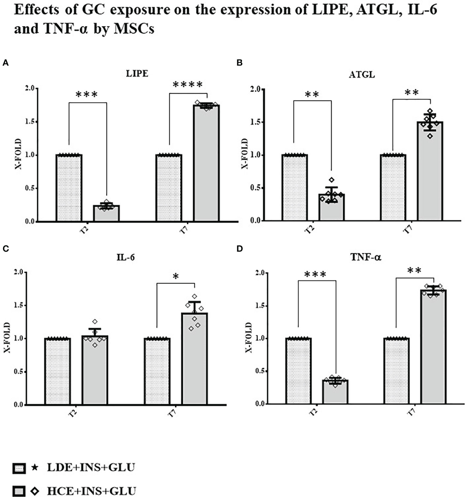
    • LINC 4 met the primary endpoint: Isturisa was significantly superior to placebo at normalising mUFC at the end of a 12-week randomised, double-blind period (77% vs 8%; P<0.0001).

    • Effects of Isturisa were rapid. Over one-quarter of patients randomised to Isturisa achieved normal mUFC as early week 2 and 58% achieved control by week 5.

    • The key secondary endpoint was also met, with 81% of all patients in the study having normal mUFC at week 36.

    • Improvements in cardiovascular and metabolic parameters of Cushing’s disease, including blood pressure and blood glucose metabolism, were seen by week 12 and were maintained throughout the study.

    • Physical features of hypercortisolism improved during Isturisa treatment, including fat pads, facial rubor, striae, and muscle wasting. Improvements were observed by week 12, with continued improvement throughout the study to week 48.

    • Patient-reported QoL scores (CushingQoL and Beck Depression Inventory) also improved during Isturisa treatment.

    • Isturisa was well tolerated in the majority of patients, with no unexpected adverse events (AEs). The most common AEs overall were decreased appetite, arthralgia, fatigue and nausea.

    "These results show convincingly that osilodrostat is an effective treatment for Cushing’s disease," said Peter J. Snyder MD, Professor of Medicine at the University of Pennsylvania. "Osilodrostat rapidly lowered cortisol excretion to normal in most patients with Cushing’s disease and maintained normal levels throughout the core phase of the study. Importantly, this normalisation was accompanied by improvements in cardiovascular and metabolic parameters, which increase morbidity and mortality in Cushing’s disease."

    "These compelling data build on the positive Phase III LINC 3 study, published in The Lancet Diabetes & Endocrinology in 2020,4 demonstrating that Isturisa enables most patients with Cushing’s disease to gain rapid control of their cortisol levels, which in turn provides relief from a host of undesirable symptoms," said Alberto Pedroncelli, Clinical Development & Medical Affairs Lead, Global Endocrinology, Recordati AG. "Recordati Rare Diseases is committed to improving the lives of patients with this rare, debilitating and life-threatening condition. I would like to thank everyone who has contributed to LINC 4 and the LINC clinical programme."

    "I had Cushing's disease for 8 years without being diagnosed," said Thérèse Fournier from L'association "Surrénales". "I was trapped in a vicious circle of missed diagnoses and worsening physical and psychological symptoms that became life-threatening. I lost everything – my job, my house, my partner, my friends – I was isolated. When I finally received my diagnosis, I was relieved because I knew the truth. Since my surgery, I have been learning to live again, enjoying the moments that make a life. I am still on the path to remission, but I feel deeply happy, even if I carry this journey that nobody can understand."

    About Cushing’s syndrome
    Cushing’s syndrome is a rare disorder caused by chronic exposure to excess levels of cortisol from either an exogenous (eg medication) or an endogenous source.5 Cushing’s disease is the most common cause of endogenous Cushing’s syndrome and arises as a result of excess secretion of adrenocorticotropic hormone from a pituitary adenoma, a tumour of the pituitary gland.5,6 There is often a delay in diagnosing Cushing’s syndrome, which consequently leads to a delay in treating patients.7 Patients who are exposed to excess levels of cortisol for a prolonged period have increased comorbidities associated with the cardiovascular and metabolic systems, which consequently reduce QoL and increase the risk of mortality.3,6 To alleviate the clinical signs associated with excess cortisol exposure, the primary treatment goal in Cushing’s syndrome is to reduce cortisol levels to normal.8

    About LINC 4
    LINC 4 is a multicentre, randomised, double-blind, 48-week study with an initial 12-week placebo-controlled period to evaluate the safety and efficacy of Isturisa® in patients with Cushing’s disease. The LINC 4 study enrolled patients with persistent or recurrent Cushing’s disease or those with de novo disease who were ineligible for surgery; 73 randomised patients were treated with Isturisa® (n=48) or placebo (n=25).1 The primary endpoint of the study is the proportion of randomised patients with a complete response (mUFC ≤ULN) at the end of the placebo-controlled period (week 12). The key secondary endpoint is the proportion of patients with an mUFC ≤ULN at week 36.1,9

    About Isturisa®
    Isturisa® is an oral inhibitor of 11β-hydroxylase (CYP11B1), which catalyses the final step of cortisol synthesis in the adrenal glands.2 Isturisa® is available as 1 mg, 5 mg and 10 mg film-coated tablets.2 Isturisa® is approved for the treatment of adult patients with endogenous Cushing’s syndrome in the EU and is now available in France, Germany, Greece and Austria.2

    Isturisa® was granted marketing authorisation by the European Commission on 9 January 2020. For detailed recommendations on the appropriate use of this product, please consult the summary of product characteristics.2

    References

    1. Gadelha M, Bex M, Feelders RA et al. Randomised trial of osilodrostat for the treatment of Cushing's disease. J Clin Endocrinol Metab 2022; dgac178, https://doi.org/10.1210/clinem/dgac178.
    2. Isturisa® summary of product characteristics. May 2020.
    3. Ferriere A, Tabarin A. Cushing's syndrome: Treatment and new therapeutic approaches. Best Pract Res Clin Endocrinol Metab 2020;34:101381.
    4. Pivonello R, Fleseriu M, Newell-Price J et al. Efficacy and safety of osilodrostat in patients with Cushing's disease (LINC 3): a multicentre phase III study with a double-blind, randomised withdrawal phase. Lancet Diabetes Endocrinol 2020;8:748-61.
    5. Lacroix A, Feelders RA, Stratakis CA et al. Cushing's syndrome. Lancet 2015;386:913-27.
    6. Pivonello R, Isidori AM, De Martino MC et al. Complications of Cushing's syndrome: state of the art. Lancet Diabetes Endocrinol 2016;4:611-29.
    7. Rubinstein G, Osswald A, Hoster E et al. Time to diagnosis in Cushing's syndrome: A meta-analysis based on 5367 patients. J Clin Endocrinol Metab 2020;105:dgz136.
    8. Nieman LK, Biller BM, Findling JW et al. Treatment of Cushing's syndrome: An Endocrine Society clinical practice guideline. J Clin Endocrinol Metab 2015;100:2807-31.
    9. ClinicalTrials.gov. NCT02697734; available at https://clinicaltrials.gov/ct2/show/NCT02697734 (accessed March 2021).

    Recordati Rare Diseases, the company’s EMEA headquarters are located in Puteaux, France, with global headquarter offices in Milan, Italy.

    For a full list of products, please click here: www.recordatirarediseases.com/products.

    Recordati, established in 1926, is an international pharmaceutical group, listed on the Italian Stock Exchange (Reuters RECI.MI, Bloomberg REC IM, ISIN IT 0003828271), with a total staff of more than 4,300, dedicated to the research, development, manufacturing and marketing of pharmaceuticals. Headquartered in Milan, Italy, Recordati has operations in Europe, Russia and the other C.I.S. countries, Ukraine, Turkey, North Africa, the United States of America, Canada, Mexico, some South American countries, Japan and Australia. An efficient field force of medical representatives promotes a wide range of innovative pharmaceuticals, both proprietary and under license, in several therapeutic areas including a specialized business dedicated to treatments for rare diseases. Recordati is a partner of choice for new product licenses for its territories. Recordati is committed to the research and development of new specialties with a focus on treatments for rare diseases. Consolidated revenue for 2021 was € 1,580.1 million, operating income was € 490.2 million and net income was € 386.0 million.

    For further information:

    Recordati website: www.recordatirarediseases.com

    This document contains forward-looking statements relating to future events and future operating, economic and financial results of the Recordati group. By their nature, forward-looking statements involve risk and uncertainty because they depend on the occurrence of future events and circumstances. Actual results may therefore differ materially from those forecast as a result of a variety of reasons, most of which are beyond the Recordati group’s control. The information on the pharmaceutical specialties and other products of the Recordati group contained in this document is intended solely as information on the Recordati group’s activities and therefore, as such, it is not intended as medical scientific indication or recommendation, nor as advertising.

    View source version on businesswire.com: https://www.businesswire.com/news/home/20220325005169/en/

    Contacts

    Celine Plisson, MD
    Medical Affairs Director
    Telephone: +33(0)147739463
    Email: PLISSON.C@recordati.com


    • Rie Hagiwara

      Department of Endocrinology and Metabolism, Hirosaki University Graduate School of Medicine, Hirosaki 036-8562, Japan

    • Kazunori Kageyama

      Department of Endocrinology and Metabolism, Hirosaki University Graduate School of Medicine, Hirosaki 036-8562, Japan

    • Yasumasa Iwasaki

      Suzuka University of Medical Science, Suzuka 510-0293, Japan

    • Kanako Niioka

      Department of Endocrinology and Metabolism, Hirosaki University Graduate School of Medicine, Hirosaki 036-8562, Japan

    • Makoto Daimon

      Department of Endocrinology and Metabolism, Hirosaki University Graduate School of Medicine, Hirosaki 036-8562, Japan


    Abstract


    Cushing’s disease is an endocrine disorder characterized by hypercortisolism, mainly caused by autonomous production of ACTH from pituitary adenomas.

    Autonomous ACTH secretion results in excess cortisol production from the adrenal glands, and corticotroph adenoma cells disrupt the normal cortisol feedback mechanism. Pan-histone deacetylase (HDAC) inhibitors inhibit cell proliferation and ACTH production in AtT-20 corticotroph tumor cells. A selective HDAC6 inhibitor has been known to exert antitumor effects and reduce adverse effects related to the inhibition of other HDACs.

    The current study demonstrated that the potent and selective HDAC6 inhibitor tubastatin A has inhibitory effects on proopiomelanocortin (Pomc) and pituitary tumor-transforming gene 1 (Pttg1) mRNA expression, involved in cell proliferation. The phosphorylated Akt/Akt protein levels were increased after treatment with tubastatin A.

    Therefore, the proliferation of corticotroph cells may be regulated through the Akt-Pttg1 pathway. Dexamethasone treatment also decreased the Pomc mRNA level. Combined tubastatin A and dexamethasone treatment showed additive effects on the Pomc mRNA level.

    Thus, tubastatin A may have applications in the treatment of Cushing’s disease.


    Access the PDF at https://www.jstage.jst.go.jp/article/endocrj/advpub/0/advpub_EJ21-0778/_pdf/-char/en


    • Like 1
  23. https://doi.org/10.1016/j.ajoc.2022.101455

    Abstract

    Purpose

    To report the clinical course of a patient with central serous chorioretinopathy (CSCR) secondary to subclinical hypercortisolism before and after adrenalectomy.

    Observations

    A 50-year-old female patient with multifocal, chronic CSCR was found to have an adrenal incidentaloma and was diagnosed with subclinical hypercortisolism. Patient elected to undergo minimally-invasive adrenalectomy and presented at 3 months after surgery without subretinal fluid.

    Conclusions and Importance

    Subclinical Cushing's Syndrome (SCS) may present an underrecognized risk factor for developing chronic CSCR. Further investigation is needed to determine the threshold of visual comorbidity that may influence surgical management.

    Keywords

    Central serous chorioretinopathy
    Subclinical Cushing's syndrome
    Hypercortisolism
    Adrenalectomy
    Retina
    Surgical intervention

    1. Introduction

    Central serous chorioretinopathy (CSCR) is characterized by accumulation of fluid in the subretinal or sub-RPE space, often with consequential visual impairment. Chronic CSCR has been reported as a manifestation of hypercortisolism due to Cushing's syndrome and subclinical hypercortisolism.1,2 However, the latter is often underrecognized owing to its inherently subtle nature and the optimal threshold for intervention based on associated comorbidities remains unclear. Herein we report the clinical course of a patient with CSCR secondary to subclinical hypercortisolism before and after adrenalectomy.

    2. Case report

    A 50-year-old female presented with blurred, discolored spots in the right eye for several months. Her past medical history included mild hypertension treated with amlodipine. Past ocular and family history were noncontributory.

    On exam, Snellen visual acuity was 20/50 OD, 20/25 OS. Her pupils, intraocular pressure, and anterior segment exam were within normal limits. Dilated fundus exam revealed bilateral, multifocal areas of subretinal fluid and mottled pigmentary changes (Fig. 1A). Optical coherence tomography confirmed areas of subretinal fluid and other areas of outer retinal atrophy (Fig. 1B). Fundus autofluorescence revealed areas of hyperautofluorescence that highlighted the fundoscopic findings (Fig. 1C). Fluorescein angiography showed multifocal areas of expansile dot leakage (Fig. 1D). Altogether these findings were consistent with multifocal, chronic CSCR.

    Fig. 1
    1. Download : Download high-res image (1MB)
    2. Download : Download full-size image

    Fig. 1. Multimodal imaging of bilateral multifocal central serous chorioretinopathy. Fundus photographs reveal multifocal subretinal fluid and pigmentary changes (Fig. 1A). Optical coherence tomography demonstrates subretinal fluid and outer retinal atrophy (Fig. 1B). Areas of hyperautofluorescence highlight the fundoscopic findings of subretinal fluid (Fig. 1C). Fluorescein angiography showing multiple areas of expansile dot leakage (Fig. 1D).

    On further clinical follow-up, an adrenal incidentaloma (AI) was detected when the patient underwent imaging for back pain. Subsequently she saw an endocrinologist; she had a normal serum cortisol, but low ACTH and an abnormal dexamethasone suppression test. This led to a diagnosis of subclinical hypercortisolism and provided a reason for her hypertension and chronic CSCR.

    Since the blur and relative scotomata interfered with her daily activities, she elected to try eplerenone, which yielded a moderate but suboptimal therapeutic response at 50 mg daily. While contemplating photodynamic therapy, in discussion with her endocrinologist, the patient opted to undergo minimally-invasive adrenalectomy. At last follow-up 3 months after surgery and 6 years after her initial presentation, she has been off eplerenone and without subretinal fluid (Fig. 2).

    Fig. 2
    1. Download : Download high-res image (1MB)
    2. Download : Download full-size image

    Fig. 2. Optical coherence tomography imaging at presentation and at last follow-up 3 months after adrenalectomy. There is a significant improvement in subretinal fluid in both eyes, though outer retinal irregularity remains.

    3. Discussion

    CSCR has previously been associated with many risk factors including exposure to excess steroid. A recent study analyzing a nationally representative dataset of 35,000 patients found that patients with CSCR had a higher odds of Cushing's syndrome (OR 2.19, 95% CI 1.33 to 3.59, p = 0.002) than exposure to exogenous steroids (OR 1.14, 95% CI 1.09 to 1.19, p < 0.001)1 Our case highlights the importance of thorough medication reconciliation and careful consideration of comorbid conditions in patients with chronic CSCR.

    In recent years, subtle endogenous hypercortisolism, termed subclinical Cushing's syndrome (SCS), has been of particular interest in the endocrinology literature because it can be a challenging diagnosis and the most appropriate management remains controversial.3 In general, SCS is comprised of: 1) the presence of an adrenal incidentaloma or mass, 2) biochemical confirmation of cortisol excess, and 3) no classic phenotypic manifestations of Cushing's syndrome.4 Since adrenal incidentaloma has an estimated prevalence of 1–8% of the population,5 it is quite possible that SCS is an underrecognized risk factor for CSCR.

    SCS may potentiate metabolic syndrome and osteoporosis; studies have found that surgical resection of adrenal incidentalomas improve weight, blood pressure, and glucose control. Consequently, some authors recommend those with SCS-associated comorbidities be considered for resection.6 An important consideration in these patients is how visual comorbidity factors into intervention. In our patient's case, the recurrent CSCR, hypertension, and increased risk of metabolic syndrome were sufficient reasons to elect to have surgery.

    4. Conclusion

    In summary, SCS is a condition of subtle cortisol dysregulation that may represent an underrecognized risk factor for chronic CSCR. Further investigation is needed to determine the threshold of visual comorbidity that may influence surgical management.

    Patient consent

    Consent to publish the case report was not obtained. This report does not contain any personal information that could lead to the identification of the patient.

    Acknowledgments and Disclosures

    Grant support was from the J. Arch McNamara Retina Research Fund. The following authors have no financial disclosures: RRS, AS, AC All authors attest that they meet the current ICMJE criteria for Authorship. No other contributions to acknowledge.


    References

    © 2022 The Authors. Published by Elsevier Inc.
     
    • Like 1
×
×
  • Create New...