Jump to content

MaryO

~Chief Cushie~
  • Posts

    8,082
  • Joined

  • Last visited

  • Days Won

    556

Posts posted by MaryO

  1. From another patient...

    Adapted from a thread on the message boards.

    I'm going to try to keep all of my post-op BLA updates in this thread. I am hoping it will eventually show positive progression and be a realistic and inspirational thread for others.

    Today I am two weeks post-op BLA. So far, no scares. I am on 30/20/20 of hydrocortisone and weaning by 10 mg every four days. I am sleeping a good bit during the day and resting a lot to get my strength back. If I am upright too long my abdominal area aches and I get fatigued, sometimes it still aches even if I am not upright. My nighttime sleep has been good. I'm waking up only 1-2 times to go to the bathroom (I think the meds are making my bladder more active than normal), but otherwise am sleeping through the night which is a huge change from Cushing's. I am hoping this is due to being Cushing's-free rather than just due to the pain meds I am taking right now. We'll see if this lasts as I drop the pain meds and hopefully the nighttime urination will let up as the hydro levels drop.

    Also, and I don't think its my imagination, but some of my stretch marks are getting lighter. In particular, the ones that formed on my legs after my pit surgery. This is a positive sign! I showed my mom and hubbie and they could both see the change too. Unfortunately, my hump is bigger right now than pre-BLA and my cheeks are still pretty red, but I bet this will change as I wean down.

    No weight changes as of yet, but not expecting any because I am still on such a high dose of hydrocortisone. I was 198 the day of the BLA, which was about 15 pounds heavier than the day of my pit surgery seven months ago. For the first week and a half after the BLA I was really, really bloated - and it was all in the stomach area. Most of this bloating has gone down in the past two days.

    I've watched my calorie intake throughout the battle with Cushing's but I started a food journal yesterday just to make sure I am keeping myself in check. I'm eating 1500 calories a day. I noticed right away that I haven't even been eating that much on a normal basis because I actually had to eat more than normal to meet the 1500 calories. So that's also a good sign that watching my food intake won't be a big change in order to help the weight to come off.

    So that's really the main things happening right now. Just taking things slow and steady and trying to have realistic expectations!


    I had my six week post op appointment in Seattle last week. My weight is actually up (204, I was so bummed that I went over 200). But Dr. L said not to worry, that its normal to gain weight during the weaning process. I am still on a 1500 calorie a day diet and will stay there until I start to see weight loss and then I'll reassess calories then. I was advised that weight will probably start to fall off when I'm six months out from surgery, so I am trying not to focus too much on it or get discouraged.

    My nighttime sleep is weird right now. I'm not waking up all night long like I was before the BLA, but I can't fall asleep at night either. I lay awake until 1 or 2 am. On the flip side, I am waking up at a normal hour - 7 am.

    I just started weaning to 20/5 of hydrocortisone. It is pretty rough. The wean from 20/10/10 to 20/10 was hard, but this is even harder. Feels like the flu, achey all over, headaches, sleeping all day (which probably doesn't help me fall asleep at a good time at night!). I have realized that I must take the wean really slowly now. The goal is to get to 20 or maybe just a little less and hopefully that dose will work for me.

    In other news, I got the path report back on my adrenals - my adrenals combined weighed in at 30 gm (normal combined weight of adrenals should be between 8-12 gm). The left one was twice the weight of the right one, and they had "subtle vague expansion" and "microscopic nodularity" suggestive of adrenocortical hyperplasia.

    So I am feeling very validated at this point and I know I made the right decision to have the BLA.


    I'm just past the 3 month post-op anniversary. Some things are better and others are still the same. But more positive changes than anything.

    We'll get the negative overwith first - my stretch marks did an about-face and actually got a lot worse about a week after I got down to my physiological dose (20mg). Dr. L said not to worry, they're just showing up now due to past cortisol exposure. Still, they're pretty bad. So I was disappointed in that. My period still has not come back since I had the pit surgery. All my hormones are fine except the progesterone, but progesterone supplements are not helping. We're taking a wait and see approach to give my body some time to get over the shock of two major surgeries.

    Other than the stretch marks, the other Cushing's symptoms are slooooowly getting better. I am sleeping pretty well now, able to fall asleep in the evening and sleep until 5:30 or 6 am until waking up. Its a lot better than waking up at 3 am every night for sure. My hump looks a little smaller (I think). My cheeks are still red, but my face is maybe slightly slimmer (I think). I've lost six pounds (with 80 more to lose), but am still heavier than I was the day of my BLA. Although my stomach doesn't pooch out so much anymore, so I look less pregnant. My hair has stopped falling out.

    I have been working out for a few weeks now and my strength is really starting to improve. Walking is very good for me. I'm eating about 1200 calories a day and dropping down this low seemed to jumpstart some weight loss. I am hoping it continues. I'm certainly doing nothing food-wise to keep the weight from coming off.

    I was tested for insulin resistance and any thyroid problems - everything came back normal. My ACTH was super low when it was last checked - came back at 3. (yay!!!)

    I went back to work 80% time this week. I'm trying to work short days but my work is very demanding so I will probably have to end up working 4 days a week and taking off one day a week to rest. I am very tired at the end of the work day. Exposure to stress is also very hard on my body - the stress I have encountered this week has caused nausea, diarrhea and one time I had to take straight to the bed and lay down all evening. Right now I feel like I am not as sharp and "on the ball" as I used to be.

    My sinus infections from the pit surgery keep continuing about every 6-8 weeks. I've probably had at least 4-6 sinus infections since March. At the last visit to the ENT doc, she said she thinks I have a deviated septum from the pit surgery and may need surgery to correct it. I have a CT scan on Tuesday so hopefully we'll know more soon on whether I am having another surgery.

    But overall, I just feel better. The Cushing's symptoms are slowly fading, but at least we're going in the right direction. I am trying to be patient, and trying to remain motivated. I have to admit I am becoming very impatient for the weight to come off and still harbor fears that it won't. I am considering throwing my scale in the spare bathroom and forgetting its existence for a while.

    I hope my next update will have tons more good things to share.


    So I am 6 months post BLA today. Yay! This is the magical date - things are supposed to start changing more quickly after passing this milestone. Here's the stats so far:

    20 mg hydrocortisone per day

    0.1 mg florinef per day

    Had thyroid checked in January - fine

    Had glucose tolerance test in January - fine, no insulin resistance

    Dr. L didn't think I had GH issues at my 3 month post op appointment

    Estrogen and all other female hormones fine except progesterone, taking prometrium to try to induce period with no success so far

    I started losing weight at the end of January through mid March. I lost 10 pounds. But now, I haven't lost any weight in over a month and I've actually regained two pounds. I am exactly what I weighed the day of my BLA now.

    I've been working out 90 minutes 4-5 days a week (elliptical machine and weights). I'm eating net 1200 calories a day (which means I am actually eating more than 1200 because of all the exercise I am doing) and very closely tracking calories on livestrong.com.

    I have to say I am very frustrated at this point because I'm working so hard and not losing weight. I'm going to bring this up with Dr. L at our six month post op appointment. If some other BLArs could chime in and tell me what to expect for the next six months, I would greatly appreciate it. Just starting to get a little nervous here.

    As far as the Cushing's goes, I have more energy and I am sleeping better. Most nights I sleep through the night and if I wake up, its only once and closer to 6 am than 3 am like it used to be. Hump is still there, hasn't gone away but is a tad smaller. Hair stopped falling out a while ago and has stayed just fine, no relapse.

    The stretch marks (which had gotten worse after the BLA) are getting much better, at least the ones on my legs. Those are noticeably better. I've gotten comments that my face is slimmer and I look like I've lost weight. I've gone down from third trimester maternity pants to second trimester pants. That is some progress because I look less pregnant.

    Since my last update, I have had three severe episodes of AI. All occurred late at night following a week of being pushed beyond my medical restrictions at work. 32 hours a week seems to be a good balance though, more than that causes me to be really tired and at risk for AI.

    I'll close out with a great accomplishment story. Hubbie and I went on a cruise to Mexico and Belize. I was able to do a hike through the jungle (which was relatively level, for a jungle). But the best part was when we got to a clearing and saw the Mayan temples. You could climb one that was about 45 meters high with very steep stairs to the top. Of course my hubbie was the first in the group to take off up the temple. The stairs were so steep they had a rope that came from the top all the way to the bottom to pull on to help yourself get up. This was the type of thing that, before Cushing's, I would have been right there with my husband.

    He was about halfway to the top when I said, "Heck, I'm going too." Probably shouldn't have, but I took off up the temple stairs after him. I climbed up and up in the Belize heat and made it to the top. The view was rewarding, but the greater reward was that I could DO it. I was getting part of my life back - the adventurous, hiking, exploring, running-being-free part.

    That part was the best. :D


    I am weight training, 4x a week for 20-25 minutes per session, on machines, not free weights. I want to make sure I'm not getting the wrong form. I am pushing myself, sometimes only able to do five reps at a time because of the heaviness of the weight. I do a total of 3 sets of 10 reps per exercise. I'm doing upper body and lower body on different days, so 2 days a week of upper and 2 days of lower, never back to back.

    My diet is good. Short of starting to cut out food groups altogether, there's not much else I can do. I eat either whole grain cereal and skim milk or two boiled eggs and skim milk for breakfast. My mid-morning snack is fruit - usually a cup of red grapes or an apple. Lunch is a salad with grilled chicken or a Lean Cuisine or Smart Ones that has fish as the main entree - nothing over 300 calories. Mid afternoon before working out I have fat free yogurt or 30 almonds. Dinner is normally something like stuffed green peppers or chicken fajitas - usually about 500-600 calories.

    I weigh/measure just about everything . . .


    I'm 7 months 9 days post op today. The weight has changed a little, but only a little. At least its going down and not up, but I admit I am frustrated with my progress. I told Dr. L about my intense working out and dieting and he suggested I wean some more. So I weaned to 17.5 mg of hydro first and then down to 15 mg. I've been at 15 mg for 3 weeks now. The past week I started to see some progress - I lost 2.5 pounds this past week, so now for a weight loss total of 12 pounds since January. This is in conjunction with a 1200 calorie a day diet. I've now gone to a combination Zone diet (30 protein, 30 fat and 40 carbohydrates) and sort of low glycemic index - just as little sugar as possible. So I am eating a lot of bran, salads, chicken and fish. I've instituted a "salad for dinner two times a week" rule at home, which my lovely, Southern-food loving husband has generously agreed to go along with.

    May was not as good a workout month as March and April. However, we did a one week vacation with LOTS of activity - hiking every other day for 2-3 miles, and we did a 14.5 mile bike ride at the end - it was mostly flat, but still! That was a long way and I was so proud of myself when I finished it. It was a struggle, but I did it.

    I also got my period for the first time in over a year in May. I wonder if it is related to weaning to 15 mg? We will see if it comes back in June . . . .

    Other things have gotten much better - sleeping well through the night, feeling better in general. My hair was much better until the past two weeks or so when I've seen more of it coming out in the shower than normal (what is that all about?!?!?) but not falling out on a regular basis like it was with Cushing's there at the end.

    I am losing some inches for sure and I don't look as pregnant as I used to, I was able to drop from my maternity black dress pants to a size 18 pants (although the legs are still huge). I am still in my maternity jeans but I did go from trimester three to trimester two! I picked up prescriptions at the pharmacy today and my pharmacist said "You are looking great!" So that was nice to hear :)

    So all in all, very very slow but seeing some progress now. I think its going to be a very long process with lots of hard work and healthy eating. It may take some more weans too, depending on whether I hit a wall again.

    I know you and a lot of other BLA-ers are struggling right now. Its hard. I feel good right now because the scale went down this week and I've seen some physical changes in how my clothes are fitting. I know its depressing when you are not seeing that. But for you and everyone else, just hang in there. Do as much physical activity as you can, and at least control your diet, because that is in your control. I know we're told the weight is supposed to come off on its own but I can tell a difference when I'm eating right and when I'm not. At least for me, I think it does help with the weight loss. At least psychologically I know I'm doing everything I can to make it come off.


    By way of a mini update, I have lost another 2 pounds since I posted three days ago. This is getting exciting! And its not just water, you know the size 18 blank dress pants I just talked about in my last post? They are now TOO BIG!!! A friend of mine hadn't seen me in two weeks and she was shocked today just to see the changes that have happened in two weeks. It really is noticeable.

    Ok, hope I am not jinxing myself. When I update again in a few weeks hopefully I can report a very large weight loss and even more changes!


    So, today I am 9 months post-op BLA. Its been almost two months since my last update. There's been a lot of developments:

    - In July, I got the results of my bone density scan: I have osteopenia and a severe vitamin D deficiency. I am now on 1200 mg of calcium a day and 50,000 IUs of Vitamin D a week.

    - Hair is doing great! Not falling out, shiny, less frizzy.

    - Energy is ok. Work is wearing me out, still working me beyond my medical restrictions, but I am supposed to be moving into a new job at the end of next month that will hopefully take care of some of that. I tend to get sleepy during the day and stressful days make me weak. I've also started waking up in the middle of the night again (NO!!! Why is this happening?!?!?) and there for a while I was waking up to pee in the middle of the night again. I wish that would stop because I was enjoying sleeping all the way to the morning.

    - Stamina is great. I did a two-hour workout last week (weights and cardio) that was intense and awesome. I was so proud when I was done. I am considering returning to kickboxing in a few months if my Vitamin D levels go up and I have some confidence that my bones have gotten stronger.

    - The weight is stalled out. I have lost 16 pounds now, but I haven't lost a pound since mid-June. I weaned to 10 mg of hydro about three weeks ago and no results even with doing that. I don't feel comfortable going any lower than that. Still at 1200 calories a day and low glycemic diet, heavy on protein, very little to no bread or cereal products. Husband and I met with reproductive endo here in Atlanta today (who I love!) and he expressed concern. My thyroid and insulin resistance tests are normal but he's putting me on some Synthroid and Glucophage and some Prometrium. When I got my period in May the weight was just falling off . . . he thinks its PCOS-like issues and this combo of meds might help. So we're going to try that and see how it goes.

    - Stretch marks are much, much better - I noticed a marked difference after I weaned to 10 mg hydro. BLA scars are lightening too, especially with help of some new special cream from my dermotologist.

    - Haven't gotten my period again :( Boo. Hopefully the above cocktail will help with that.

    - Had lasik surgery!!!! I love it. I did stress dose 30 mg extra for that. I did just fine.

    So, positives yes but still very bummed about the struggles with the weight. I am hoping the new medicines will give me some results. I also feel like I've become more emotional lately because I'm tired of eating lettuce, spinach and egg whites (yes, that makes up a large portion of my diet) and working out and getting no relief. I hate being emotional and moody and feeling like I just can't take it anymore. So I certainly do have those days. But thankfully they are just days - usually just one - and it passess and the next day I'm back in the battle. Because really - what else can you do?


    I'm 10 months post-BLA today. Unfortunately, this update is not going to be as positive as some of my past updates.

    The weight loss stands at 20 lbs now. I did start on Metformin and Synthroid at the beginning of August. I lost five pounds right away the first week, and then the weight loss stopped and I have gained back one pound. Nothing else has happened since then (despite doubling the dose of Metformin).

    I can't deny that I have become extremely depressed. Its been building for several months now. Its not just having the extra weight, but the weight keeping me from what I want to do - principally, have a baby. I've just lost interest in so many things and I am very down, despite the progress I have made in other areas of recovery.

    I have discussed this with both Dr. L and my reproductive endo. I am going to Seattle in two weeks and we're doing a round of labs and a growth hormone stim test. GH deficiency would explain a lot of things - the large amount of weight around the middle, the Cushie-like shape I still have. I still have a bit of a hump too.

    My reproductive endo is re-testing all my thyroid hormones, estrogen, progesterone and a few others soon as well.

    I am beginning to suspect I have slowly been becoming hypo-pit. Or perhaps hypo-pit in an intermittent way. I have no menstrual cycle anymore. I have ostepenia. I have energy to do stuff but then I get exhausted and sometimes it takes me days to recover. I have hot flashes, memory issues, loss of libido and insulin resistance. And, again, super slow weight loss that seems to go up every time I eat anything other than raw vegetables. I also have on and off DI.

    So, I guess I am just at the end of my rope. I hope that someone can fix me. Because something is still clearly wrong.


    I'm now 10 1/2 months post-op BLA. I just completed a visit to Dr. L in Seattle. I did the GH stim test and labs for thyroid, ACTH and some other things.

    As I suspected, I do have some continuing issues - I am severely GH deficient. I didn't stim above 0.9 during the entire stim test. I'll be starting on GH as soon as possible.

    My thyroid numbers are all in the normal range but they are low normal. We're upping the Synthroid to 125 mg per day.

    My MRI was clear - no new tumor (yay!) and my ACTH was 40. So that is all good. I feel hopeful that I am doing good in some areas and now we have identified the areas that are causing me problems.

    I also had estrogen, FSH and LH tested today. I am hoping to find out if I am deficient there even though I haven't been in the past - I have a suspicion the estrogen may be low now.

    So, we'll see where we stand in a few months when this medicine has had some time to kick in.


    Today is the one year anniversary of my BLA. I am doing well. I'll update here and post a separate 1 year post-op BLA thread so those who don't follow here can be encouraged by my, dare I say it, success story?

    The past month and a half I have seen some significant improvement. Here's the breakdown:

    Medicine every day:


    12.5 mg of hydro (all taken in the morning)

    0.1 mg florinef

    1500 mg Metformin at night

    125 mg of levothyroxine

    Calcium pill and daily multivitamin

    Progestrone pills on days 1-10 of each month

    To start 0.2 mg of Genotropin in next few weeks


    Energy: The thyroid medicine has helped a lot with energy. My thyroid numbers were all normal but just a bit on the low normal, so the docs didn't think I needed meds. But I did, it has helped a lot. I am still tired but I am a lot better than I was. My GH is supposed to arrive today (yay!) so that should also help me on my path to recovery.


    Weight loss: I haven't really lost weight in the past few weeks but inches, oh my! I have lost inches. I have gotten tons of comments from friends, family, coworkers, etc on the change all over - face, body, etc. I am now down to a size 14. That is down from being mostly in maternity clothes and barely squeezing into a few size 18 pants a year ago. No more maternity clothes for me (for now!). Its so nice to be shopping in the regular clothes again. I have gone a bit crazy buying some new things - skinny jeans, sweaters, ballet flats, boots. I am all decked out for fall in the latest styles. It feels so good to be stylish and to have choices again.


    The pregnancy look is gone. No more comments on when I am due or what sex the baby is. That is an awesome feeling. I've lost 20 solid pounds, some days a little more but it seems to always go back to that 20 number. I am trying not to weigh too much until the GH has a chance to start working.


    Stretch marks: My stetch marks have really done some fading. Somedays they are more noticeable than others, but they are so so so much lighter than they were.


    Hump: My hump is much smaller - its barely there at all now, I probably see it only because I am paranoid. But I have no issue wearing tank tops or anything that shows the back of my neck.


    Hair: My hair has grown long and thicker than it used to be. Much less oily! I don't have to wash it every single day now. I can put it in a ponytail on the weekends and it looks cute and not greasy.


    Sleep: I am sleeping great. I sleep all through the night and don't wake up anymore. That has become very consistent, which is a wonderful thing. I still feel tired though because of the GH but hopefully that will improve.


    Activities: I work 32 hours a week. This works well, it gives me an extra day a week to rest and recover from the work week. I exercise often, I went back to kickboxing this month which is kicking my butt but I need to rebuild muscle. Its also nice to get back to your old hobbies.


    Attention and Memory: This isn't always as great, hoping the GH will help. I don't focus as well as I used to or catch spelling details at work like I used to. Sometimes my memory is sharp and other times I forget something someone just told me or how to spell a word or the names of objects (or even people at times). Again, hoping GH will help here.


    Female stuff: No period still, progesterone is not really helping. I think if the GH doesn't help here, we're going to move on to estrogen therapy in a few months. We're hoping to start trying for a baby at the end of next year.


    Other health issues: High blood pressure went away immediately after the BLA. I never did have a blood sugar problem so no issues there. I do have osteopenia which we are working on with more calcium and Vitamin D and weightlifting exercies. Again, hoping GH will help here.


    Emotional: Really doing much better. I was getting really depressed for a while there, about 7-10 months post op. Even though I thought my expectations of recovery were reasonable, I was frustrated with my progress. I had hoped to lose more like 30 or 40 pounds in my first year. But, I found out I had other issues (thyroid and Gh deficiency) that were messing with that goal. So I only got halfway there but it wasn't my fault. I also thought the weight would "fall off" more than it has, but it hasn't. I've had to diet and exercise hard for every pound lost. Don't know if that's normal or if its just me or because of the thyroid/Gh issues we are still working on. But it helped me to know that at least there was a medical reason for my frustrations!


    But overall I am really doing well. No one who meets me for the first time has any idea that I've been sick. I recently started a new position with my same employer (which has been going well) and I met my new team and everyone commented on my "glowing skin" and "happy nature." I have no regrets about the BLA.


    My advice to anyone considering it or just having had the BLA is: patience, patience, patience. Realistic expecatations. Then, hard work on controlling your diet and being physically fit. Do everything you can towards getting better, and then if time and hard work don't pay off, don't hesitate to detail your hard work and patience to your doctor and tell them to find out what else is holding you back.


    I hope this is an inspiration to anyone out there who is struggling right now.


    Wow, I am way overdue for an update! I've been out working, having fun and living my life!

    Its been a little over three months since my last post here. I am now 1 year, three months and ten days post op BLA. Here's the breakdown of where I am now:

    Medicine every day:


    12.5 mg of hydro (all taken in the morning)

    0.1 mg florinef

    1500 mg Metformin at night

    150 mg of levothyroxine

    Calcium pill and daily multivitamin

    Progestrone pills on days 1-10 of each month

    0.2 mg of Genotropin 7 days a week (started in November)


    Energy: I am doing great here. I am tired sometimes and traveling or working long hours wears me out, but I have limited that in my life with my new job. At my new job I am working full time now, 40 hours a week. I also work out 5-6 days a week now at very energetic things like kickboxing (with punching bags) or the elliptical machine.


    Weight loss: I didn't lose anything between my last post and the end of December. When my thyroid medicine was raised to 150 mg at Christmas, combined with continuing my workout and diet, I really started to see results. I have lost 10 lbs since then, for a total of 31 lbs now. Still, I am not losing at the rate I should be for the math of the intake/output of my diet and working out. We are working on that, possibly some more meds to come soon. But it is much, much improved! I have about 35 pounds to go until I am at a good weight for me. Ideally I'd like to lose 45 more but 35 more would be a healthy weight for me.


    The best thing I did was have my husband hide the scale. I only weigh every six weeks now. Now I can focus on the process and not focus on how hard it is to get the scale to go down or get depressed when it doesn't budge. I am now in size 12 clothes. I was a 6-8 before Cushing's, sometimes I could wear a 4. I have a few more sizes till I can wear most of the clothes in my closet.


    Stretch marks: This is about the same since my last post. My stetch marks are almost all white. Somedays they are more noticeable than others or pinker than usual, but they are so so so much lighter than they were.


    Hump: Same as last post - much smaller and hardly there at all.


    Hair: Same as last post - doing great.


    Sleep: Same as last post - doing great.


    Activities: Like I said above, working 40 hours a week, kickboxing probably 3-4 times a week, other days I work out on the elliptical machine and lifting weights. I go walking or hiking with my husband on the weekends if the weather is nice, but this low impact working out didn't do much for the weight loss. The kickboxing has really helped.


    Attention and Memory: This is about the same as last time. I can't tell that it has improved all that much. I forget things (like reminding my husband to do something when he has asked me to remind him) all the time.


    Female stuff: No period still, had blood drawn for estrogen today. Will see whether I am going on that or not.


    Other health issues: High blood pressure went away immediately after the BLA. No return of that, blood pressure is very good. I never did have a blood sugar problem so no issues there. I do have osteopenia which we are working on with more calcium and Vitamin D and weightlifting exercies. Again, hoping GH will help here. My sinus issues have really escalated and just never got better after surgery. I've had a persistent sinus infection for two years. I have mold and some other bacteria in there that countless treatments have not killed. I am having the sinuses washed in a surgery at the end of the month and am now working with an infectious disease doctor to try to kill it. Its too gross to talk about!


    Emotional: I am really doing well. The recent weight loss has really pleased me. I don't think I am at the maximum improvement for my weight loss rate yet, but hopefully we are getting there. I am pulling out old clothes I haven't worn in years out of my closet. I now officially weigh less than my husband for the first time in over two years, which is also wonderful.


    So, that is about it for now. I will update again when there are more developments!


    P.S. - Notable fitness accomplishment! Six weeks ago throughout a kickboxing class I could do about 5 girlie push-ups (on knees). Last night at kickboxing class I did a total of 5 interspaced intervals of 10 for a total of . . . 50 push-ups!


    The power of regular exercise and GH unites!


    Today is the two year anniversary of my BLA. It is hard to believe that much time has passed. I can say with 100% confidence that I am doing so much better and that the BLA was the right thing for me.

    I'll update this along the same lines as my one year update, just in the name of consistency:


    Here's the breakdown on my meds:


    Medicine every day:


    7.5 mg of hydro (all taken in the morning)

    0.1 mg florinef

    125 mg of levothyroxine

    Calcium pill and daily multivitamin

    Prenatal vitamin

    0.6 mg of Neutropin (next month will be going up to 1 mg Neutropin)

    Birth control pills (formerly was taking 0.2 mg estrogen supplement and progesterone on days 1-10 of month)

    2 tsp. of Royal Jelly and Bee Pollen in honey daily

    Flonase


    Energy: The thyoid and GH have helped a lot in this area. I could still use a little help because my GH is still very low, but I really am doing great anyways. Getting the thyroid dose right has been a battle, but I think we finally found the right dose.


    Weight loss: I have now lost a total of 34 lbs, down from high of 206 to 172. At 5'5 I am a normal size 12 and its great. I look and feel like a normal person again (my mom even says I am "skinny" but I don't know about that!) I am losing more inches now than I am weight. This is partly due to the need for higher GH, and partly because I am not doing the hard working out and strict dieting because my hubbie and I are working on Baby #1!!! I have fought hard with diet and exercise for every pound lost - nothing has come off easily for me.


    So, the pregnancy look may be back in a few months, but this time it will be because I am actually pregnant


    Stretch marks: i barely notice them at all now. My BLA surgical incisions have done a great job fading as well. I don't know if a bikini is ever in my future, but if I am in that great shape again I might wear one around family and friends despite the scars.


    Hump: Gone


    Hair: Doesn't fall out anymore, its grown long and thicker, less oily. I think the prenatal vitamins have helped in that area too.


    Sleep: I sleep like a baby every night. I have been for a while. No more waking up, no more problems falling asleep. I do need more sleep than most people, and I am wondering if this is still due to the GH deficiency.


    Activities: I work 40 hours a week and have been since probably the beginning of the year. I've been in my new job now for a year and it has been such a blessing. The reduced stress makes it possible for me to work full time.


    Attention and Memory: This is the same as last year. It isn't always as great, hoping the GH will help. I don't focus as well as I used to or catch spelling details at work like I used to. Sometimes my memory is sharp and other times I forget something someone just told me or how to spell a word or the names of objects (or even people at times). Again, hoping GH will help here.


    Female stuff: I need a combination of estrogen and progesterone in order to have a period. This still does not cause ovulation. So, we are using fertility mediation to induce ovlutation in order to get pregnant.


    Other health issues: Same - High blood pressure went away immediately after the BLA. I never did have a blood sugar problem so no issues there. I do have osteopenia which we are working on with more calcium and Vitamin D and weightlifting exercies. Again, hoping GH will help here. I had some problems with my gums recessing and GH and better female hormones have helped there too.


    My sinus recovery from the pit surgery has really been hard, perhaps my worst problem of all. I had surgery in April to correct the deviated septum caused by the pit surgery. I have been on and off antibiotics like crazy. I was a habitual Neti-Pot user with no improvement. Finally, I started using those spray irrigation cans twice a day, combined with Flonase to lessen the mucus, and that has helped for the past 8 weeks. I've seen my best improvement since by pit surgery 2.5 years ago. So let's hope that continues.


    Emotional: i am really very happy in my life. I am not depressed anymore and so many good things are happening to me. I thought I would have lost more weight by now but solving the GH deficiency has really taken a long time (and its still not resolved yet). Also, its important when using fertility medications to take it easy and not eat a restrictive diet, so I've been focusing more on the things to help us have a baby more than weight loss. I pray we are successful in having kids, and I will get back on the weight loss track after that. But its so positive to shop in normal clothes and not even be considered plus size anymore!


    My relationship with my husband is great, unlike so many relationships we pulled together through Cushing's and it made us stronger.


    I am still working to have patience in the recovery and just to recognize that it goes on for a long while. I am two years out and things improve all the time. Its just good to be in a place where things are getting better rather than worse, and I can eat a piece of pizza and not gain 5 lbs, and actually be out enjoying life. Hopefully this next year I can tackle motherhood too :)


    So far the BLA hasn't been the doctor's concern at all for getting pregnant. The problem has been the lost pituitary hormones from the pituitary surgery. If I get pregnant, there will be focus on keeping the cortisol levels appropriate, as they rise naturally during pregnancy and my meds will have to do that. But I would guess someone who did not have a BLA and had pit surgery and is still reliant on cortisol replacement would have the same issue.

    There is also some focus on cortisol dosage if I have morning sickness in order to avoid AI, but the docs don't seem too concerned and feel confident we can handle it.

    PS- this was why I chose the BLA over the second pit surgery, although I lost ovulation with the first pit surgery, so fertility meds were unavoidable.


    Wow, I can't believe it, but yesterday was the three month (year!) anniversary of my BLA. I am doing awesome. Honestly, I hardly come on the boards anymore but I am trying to update this thread at least yearly in the hopes that it will help someone. Here is an update on the areas I have traditionally noted:

    Here is the breakdown on my meds:


    Hydrocortisone: There is controversy here. Technically, I am supposed to be taking 7.5 mg a day as the minimun. But its too much for me. I can live without it. I have gone months living without it. Every now and then if I feel bad I will take 5 mg. The rest tissue testing I have done at Vanderbilt has been negative for rest tissue, but clearly something is going on. I've also lost weight being off of the hydro.


    Fludrocortine: Again, I am supposed to be on 0.1 mg a day, but I can live without it. I may need to take a pill once every three or four weeks, but otherwise I am fine right now.


    125 mg of levothyroxine


    0.6 mg of Nutropin


    Calcium, multivitamins


    Vaginal progestrone suppositories - these, combined with no hydro, have really helped the weight peel off


    Estrogen patch - same, have helped the weight come off, because oral meds interfere with GH


    Energy: I am doing great, working 40+ hours a week. Sometimes pain in my knees interferes with my workouts, but otherwise I am doing fine as long as I get 8-9 hours of sleep a night.


    Sleep: doing great, fall asleep and usually no waking up.


    Weight: Awesome, i made huge strides this year with the change in the manner in which female hormones are put into my body and going off the hydro. I lost 30 lbs this year, and I have now lost 64 of the 66 I gained with Cushing's. I am wearing a size 6 or 8 depending on the store.brand. Before Cushing's it was a 6 or a 4. But after all this, I consider this a huge success story :)


    Hump: still gone, and man, do I have collar bones now!


    Hair: still doing great


    Stretchmarks: Not very noticeable, and the BLA scars are very faint. A friend of mine (who saw them after surgery) saw them yesterday for the first time in three years and was amazed.


    Other health issues: High blood pressure gone, high cholesterol gone, sinus issues are still present but I have now had two sinus surgeries. I may be going into IV antibiotic therapy next.


    As far as Baby #1, I had a miscarriage in March but we determined the reason was not Cushing's related and another fixable problem I had. So, hopefully in the future I will get my bundle of joy. I am much happier that I am now at a healthier weight for it (142 lbs at 5'5).


    Again, so happy I made this decision. I consider myself fully cured, and I am still losing weight now without much effort. Before this year, I was fighting against unbalanced hormones and while I did lose 34 lbs during that time, it took me two years! This year, only one year and 30 lbs. Balanced hormones are totally necessary, but you also need the proper manner of distribution to your body, and healthy eating and exercise.


    I hope this helps someone along their Cushing's journey! There is hope and light at the end of the tunnel.


    Time for another update I guess.  I am continuing to do really well.  I am down to 118lbs at 5'5.  I am a size 4, sometimes a size 2.  I never thought I would see any of those numbers again, but here I am!  I am feeling good in pretty much all respects.  The only bad thing is that I seem prone to sports injuries. I don't know if its because I'm post-Cushings or if its just me.  I've been in physical therapy twice in the past year now.  But I am continuing to be active and have a healthy lifestyle.

    I hope everyone is doing well.  As always, let me know if you have questions about anything in my journey.


    Wow I didn't realize how long it had been since my last update! So much has happened in the last 8 years. I've gotten divorced and since remarried. The biggest update is that I am pregnant from IVF and expecting my first child. There was always a question after my pituitary surgery on whether this would be possible. But I froze my eggs in 2013 and 2014 and finally can say that investment paid off :) 

    The pregnancy has put a lot of stress on my body so I've had to go back on hydrocortisone and fludro. I've been off of both for about ten years now and surviving just on my rest tissue.  I've done incredibly well! So far I've only gained a little more than what you are supposed to while pregnant so losing the weight will be my next project once this baby is born. I'm in my third trimester now. 

    Its been an incredible journey. I remember reading these boards and struggling to find anyone who had had a BLA and then gotten pregnant. I hope my journey will continue to help and inform others. 

    • Like 1
  2. Authors Nisticò D , Bossini B, Benvenuto S, Pellegrin MC, Tornese G 

    Received 29 October 2021

    Accepted for publication 28 December 2021

    Published 11 January 2022 Volume 2022:18 Pages 47—60

    DOI https://doi.org/10.2147/TCRM.S294065

    Checked for plagiarism Yes

    Review by Single anonymous peer review

    Peer reviewer comments 2

    Editor who approved publication: Professor Garry Walsh

    Download Article [PDF] 
     

    Daniela Nisticò,1 Benedetta Bossini,1 Simone Benvenuto,1 Maria Chiara Pellegrin,1 Gianluca Tornese2

    1University of Trieste, Trieste, Italy; 2Department of Pediatrics, Institute for Maternal and Child Health IRCCS Burlo Garofolo, Trieste, Italy

    Correspondence: Gianluca Tornese
    Department of Pediatrics, Institute for Maternal and Child Health IRCCS Burlo Garofolo, Via dell’Istria 65/1, Trieste, 34137, Italy
    Tel +39 040 3785470
    Email gianluca.tornese@burlo.trieste.it

    Abstract: Adrenal insufficiency is an insidious diagnosis that can be initially misdiagnosed as other life-threatening endocrine conditions, as well as sepsis, metabolic disorders, or cardiovascular disease. In newborns, cortisol deficiency causes delayed bile acid synthesis and transport maturation, determining prolonged cholestatic jaundice. Subclinical adrenal insufficiency is a particular challenge for a pediatric endocrinologist, representing the preclinical stage of acute adrenal insufficiency. Although often included in the extensive work-up of an unwell child, a single cortisol value is usually difficult to interpret; therefore, in most cases, a dynamic test is required for diagnosis to assess the hypothalamic-pituitary-adrenal axis. Stimulation tests using corticotropin analogs are recommended as first-line for diagnosis. All patients with adrenal insufficiency need long-term glucocorticoid replacement therapy, and oral hydrocortisone is the first-choice replacement treatment in pediatric. However, children that experience low cortisol concentrations and symptoms of cortisol insufficiency can take advantage using a modified release hydrocortisone formulation. The acute adrenal crisis is a life-threatening condition in all ages, treatment is effective if administered promptly, and it must not be delayed for any reason.

    Keywords: adrenal gland, primary adrenal insufficiency, central adrenal insufficiency, Addison disease, children, adrenal crisis, hydrocortisone

    Introduction

    Primary adrenal insufficiency (PAI) is a condition resulting from impaired steroid synthesis, adrenal destruction, or abnormal gland development affecting the adrenal cortex.1 Acquired primary adrenal insufficiency is termed Addison disease. Central adrenal insufficiency (CAI) is caused by an impaired production or release of adrenocorticotropic hormone (ACTH). It can originate either from a pituitary disease (secondary adrenal insufficiency) or arise from an impaired release of corticotropin-releasing hormone (CRH) from the hypothalamus (tertiary adrenal insufficiency). An underlying genetic cause should be investigated in every case of adrenal insufficiency (AI) presenting in the neonatal period or first few months of life, although AI is relatively rare at this age (1:5.000–10.000).2

    Physiology of the Adrenal Gland

    The adrenal cortex consists of three zones: the zona glomerulosa, the zona fasciculata, and the zona reticularis, responsible for aldosterone, cortisol, and androgens synthesis, respectively.3 Aldosterone production is under the control of the renin-angiotensin system, while cortisol is regulated by the hypothalamic-pituitary-adrenal axis (HPA).4 This explains why patients affected by CAI only manifest glucocorticoid deficiency while mineralocorticoid function is spared. CRH is secreted from the hypothalamic paraventricular nucleus into the hypophyseal-portal venous system in response to light, stress, and other inputs. It binds to a specific cell-surface receptor, the melanocortin 2 receptor, stimulating the release of preformed ACTH and the de novo transcription of the precursor molecule pro-opiomelanocortin (POMC). ACTH is derived from the cleavage of POMC by proprotein convertase-1.5–9 ACTH binds to steroidogenic cells of both the zona fasciculata and reticularis, activating adrenal steroidogenesis. It also has a trophic effect on adrenal tissue; therefore, ACTH deficiency determines adrenocortical atrophy and decreases the capacity to secrete glucocorticoids. Circulating cortisol is 75% bound to corticosteroid-binding protein, 15% to albumin, and 10% free. The endogenous production rate is estimated between 6 and 10 mg/m2/day, even though it depends on age, gender, and pubertal development. Glucocorticoids have multiple effects: they regulate immune, circulatory, and renal function, influence growth, development, energy and bone metabolism, and central nervous system activity. Several studies reported higher cortisol plasma concentrations in girls than in boys and younger children.3,4,8

    Cortisol secretion follows a circadian and ultradian rhythm according to varying amplitudes of ACTH pulses. Pulses of ACTH and cortisol occur every 30–120 minutes, are highest at about the time of waking, and decline throughout the day, reaching a nadir overnight.3,8,9 This pattern can change in the presence of serious illness, major surgery, and sleep deprivation. During stressful situations, glucocorticoid secretion can increase up to 10-fold to enhance survival through increased cardiac contractility and cardiac output, sensitivity to catecholamines, work capacity of the skeletal muscles, and availability of energy stores.3

    The interaction between the hypothalamus and the two endocrine glands is essential to maintain plasma cortisol homeostasis (Figure 1). Cortisol exerts double-negative feedback on the HPA axis. It acts on the hypothalamus and the corticotrophin cells of the anterior pituitary, reducing CRH and ACTH synthesis and release.6 ACTH inhibits its secretion through a feedback effect mediated at the level of the hypothalamus.3 Increased androgen production occurs in the case of cortisol biosynthesis enzymatic deficits.

     
    TCRM_A_294065_O_F0001g_Thumb.jpg

    Figure 1 The hypothalamic–pituitary–adrenal axis.


    Primary Adrenal Insufficiency

    PAI affects 10–15 per 100,000 individuals and recognizes different classes of genetic causes (Table 1). Congenital adrenal hyperplasia (CAH) is the main cause of PAI in the neonatal period, being included among the disorders of steroidogenesis secondary to deficits in enzymes. It has an autosomal recessive transmission.1,10,11 The estimated incidence ranges between 1:10,000 and 1:20,000 births. CAH phenotype depends on disease-causing mutations and residual enzyme activity. 21-hydroxylase deficiency (21OHD) accounts for more than 90% of cases, 21-hydroxylase converts cortisol and aldosterone precursors, respectively 17-hydroxyprogesterone (17-OHP) to 11-deoxycortisol and progesterone to deoxycortisone. Less frequent forms of CAH include 11 β -hydroxylase deficiency (11BOHD, 8% of cases), 17α-hydroxylase/17–20 lyase deficiency (17OHD), 3β-hydroxysteroid dehydrogenase deficiency (3BHDS), P450 oxidoreductase deficiency (PORD).12 Steroidogenesis may also be impaired by steroidogenic acute regulatory (StAR) protein deficiency, which is involved in cholesterol transport into mitochondria, or P450 cytochrome side-chain cleavage (P450scc) deficiency, that converts cholesterol into pregnenolone.12,13 Of these conditions, 21OHD and 11BOHD only affect adrenal steroidogenesis, whereas the other deficits also impact gonadal steroid production. In classic CAH, enzyme activity can be absent (salt-wasting form) or low (1–2% enzyme activity, simple virilizing form). The salt-wasting form is the most severe and affects 75% of patients with classic 21OHD.1,10,12,14 Non-classic CAH (NCCAH) is more prevalent than the classic form, in which there is 20–50% of residual enzymatic activity. Two-thirds of NCCAH individuals are compound heterozygotes with different CYP21A2 mutations in two different alleles (classic severe mutation plus mild mutation in two different alleles or homozygous with two mild mutations). Notably, 70% of NCCAH patients carry the point mutation Val281Leu.

     
    TCRM_A_294065_t0001_Thumb.jpg

    Table 1 Causes of Primary Adrenal Insufficiency (PAI)


    Central Adrenal Insufficiency

    CAI incidence is estimated between 150 and 280 per million, and it should be suspected when mineralocorticoid function is preserved. When, rarely, isolated is due to iatrogenic HPA suppression secondary to prolonged glucocorticoid therapy or the removal of an ACTH- or cortisol-producing tumor (Cushing syndrome).15 Defects in POMC,16 characterized by red or auburn-haired children, pale skin (due to melanocyte stimulating hormone [MSH] - deficiency) and hyperphagia later in life, and in transcription factor TPIT,17 which regulates POMC synthesis in corticotrope cells, are the two leading genetic causes of isolated ACTH deficiency (Table 2). Mainly, it occurs as part of complex syndromes in which a combined multiple pituitary hormone deficiency (CMPD) is associated with craniofacial and midline defects, such as Prader-Willi syndrome, CHARGE syndrome, Pallister-Hall syndrome (anatomical pituitary abnormalities), white vanishing matter disease (progressive leukoencephalopathy).5 Individuals with an isolated pituitary deficiency, usually a growth hormone deficiency (GHD), may develop multiple pituitary hormone deficiencies over the years. Therefore, excluding a latent CAI at GHD onset and periodically monitoring of HPA axis is of utmost importance. Notably, cortisol reduction secondary to an increased basal metabolism when starting GHD or thyroxin substitutive therapy may unleash a misdiagnosed CAI. CMPD can be caused by several defective genes, such as GLI1, LHX3, LHX4, SOX2, SOX3, HESX1: in such cases, hypoglycemia or small penis with undescended testes may respectively suggest concomitant GH and gonadotropins deficits.18

     
    TCRM_A_294065_t0002_Thumb.jpg

    Table 2 Causes of Central Adrenal Insufficiency (CAI)


    Clinical Manifestations of Adrenal Insufficiency

    AI is an insidious diagnosis presenting non-specific symptoms and may be mistaken with other life-threatening endocrine conditions (septic shock unresponsive to inotropes or recurrent sepsis, acute surgical abdomen).1,19 Children can be initially misdiagnosed as having sepsis, metabolic disorders, or cardiovascular disease, highlighting the need to consider adrenal dysfunction as a differential diagnosis for an unwell or deteriorating infant. With age-related items, clinical features depend on the type of AI (primary or central) and could manifest in an acute or chronic setting (Table 3).

     
    TCRM_A_294065_t0003_Thumb.jpg

    Table 3 Features of Isolated Adrenal Insufficiency in Pediatric Age


    Clinical signs of PAI are based on the deficiency of both gluco- and mineralocorticoids. Signs due to glucocorticoid deficiency are weakness, anorexia, and weight loss. Hypoglycemia with normal or low insulin levels is frequent and often severe in the pediatric population. Mineralocorticoid deficiency contributes to hyponatremia, hyperkalemia, acidosis, tachycardia, hypotension, and salt craving. The lack of glucocorticoid-negative feedback is responsible for the elevated ACTH levels. The high levels of ACTH and other POMC peptides, including the various forms of MSH, cause melanin hypersecretion, stimulating mucosal and cutaneous hyperpigmentation. Searching for an increased pigmentation may represent an essential diagnostic tool since all the other symptoms of PAI are non-specific. However, hyperpigmentation is variable, dependent on ethnic origin, and more prominent in skin exposed to sun and in extension surface of knees, elbows, and knuckles.15 In autoimmune PAI, vitiligo may be associated with hyperpigmentation.

    In the classic CAH simple virilizing form, salt wasting is absent due to the presence of aldosterone production. In males, diagnosis typically occurs between 3 and 4 years of age with pubarche, accelerated growth velocity, and advanced bone age at presentation.1,10,12,14

    NCCAH may occur in late childhood with signs of hyperandrogenism (premature pubarche, acne, adult apocrine odor, advanced bone age) or be asymptomatic. In adolescents and adult women, conditions of androgen excess (acne, oligomenorrhea, hirsutism) may underlie an NCCAH.20,21

    The clinical presentation of CAI may be more complex when caused by an underlying central nervous system disease or by CMPD. In the case of a pituitary or hypothalamic tumor, patients may present headache, vomiting, visual disturbances, short stature, delayed or precocious puberty. In the case of CMPD, manifestations vary considerably and depend on the number and severity of the associated hormonal deficiencies. In CAI, aldosterone production is spared, which means that serum electrolytes are usually normal. However, cortisol contributes to regulating free water excretion, so patients with CAI are at risk for dilutional hyponatremia, with normal serum potassium levels. Since adrenal androgen secretion is under the control of ACTH, girls with ACTH deficiency may present light pubic hair. Patients with partial and isolated ACTH defects can be “asymptomatic”, and adrenal crisis appears during stress or in case of major illness (high fever, surgery).

    The acute adrenal crisis is a life-threatening condition in all ages. Patients present with profound malaise, fatigue, nausea, vomiting, abdominal or flank pain, muscle pain or cramps, and dehydration, which lead to hypotension, shock, and metabolic acidosis. Hyponatremia and hyperkalemia are less common in CAI than in PAI, but possible in acute AI. Severe hypoglycemia causes weakness, pallor, sweatiness, and impaired cognitive function, including confusion, loss of consciousness, and coma. Immediate treatment is required (see below).

    Children and adolescents affected by autoimmune primary adrenal insufficiency develop a chronic AI, with an insidious onset and slow progress to an acute adrenal crisis over months or even years. Initial symptoms are decreased appetite, anorexia, nausea, abdominal pain, unintentional weight loss, lethargy, headache, weakness, and fatigue, with prominent pain in the joints and muscles. Due to salt loss through the urine and the subsequent reduction in blood volume, blood pressure decreases, and orthostatic hypotension develops together with salt craving. An increased risk of infection in AI patients is reported only in those exposed to glucocorticoids. However, in APECED (Autoimmune Polyendocrinopathy-Candidiasis- Ectodermal-Dystrophy) patients, there is an increased risk of candidiasis and splenic atrophy increases the likelihood for severe infections.

    In neonates, AI classically presents with failure to thrive and hypoglycemia, commonly severe and associated with seizures. The condition can be life-threatening and, if misdiagnosed, may result in coma and unexplained neonatal death. In newborns, cortisol deficiency causes delayed bile acid synthesis and transport maturation, determining prolonged cholestatic jaundice with persistently raised serum liver enzymes. The cholestasis can be resolved within ten weeks of correct treatment. StAR deficiency and P450scc cause salt-losing AI with female external genitalia in genetically male neonates.22 In the classic CAH salt-wasting form, the mineralocorticoid deficiency presents with the adrenal crisis at 10–20 days of life. Females show atypical genitalia with signs of virilization (clitoral enlargement, labial fusion, urogenital sinus), whereas males have normal-appearing genitalia, except for subtle signs as scrotal hyperpigmentation and enlarged phallus.1,10,12,14 Neonates with CMPD may display non-specific symptoms including hypoglycemia, lethargy, apnea, poor feeding, jaundice, seizures, hyponatremia without hyperkalemia, temperature and hemodynamic instability, recurrent sepsis, and poor weight gain. A male with hypogonadism may have undescended testes and micropenis. Infants with optic nerve hypoplasia or agenesis of the corpus callosum may present with nystagmus. Furthermore, infants with midline defects may have various neuro-psychological problems or sensorineural deafness.

    Genetic Disorders and Other Conditions at Increased Risk for Adrenal Insufficiency

    Among the cholesterol biosynthesis disorder, there is the Smith-Lemli-Opitz syndrome,23 where microcephaly, micrognathia, low-set posteriorly rotated ears, syndactyly of the second and third toes, and atypical genital may, although rarely, combine with AI; this autosomal recessive disorder is due to defective 7-dehydrocholesterol reductase so that elevated 7-dehydrocholesterol is diagnostic. In lysosomal acid lipase A deficiency,24 AI is due to calcification of the adrenal gland as a result of the accumulation of esterified lipids; in infantile form, that is Wolman disease, hepatosplenomegaly with hepatic fibrosis and malabsorption lead to death in the first year of life, if not treated with enzyme replacement therapy such as sebelipase alfa.25

    Adrenal development may be impaired in X-linked congenital adrenal hypoplasia (AHC),13,26 a disorder caused by defective nuclear receptor DAX-1, presenting with salt-losing AI in infancy in approximately half of the cases, but also later in childhood or adolescence with two other key features such as hypogonadotropic hypogonadism and impaired spermatogenesis. Two syndromes combine adrenal hypoplasia with intrauterine growth restriction (IUGR): in IMAGe syndrome,27 caused by CDKN1C gain-of-function mutations, IUGR and AI present with metaphyseal dysplasia and genitourinary anomalies; MIRAGE syndrome28 is instead characterized by myelodysplasia, infections, genital abnormalities, and enteropathy, as a result of gain-of-function mutations in SAMD9, with elevated mortality rates.

    In some other conditions, AI is due to ACTH resistance. Familial Glucocorticoid Deficiency type 1 (FGD1)13,29 and type 2 (FGD2)30 derive from defective ACTH receptor (MC2R) or its accessory protein MRAP, and both present with early glucocorticoid insufficiency (hypoglycemia, prolonged jaundice) and pronounced hyperpigmentation; there is usually an excellent response to cortisol replacement therapy, even though ACTH levels remain elevated.

    In Allgrove or Triple-A Syndrome,13,31 defective Aladin protein (an acronym for alacrimia-achalasia-adrenal insufficiency) leads to primary ACTH-resistant adrenal insufficiency with achalasia and absent lacrimation, often combined with neurological dysfunction, either peripheral, central, or autonomic. It is an autosome recessive condition, phenotypically characterized by microcephaly, short stature, and skin hyperpigmentation.32,33

    Among metabolic disorders associated with AI, Sphingosine-1-Phosphate Lyase (SGPL1) Deficiency34 is a sphingolipidosis with various features such as steroid-resistant nephrotic syndrome, primary hypothyroidism, undescended testes, neurological impairment, lymphopenia, ichthyosis; interestingly, in cases where nephrotic syndrome develops before AI, the latter may be masked by glucocorticoid treatment.

    Adrenoleukodystrophy (ALD)35–37 is an X-linked recessive proximal disorder of beta-oxidation due to defective ABCD1, where the accumulation of very-long-chain fatty acids (VLCFA) affects in almost all cases adrenal gland among other tissues. Most patients present with progressive neurological impairment, but in some, AI is the only (approximately 10%) or first manifestation, so that every unexplained AI in boys should receive plasma VLCFA evaluation to diagnose ALD and reduce cerebral involvement through a low VLCFAs diet (Lorenzo’s oil) and allogeneic bone marrow transplantation. Early disease-modifying therapies have been developed. Gene therapy adds new functional copies of the ABCD1 gene in hematopoietic stem cells through a lentiviral vector reinfusing the modified cells in the patient’s bloodstream. Recent trials show encouraging results.38

    In Zellweger syndrome, caused by mutations in peroxin genes (PEX), peroxisomes are absent, and disease presentation occurs in the neonatal period, with low survival rates after the first year of life. Finally, mitochondrial disorders have been described to occasionally develop AI: Pearson syndrome (sideroblastic anemia, pancreatic dysfunction), MELAS syndrome (encephalopathy with stroke-like episodes), and Kearns-Sayre syndrome (external ophthalmoplegia, heart block, retinal pigmentary changes) belong to this class.39

    Autoimmune pathogenesis (Addison disease) accounts for approximately 15% of cases of primary AI in children, in contrast with adolescents and adults where it is the most common mechanism; half of these children present other glands involvement as well. Two syndromes recognize specific combinations: in Autoimmune Polyglandular Syndrome Type 1 (APS1, or APECED)40 defective autoimmune regulator AIRE causes AI, hypoparathyroidism, hypogonadism, malabsorption, chronic mucocutaneous candidiasis; APS2 usually present later in life (third-fourth decades) with AI, thyroiditis, and type 1 diabetes mellitus (T1DM). Antibodies against 21-hydroxylase enzyme are the hallmark of APS.

    Apart from a genetic disorder, a strong link between autoimmune conditions and autoimmune primary AI has been established, with more than 50% of patients with the latter also having one or more other autoimmune endocrine disorders; on the other hand, only a few patients with T1DM or autoimmune thyroiditis or Graves’ disease develop AI. As an example, in a study of 629 patients with T1DM, only 11 (1.7%) presented 21-hydroxylase autoantibodies, with three of them having AI.41 Nevertheless, these patients are to be considered at increased risk for a condition that is potentially fatal yet easy to diagnose and treat; that is why it is reasonable to screen for autoimmune AI at least patients with T1DM, significantly if associated with DQ8 HLA combined with DRB*0404 HLA alleles, who have been observed to develop AI in 80% of cases if also 21-hydroxylase autoantibodies positive.42

    Regarding immunological disruption, the link with celiac disease is instead well established: celiac patients have an 11-fold increased risk for AI, while in a study, 6 of 76 patients with AI had celiac disease, so that mutual evaluation should be granted in these patients.43,44

    Subclinical Adrenal Insufficiency

    Subclinical AI is a particularly insidious challenge for a pediatric endocrinologist. It represents the preclinical stage of Addison disease when 21-hydroxylase autoantibodies are already detectable but still absent from evident symptoms. 21-hydroxylase autoantibodies positivity carries a greater risk to develop overt AI in children than in adults: in a study, estimated risk was 100% in children versus 32% in adults on a medium six-year period of follow-up.45 As the adrenal crisis is a potentially lethal condition, it is essential to recognize and adequately manage subclinical AI.

    Although asymptomatic by definition, subclinical AI may present with non-specific symptoms such as fatigue, lethargy, gastrointestinal symptoms (nausea, vomiting, diarrhea, constipation), hypotension; physical or psychosocial stresses may sometimes exacerbate these symptoms. When symptoms lack, subclinical AI may be identified thanks to the co-occurrence with other autoimmune endocrinopathies.46

    21-hydroxylase autoantibodies titer is considered a marker of autoimmune activity and correlates with disease progression.47 Other reported risk factors for the disease evolution include young age, male sex, hypoparathyroidism or candidiasis coexistence, increased renin activity, or an altered synacthen test with normal baseline cortisol and ACTH.45 ACTH elevation has been reported as the best predictor of progression to the clinical stage in 2 years (94% sensitivity and 78% specificity).48

    Management of patients with subclinical AI should include serum cortisol, ACTH, renin measurement, and a synacthen test. If normal, cortisol and ACTH should be repeated in 12–18 months, while synacthen test every two years. After synacthen test results are subnormal, cortisol and ACTH should be assessed every 6–9 months if ACTH remains in range or every six months if ACTH becomes elevated.49 In the latter case, therapy with hydrocortisone should be started.19 This strategy will prevent acute crises and possibly improve the quality of life in patients reporting non-specific symptoms.

    Diagnosis

    Laboratory evaluation of a stable patient with suspected AI should start with combined early morning (between 6 and 8 AM) serum cortisol and ACTH measurements (Figure 2).

     
    TCRM_A_294065_O_F0002g_Thumb.jpg

    Figure 2 Diagnostic algorithm for adrenal insufficiency.


    Although often included in the extensive work-up of an unwell child, a single cortisol value is usually challenging to interpret: circadian cortisol rhythm is highly variable and morning peak is unpredictable; morning cortisol levels in children with diagnosed AI may range up to 706 nmol/L (97th percentile); several factors, such as exogenous estrogens, may alter total serum cortisol values by influencing the free cortisol to cortisol binding globulin or albumin-bound cortisol ratio.7

    Significant variability is also observed depending on the specific type of cortisol assay; therefore, it is recommended to check the reference ranges with the laboratory. Mass spectrometry analysis and the new platform methods (Roche Diagnostics Elecsys Cortisol II)50 have more specificity because it detects lower cortisol concentrations than standard immunoassays.15 Low serum cortisol with normal or low ACTH levels is compatible with CAI. In such cases, morning serum cortisol levels below 3 µg/dL (83 nmol/L) best predict AI, while greater than 13 µg/dL (365 nmol/L) values tend to exclude it.51 This is why in most cases, a dynamic test is required for diagnosis and has been introduced to assess the hypothalamic-pituitary-adrenal (HPA) axis in case of intermediate values.5

    The insulin tolerance test (ITT) is considered the gold standard for CAI diagnosis as hypoglycemia results in an excellent HPA axis activation; moreover, it allows simultaneous growth hormone evaluation in patients with suspected CPHD. Serum cortisol is measured at baseline and 15, 30, 45, 60, 90, and 120 minutes after intravenous administration of 0.1 UI/Kg regular insulin; the test is valid if serum glucose is reduced by 50% or below 2.2 mmol/L (40 mg/dL).52 CAI is diagnosed for a <20 µg/dL (550 nmol/L) cortisol value at its peak.15 Hypoglycemic seizures and hypokalemia (due to glucose infusion) are the main risks of this test so that it is contraindicated in case of a history of seizures or cardiovascular disease.

    Glucagon stimulation test (GST, 30 µg/Kg up to 1 mg i.m. glucagon with cortisol measurements every 30 min for 180 min) allows both CAI and growth hormone deficiency evaluation as well but is characterized by frequent gastrointestinal side effects and poor specificity.8

    Metyrapone is an 11-hydroxylase inhibitor, thereby decreasing cortisol synthesis and removing its negative feedback on ACTH release. Overnight metyrapone test is based on oral administration of 30 mg/Kg metyrapone at midnight, and 11-deoxycortisol measurement on the following morning: in case of CAI, its level will not reach 7 µg/dL (200 nmol/L). This test may, however, induce an adrenal crisis so that it is rarely performed.

    Given their safety profile and accuracy, corticotropin analogs such as tetracosactrin (Synacthen®) or cosyntropin (Cortrosyn®) are recommended as first-line stimulation tests. Nevertheless, false-negative results are probable in the case of recent or moderate ACTH deficiency, which would not have induced adrenal atrophy. The standard dose short synacthen test (SDSST) is based on a 250 µg Synacthen vial administration with serum cortisol measurement at baseline and 30 and 60 minutes after. CAI is diagnosed if peak cortisol level is <16 µg/dL (440 nmol/L), or excluded if >39 µg/dL (1076 nmol/L). However, the cut-offs for both the new platform immunoassay and mass spectrometry serum cortisol assays are 13.5 to 14.9 mcg/dL (373 to 412 nmol/L).53 The 250 µg Synacthen dose is considered a supraphysiological stimulus since it is 500 times greater than the minimum ACTH dose reported to induce a maximal cortisol response (500 ng/1.73 m2). The low dose short synacthen test (LDSST) has been introduced as a more sensitive first-line test in children greater than two years.54 The recommended dose is 1 µg55, which is contained in 1 mL of the solution obtained by diluting a 250 µg vial into 250 mL saline. Serum cortisol level is then measured at baseline and after 30 minutes, resulting in diagnose of CAI if <16 µg/dL (440 nmol/L), otherwise ruling it out if >22 µg/dL (660 nmol/L). Using these thresholds, LDSST is more precise than SDSST in children, with an area under the ROC curve of 0.99 (95% CI 0.98–1.00).56 LDSST has not been validated in acutely ill patients, pituitary acute disorders or surgery or radiation therapy, and impaired sleep-wake cycle. Patients with an indeterminate LDSST result should be furtherly studied with ITT or metyrapone test.

    Finally, the CRH test is based on 1 µg/Kg human CRH (Ferring®) administration and may differentiate secondary from tertiary AI, but its thresholds are still not precisely defined.57

    Once CAI is diagnosed, other pituitary hormones should be assessed (prolactin, IGF1, LH, FSH, fT4, TSH), and an MRI of the pituitary region should be performed to exclude neoplastic or infiltrative processes.

    Primary adrenal insufficiency (PAI) should be suspected in case of low serum cortisol with elevated ACTH levels. When hypocortisolemia has been confirmed, ACTH levels >66 pmol/L or greater than twice the upper limit best predict PAI. Nevertheless, a confirmatory dynamic test is always recommended for diagnosis.19 Given the comparable accuracy between standard and low dose SST reported in these patients, SDSST is recommended as the most feasible test.58 Moreover, suspected PAI cases should receive plasma renin activity or direct renin and aldosterone assessment to evaluate mineralocorticoid deficiency.

    Etiologic work-up of confirmed PAI should start from 21-hydroxylase antibodies assessment: if positive, differential diagnosis will include Addison disease and APS1 or APS2. Adrenal autoantibody negative patients should instead be screened for CAH by measuring 17-hydroxyprogesterone, ALD (if young male) by assessing VLCFA, and tuberculosis if endemic; adrenal glands imaging will complete the work-up in order to exclude infection, hemorrhage, or tumor.6

    While universal newborn screening is already implemented for CAH in many countries, allowing a timely replacement therapy, basal salivary cortisol, and salivary cortisone measurements could improve CAI screening in the future: this technique is simple, cost-effective, and independent of binding proteins.15

    Treatment

    All patients with adrenal insufficiency need long-term glucocorticoid replacement therapy. Individuals with PAI also require mineralocorticoids replacement, together with salt intake as required (Table 4). Otherwise, guidelines do not recommend androgen replacement.5,9,19

     
    TCRM_A_294065_t0004_Thumb.jpg

    Table 4 Management of Adrenal Insufficiency (AI)


    Oral hydrocortisone is the first-choice replacement treatment in children due to its short half-life, rapid peak in plasma concentration, lower potency, and fewer adverse effects than prednisolone and dexamethasone.5,8 Based on endogenous production, dosing replacement regimens vary from 7.5 to 15 mg/m2/day, divided into two, three, or four doses.19 The first and largest dose should be taken at awakening, the next in the early afternoon to avoid sleep disturbances. Small and frequent dosing mimic the physiological rhythm of cortisol secretion, but high peak cortisol levels after drug assumption and prolonged periods of hypocortisolemia between doses are described.8,9 Some children experience low cortisol concentrations and symptoms of cortisol insufficiency (eg, fatigue, nausea, headache) despite modifications in dosing. This cohort of patients can take advantage of using a modified-release hydrocortisone formulation, such as Chronocort® and Plenadren®. Plenadren®, approved for adults, consists of a coating of hydrocortisone released rapidly, followed by a slow release of hydrocortisone from the tablet center. It is available as 5 and 20 mg tablets. Park et al demonstrate smoother cortisol profiles and normal growth and weight gain patterns using Plenadren® in children.59 In a few cases, the continuous subcutaneous infusion of hydrocortisone using insulin pump technology proved to be a feasible, well-tolerated and safe option for selected patients with poor response to conventional therapy.19

    Monitoring glucocorticoid therapy is based on growth, weight gain, and well-being. Cortisol measurements are usually not useful, apart from cases when a discrepancy between daily doses and patient symptoms exists.15 The concomitant use of hydrocortisone and CYP3A4 inducers, such as Rifampicin, Phenytoin, Carbamazepine, requires an increased dose of glucocorticoids. Conversely, the inhibition of CYP3A4 impairs hydrocortisone metabolism.5

    Mineralocorticoid replacement is unnecessary if the patient has a normal renin-angiotensin-aldosterone axis and, hence, normal aldosterone secretion, as well as in CAI. By contrast, patients with PAI and confirmed aldosterone deficiency need fludrocortisone at the dosage of 0.1–0.2 mg/day when given together with hydrocortisone, which has some mineralocorticoid activity. When using other synthetic glucocorticoids for replacement, higher fludrocortisone doses may be needed. Infants younger than one year should also be supplemented with sodium chloride due to their relatively low dietary sodium intake and relative renal resistance to mineralocorticoids. The dose is approximately 1 gram (17 mEq) daily.19

    Surgery and anesthesia increase the glucocorticoid requirement during the pre-, intra-, and post-operative periods (Table 4). All children with AI should receive an intravenous dose of hydrocortisone at induction (2 mg/kg for minor or major surgery under general anesthesia). For minor procedures or sedation, the child should receive a double morning dose of hydrocortisone orally.60

    Adrenal crisis is a life-threatening condition, treatment is effective if administered promptly, and it must not be delayed for any reason. Hydrocortisone should be administered as soon as possible with an intravenous bolus of 4 mg/kg followed by a continuous infusion of 2 mg/kg/day until stabilization. In the alternative, it can be administered as a bolus every four hours intravenous or intramuscular. In difficult peripheral venous access, the intramuscular route must be used as the first choice. In order to counteract hypotension, a bolus of normal saline 0.9% should be given at a dose of 20 mL/kg; it can repeat up to a total of 60 mL/kg within one hour for shock. If there is hypoglycemia, 10% dextrose at a 5 mL/kg dose should be administered.5,19,61,62

    Patients with AI require additional doses of glucocorticoids in case of physiologic stress such as illness or surgical procedures to avoid an adrenal crisis. Home management of illness with a fever (> 38°C), vomiting or diarrhea, is based on the increase from two to three times the usual dose orally. If the child is unable to tolerate oral therapy, intramuscular injection of hydrocortisone should be administered (Table 4).

    Education for caregivers and patients (if adolescent) is crucial to prevent adrenal crisis. They should recognize signs and symptoms of adrenal crisis and should receive a steroid emergency card with the sick day rules. Prescribing doctors should provide for additional oral glucocorticoids and adequate training in hydrocortisone emergency self-injection.

    Abbreviations

    AI, adrenal insufficiency; PAI, primary adrenal insufficiency; CAI, central adrenal insufficiency; HPA, hypothalamic-pituitary-adrenal axis; CRH, corticotropin-releasing hormone; ACTH, adrenocorticotropic hormone; POMC, pro-opiomelanocortin; CAH, congenital adrenal hyperplasia; STAR, steroidogenic acute regulatory; 21OHD, 21-hydroxylase deficiency; 11BOHD, 11-B-hydroxylase deficiency; P450scc, P450 cytochrome side-chain cleavage deficiency; 17-OHP, 17-hydroxyprogesterone; NCCAH, non-classic congenital adrenal hyperplasia; ALD, adrenoleukodystrophy; VLCFA, very long-chain fatty acids; CMPD, combined multiple pituitary hormone deficiency; GHD, growth hormone deficiency; MSH, melanocyte stimulating hormone; IUGR, intrauterine growth restriction; APS1, autoimmune polyglandular syndrome type 1; SDSST, standard dose short synacthen test; LDSST, low dose short synacthen test.

    Take Home Messages


    1. In neonates and infants CAH is the commonest cause of PAI, causing almost 71.8% of cases.
    2. Adrenoleukodystrophy should be considered in any male with hypoadrenalism.
    3. Unexplained hyponatremia, hyperpigmentation and the loss of pubic and axillary hair should raise the suspicion of AI.
    4. Adrenal insufficiency can present with non-specific clinical features; therefore a single cortisol measurement should be included in the biochemical work-up of an unwell child.
    5. Patients and parents should be well-trained in adrenal crisis recognition and management.


    Disclosure

    The authors report no conflicts of interest in this work.

    References

    1. Charmandari E, Nicolaides N, Chrousos G. Adrenal insufficiency. Lancet. 2021;383(9935):2152–2167. doi:10.1016/S0140-6736(13)61684-0

    2. White PC. Adrenocortical insufficiency. In: Nelson Textbook of Pediatrics. Elsevier. 2019:11575–11617.

    3. White PC. Physiology of the adrenal gland. Nelson Textbook of Pediatrics. Elsevier. 2019.

    4. Butler G, Kirk J. Adrenal gland disorders. In: Paediatric Endocrinology and Diabetes. Oxford University Press. 2020:274–288.

    5. Patti G, Guzzeti C, Di Iorgi N, Loche S. Central adrenal insufficiency in children and adolescents. Best Pract Res Clin Endocrinol Metab. 2018;32(4):425–444. doi:10.1016/j.beem.2018.03.012

    6. Martin-grace J, Dineen R, Sherlock M, Thompson CJ. Adrenal insufficiency: physiology, clinical presentation and diagnostic challenges. Clin Chim Acta. 2020;505:78–91. doi:10.1016/j.cca.2020.01.029

    7. Shaunak M, Blair JC, Davies JH. How to interpret a single cortisol measurement. Arch Dis Child Educ Pract. 2020;105:347–351. doi:10.1136/archdischild-2019-318431

    8. Park J, Didi M, Blair J. The diagnosis and treatment of adrenal insuf fi ciency during childhood and adolescence. Arch Dis Child. 2016;101:860–865. doi:10.1136/archdischild-2015-308799

    9. Husebye ES, Pearce SH, Krone NP, Kämpe O. Adrenal insufficiency. Lancet. 2021;397:613–629. doi:10.1016/S0140-6736(21)00136-7

    10. Speiser P, Azziz R, Baskin L, et al. Congenital adrenal hyperplasia due to steroid 21-hydroxylase deficiency: an Endocrine Society clinical practice guideline. J Clin Endocrinol Metab. 2010;95(9):4133–4160. doi:10.1210/jc.2009-2631

    11. Buonocore F, McGlacken-Byrne S, Del Valle I, Achermann J. Current insights into adrenal insufficiency in the newborn and young infant. Front Pediatr. 2020;8:619041. doi:10.3389/fped.2020.619041

    12. Bacila I, Elder C, Krone N. Update on adrenal steroid hormone biosynthesis and clinical implications. Arch Dis Child. 2019;104(12):1223–1228. doi:10.1136/archdischild-2017-313873

    13. Buonocore F, Maharaj A, Qamar Y, et al. Genetic analysis of pediatric primary adrenal insufficiency of unknown etiology: 25 years’ experience in the UK. J Endocr Soc. 2021;5(8):1–15. doi:10.1210/jendso/bvab086

    14. Balsamo A, Baronio F, Ortolano R, et al. Congenital adrenal hyperplasias presenting in the newborn and young infant. Front Pediatr. 2020;8:593315. doi:10.3389/fped.2020.593315

    15. Hahner S, Ross RJ, Arlt W, et al. Adrenal insufficiency. Nat Rev Dis Prim. 2021;7(1):1–24. doi:10.1038/s41572-021-00252-7

    16. Krude H, Biebermann H, Luck W, et al. Severe early-onset obesity, adrenal insufficiency and red hair pigmentation caused by POMC mutations in humans. Nat Genet. 1998;19:155–157. doi:10.1038/509

    17. Vallette-Kasic S, Brue T, Pulichino A-M, et al. Congenital isolated adrenocorticotropin deficiency: an underestimated cause of neonatal death, explained by TPIT gene mutations. J Clin Endocrinol Metab. 2005;90:1323–1331. doi:10.1210/jc.2004-1300

    18. Alatzoglou K, Dattani M. Genetic forms of hypopituitarism and their manifestation in the neonatal period. Early Hum Dev. 2009;85:705–712. doi:10.1016/j.earlhumdev.2009.08.057

    19. Bornstein SR, Allolio B, Arlt W, et al. Diagnosis and treatment of primary adrenal insufficiency: an endocrine society clinical practice guideline. J Clin Endocrinol Metab. 2016;101:364–389. doi:10.1210/jc.2015-1710

    20. Kurtoğlu S, Hatipoğlu N. Non-classical congenital adrenal hyperplasia in childhood. J Clin Res Pediatr Endocrinol. 2017;9(1):1–7. doi:10.4274/jcrpe.3378

    21. Livadas S, Bothou C. Management of the female with non-classical congenital adrenal hyperplasia (NCCAH): a patient-oriented approach. Front Endocrinol. 2019;10:366. doi:10.3389/fendo.2019.00366

    22. Miller W. Disorders in the initial steps of steroid hormone synthesis. J Steroid Biochem Mol Biol. 2017;165:18–37. doi:10.1016/j.jsbmb.2016.03.009

    23. Nowaczyk M, Irons M. Smith–Lemli–Opitz syndrome: phenotype, natural history, and epidemiology. Am J Med Genet Part C Semin Med Genet. 2012;160:250–262. doi:10.1002/ajmg.c.31343

    24. Anderson R, Byrum R, Coates P, Sando G. Mutations at the lysosomal acid cholesteryl ester hydrolase gene locus in Wolman disease. Proc Natl Acad Sci USA. 1994;91:2718. doi:10.1073/pnas.91.7.2718

    25. Jones S, Rojas-Caro S, Quinn A. Survival in infants treated with sebelipase Alfa for lysosomal acid lipase deficiency: an open-label, multicenter, dose-escalation study. Orphanet J Rare Dis. 2017;12:25. doi:10.1186/s13023-017-0587-3

    26. Muscatelli F, Strom T, Walker A, et al. Mutations in the DAX-1 gene give rise to both X-linked adrenal hypoplasia congenita and hypogonadotropic hypogonadism. Nature. 1994;372:672–676. doi:10.1038/372672a0

    27. Vilain E, Merrer M, Lecointre C, et al. IMAGe, a new clinical association of Intrauterine growth retardation, metaphyseal dysplasia, adrenal hypoplasia congenita, and Genital anomalies. J Clin Endocrinol Metab. 1999;84(12):4335–4340. doi:10.1210/jcem.84.12.6186

    28. Narumi S, Amano N, Ishii T, et al. SAMD9 mutations cause a novel multisystem disorder, MIRAGE syndrome, and are associated with loss of chromosome 7. Nat Genet. 2016;48:792–797. doi:10.1038/ng.3569

    29. Maharaj A, Maudhoo A, Chan L, et al. Isolated glucocorticoid deficiency: genetic causes and animal models. J Steroid Biochem Mol Biol. 2019;189:73–80. doi:10.1016/j.jsbmb.2019.02.012

    30. Metherell L, Chapple J, Cooray S, et al. Mutations in MRAP, encoding a new interacting partner of the ACTH receptor, cause familial glucocorticoid deficiency type 2. Nat Genet. 2005;37:166–170. doi:10.1038/ng1501

    31. Prpic I, Huebner A, Persic M, et al. Triple A syndrome: genotype-phenotype assessment. Clin Genet. 2003;63:415. doi:10.1034/j.1399-0004.2003.00070.x

    32. Kurnaz E, Duminuco P, Aycan Z, et al. Clinical and genetic characterisation of a series of patients with triple A syndrome. Eur J Pediatr. 2018;177(3):363–369. doi:10.1007/s00431-017-3068-8

    33. Brett E, Auchus R. Genetic forms of adrenal insufficiency. Endocr Pract. 2015;21(4):395–399. doi:10.4158/EP14503.RA

    34. Prasad R, Hadjidemetriou I, Maharaj A, et al. Sphingosine-1-phosphate lyase mutations cause primary adrenal insufficiency and steroid-resistant nephrotic syndrome. J Clin Invest. 2017;127:942–953. doi:10.1172/JCI90171

    35. Moser H, Moser A, Smith K, et al. Adrenoleukodystrophy: phenotypic variability and implications for therapy. J Inherit Metab Dis. 1992;15:645. doi:10.1007/BF01799621

    36. Bradbury A, Ream M. Recent advancements in the diagnosis and treatment of leukodystrophies. Semin Pediatr Neurol. 2021;37:100876. doi:10.1016/j.spen.2021.100876

    37. Engelen M, Kemp S. X-linked adrenoleukodystrophy: pathogenesis and treatment. Curr Neurol Neurosci Rep. 2014;14(10):486. doi:10.1007/s11910-014-0486-0

    38. Federico A, de Visser M. New disease modifying therapies for two genetic childhood-onset neurometabolic disorders (metachromatic leucodystrophy and adrenoleucodystrophy). Neurol Sci. 2021;42(7):2603–2606. doi:10.1007/s10072-021-05412-x

    39. Artuch R, Pavía C, Playán A, et al. Multiple endocrine involvement in two pediatric patients with Kearns-Sayre syndrome. Horm Res. 1998;50:99. doi:10.1159/000023243

    40. Peterson P, Pitkänen J, Sillanpää N, et al. Autoimmune polyendocrinopathy candidiasis ectodermal dystrophy (APECED): a model disease to study molecular aspects of endocrine autoimmunity. Clin Exp Immunol. 2004;135:348. doi:10.1111/j.1365-2249.2004.02384.x

    41. Brewer K, Parziale VS, Eisenbarth GS, et al. Screening patients with insulin-dependent diabetes mellitus for adrenal insufficiency. New Engl J Med. 1997;337:202. doi:10.1056/NEJM199707173370314

    42. Yu L, Brewer K, Gates S, et al. DRB1*04 and DQ alleles: expression of 21-hydroxylase autoantibodies and risk of progression to Addison’s disease. J Clin Endocrinol Metab. 1999;84:328. doi:10.1210/jcem.84.1.5414

    43. Myhre A, Aarsetøy H, Undlien D, et al. High frequency of coeliac disease among patients with autoimmune adrenocortical failure. Scand J Gastroenterol. 2003;38:511. doi:10.1080/00365520310002544

    44. Elfström P, Montgomery S, Kämpe O, et al. Risk of primary adrenal insufficiency in patients with celiac disease. J Clin Endocrinol Metab. 2007;92:3595. doi:10.1210/jc.2007-0960

    45. Coco G, Dal Pra C, Presotto F, et al. Estimated risk for developing autoimmune Addison’s disease in patients with adrenal cortex autoantibodies. J Clin Endocrinol Metab. 2006;91(5):1637–1645. doi:10.1210/jc.2005-0860

    46. Yamamoto YT. Latent adrenal insufficiency: concept, clues to detection, and diagnosis. Endocr Pract. 2018;24(8):746–755. doi:10.4158/EP-2018-0114

    47. Laureti S, De Bellis A, Muccitelli V, et al. Levels of adrenocortical autoantibodies correlate with the degree of adrenal dysfunction in subjects with preclinical Addison’s disease. J Clin Endocrinol Metab. 1998;83:3507–3511. doi:10.1210/jcem.83.10.5149

    48. Baker P, Nanduri P, Gottlieb P, et al. Predicting the onset of Addison’s disease: ACTH, renin, cortisol and 21-hydroxylase autoantibodies. Clin Endocrinol. 2012;76:617–624. doi:10.1111/j.1365-2265.2011.04276.x

    49. Thuillier P, Kerlan V. Subclinical adrenal diseases: silent pheochromocytoma and subclinical Addison’s disease. Ann Endocrinol. 2012;73(Suppl 1):S45–S54. doi:10.1016/S0003-4266(12)70014-8

    50. Raverot V, Richet C, Morel Y, Raverot G, Borson-Chazot F. Establishment of revised diagnostic cut-offs for adrenal laboratory investigation using the new Roche Diagnostics Elecsys® Cortisol II assay. Ann Endocrinol. 2016;77(5):620–622. doi:10.1016/j.ando.2016.05.002

    51. Grossman A. The diagnosis and management of central hypoadrenalism. J Clin Endocrinol Metab. 2010;95:4855e63. doi:10.1210/jc.2010-0982

    52. Petersenn S, Quabbe HJ, Schöfl C, et al. The rational use of pituitary stimulation tests. Dtsch Arztebl Int. 2010;107(25):437–443. doi:10.3238/arztebl.2010.0437

    53. Kline GA, Buse J, Krause RD. Clinical implications for biochemical diagnostic thresholds of adrenal sufficiency using a highly specific cortisol immunoassay. Clin Biochem. 2017;50(9):475–480. doi:10.1016/j.clinbiochem.2017.02.008

    54. Agwu JC, Spoudeas H, Hindmarsh PC, Pringle PJ, Brook CGD. Tests of adrenal insufficiency. Arch Dis Child. 1999;80(4):330–333. doi:10.1136/adc.80.4.330

    55. Maghnie M, Uga E, Temporini F, et al. Evaluation of adrenal function in patients with growth hormone deficiency and hypothalamic-pituitary disorders: comparison between insulin-induced hypoglycemia, low-dose ACTH, standard ACTH and CRH stimulation tests. Eur J Endocrinol. 2005;152:735–741. doi:10.1530/eje.1.01911

    56. Kazlauskaite R, Maghnie M. Pitfalls in the diagnosis of central adrenal insufficiency in children. Endocr Dev. 2010;17:96e107.

    57. Chanson P, Guignat L, Goichot B, et al. Group 2: adrenal insufficiency: screening methods and confirmation of diagnosis. Ann Endocrinol. 2017;78:495e511. doi:10.1016/j.ando.2017.10.005

    58. Ospina N, Al Nofal A, Bancos I, et al. ACTH stimulation tests for the diagnosis of adrenal insufficiency: systematic review and meta-analysis. J Clin Endocrinol Metab. 2016;101(2):427–434. doi:10.1210/jc.2015-1700

    59. Park J, Das U, Didi M, et al. The challenges of cortisol replacement therapy in childhood: observations from a case series of children treated with modified-release hydrocortisone. Pediatr Drugs. 2018;20(6):567–573. doi:10.1007/s40272-018-0306-0

    60. Woodcock T, Barker P, Daniel S, et al. Guidelines for the management of glucocorticoids during the peri-operative period for patients with adrenal insuf fi ciency Guidelines from the Association of Anaesthetists, the Royal College of Physicians and the Society for Endocrinology UK. Anaesthesia. 2020;75:654–663. doi:10.1111/anae.14963

    61. Rushworth R, Torpy DJ, Falhammar H. Adrenal crisis. N Engl J Med. 2019;381(9):852–861. doi:10.1056/NEJMra1807486

    62. Miller BS, Spencer SP, Geffner ME, et al. Emergency management of adrenal insufficiency in children: advocating for treatment options in outpatient and field settings. J Investig Med. 2020;68:16–25. doi:10.1136/jim-2019-000999

    Creative Commons License This work is published and licensed by Dove Medical Press Limited. The full terms of this license are available at https://www.dovepress.com/terms.php and incorporate the Creative Commons Attribution - Non Commercial (unported, v3.0) License. By accessing the work you hereby accept the Terms. Non-commercial uses of the work are permitted without any further permission from Dove Medical Press Limited, provided the work is properly attributed. For permission for commercial use of this work, please see paragraphs 4.2 and 5 of our Terms.


    Download Article [PDF] 


    From https://www.dovepress.com/pediatric-adrenal-insufficiency-challenges-and-solutions-peer-reviewed-fulltext-article-TCRM

  3. Front Endocrinol (Lausanne). 2021 Dec 24;12:805647. doi: 10.3389/fendo.2021.805647. eCollection 2021.

    ABSTRACT

    Adrenal insufficiency (AI) is a life-threatening disorder, with increased morbidity and mortality, especially in case of an acute illness that can increase the requirement of cortisol. A novel infectious disease, termed Coronavirus Disease 2019 (COVID-19), appeared in 2020.

    Therefore, AI patients are experiencing a novel challenge: the risk of infection. In our experience, a prompt contact to the Endocrine center (with a telemedicine consultation) and a full awareness of diseases (cortisol deficiency, COVID-19 and the self-management of an adrenal crisis) are important to motivate patients.

    Vaccine is an effective treatment to prevent hospitalization and aggressive course of COVID-19. Some patients manifest challenges due to inequitable access and vaccine hesitancy, resulting in a delay in the acceptance of vaccines despite the availability of vaccination services. Therefore, an effort of all physicians must be conducted in order to advise patients with AI. In this short review, we try to answer some frequently asked questions regarding the management of patients with AI.

    PMID:35002978 | PMC:PMC8739913 | DOI:10.3389/fendo.2021.805647

    From https://www.docwirenews.com/abstracts/frequently-asked-questions-in-patients-with-adrenal-insufficiency-in-the-time-of-covid-19-2/

    • Like 1
  4. Single-cell transcriptome analysis identifies a unique tumor cell type producing multiple hormones in ectopic ACTH and CRH secreting pheochromocytoma

    Abstract

    Ectopic Cushing’s syndrome due to ectopic ACTH&CRH-secreting by pheochromocytoma is extremely rare and can be fatal if not properly diagnosed. It remains unclear whether a unique cell type is responsible for multiple hormones secreting. In this work, we performed single-cell RNA sequencing to three different anatomic tumor tissues and one peritumoral tissue based on a rare case with ectopic ACTH&CRH-secreting pheochromocytoma. And in addition to that, three adrenal tumor specimens from common pheochromocytoma and adrenocortical adenomas were also involved in the comparison of tumor cellular heterogeneity. A total of 16 cell types in the tumor microenvironment were identified by unbiased cell clustering of single-cell transcriptomic profiles from all specimens. Notably, we identified a novel multi-functionally chromaffin-like cell type with high expression of both POMC (the precursor of ACTH) and CRH, called ACTH+&CRH + pheochromocyte. We hypothesized that the molecular mechanism of the rare case harbor Cushing’s syndrome is due to the identified novel tumor cell type, that is, the secretion of ACTH had a direct effect on the adrenal gland to produce cortisol, while the secretion of CRH can indirectly stimulate the secretion of ACTH from the anterior pituitary. Besides, a new potential marker (GAL) co-expressed with ACTH and CRH might be involved in the regulation of ACTH secretion. The immunohistochemistry results confirmed its multi-functionally chromaffin-like properties with positive staining for CRH, POMC, ACTH, GAL, TH, and CgA. Our findings also proved to some extent the heterogeneity of endothelial and immune microenvironment in different adrenal tumor subtypes.

    Editor's evaluation

    The study described an extremely rare type of adrenal pheochromocytoma that secretes both ACTH and CRH, in addition to catecholamines. Single-cell RNA sequencing of the tumor and other tumors revealed a group of cells that are responsible for the hormone secretion. We believe that this work will provide an interesting example of functional endocrine tumors and how they are formed.

    https://doi.org/10.7554/eLife.68436.sa0


    Introduction

    Cushing’s syndrome (CS) is a rare disorder caused by long-term exposure to excessive glucocorticoids, with an annual incidence of about 0.2–5.0 per million (Lacroix et al., 2015; Newell-Price et al., 2006; Lindholm et al., 2001; Steffensen et al., 2010; Bolland et al., 2011; Valassi et al., 2011). About 80% of CS cases are due to ACTH secretion by a pituitary adenoma, about 20% are due to ACTH secretion by nonpituitary tumors (ectopic ACTH syndrome [EAS]), and 1% are caused by corticotropin-releasing hormone (CRH)-secreting tumors (Alexandraki and Grossman, 2010; Ejaz et al., 2011; Ballav et al., 2012). Most EAS tumors (~60%) are more common intrathoracic tumors, only 2.5–5% of all EAS are caused by a pheochromocytoma (Alexandraki and Grossman, 2010; Isidori et al., 2006; Ilias et al., 2005; Aniszewski et al., 2001). Pheochromocytoma, a catecholamine-producing tumor, becomes even rarer when it is capable of both secreting ACTH and CRH (Lenders et al., 2005; Zelinka et al., 2007). By 2020, only two cases with pheochromocytoma secreted both ACTH and CRH were reported (Elliott et al., 2021; O’Brien et al., 1992; Jessop et al., 1987). As one of the largest adrenal tumor treatment centers in China, our hospital, Peking Union Medical College Hospital (PUMCH) receives more than 500 adrenal surgery performed per year, with almost 100 cases undergoing pheochromocytoma surgery. But so far, we have encountered only one case of pheochromocytoma secreting both ACTH and CRH, which was first reported in this study.

    Since the combination of dual ACTH/CRH secreting pheochromocytoma with CS is extremely rare, there is limited knowledge about the diagnosis and management of this disease. Ectopic secretion hormones ACTH and CRH may complicate the presentation of pheochromocytoma, and this tumor usually leads to CS, which can be fatal if not properly diagnosed and managed (Ballav et al., 2012; Ilias et al., 2005; Lenders et al., 2014; Lase et al., 2020). Surgical resection of the pheochromocytoma is the primary treatment option. Although previous studies have reported ectopic ACTH and CRH secreting pheochromocytomas, it was unclear whether a unique cell type that produces multiple hormones influences CS. The concept of ‘one cell, one hormone, and one neuron one transmitter,’ which is known as Dale’s Principle (Dale in 1934; for detailed discussion, see Burnstock, 1976), has dominated the understanding of neurotransmission for many years (Burnstock, 1976). Currently, single-cell RNA-sequencing (scRNA-seq) can examine the expression profiles of a single cell and is recognized as the gold standard for defining cell states and phenotypes (Tang et al., 2009; Tammela and Sage, 2020; Kolodziejczyk et al., 2015; Patel et al., 2014; Tirosh et al., 2016b; Tirosh et al., 2016a; Puram et al., 2017; Venteicher et al., 2017; Young et al., 2018; Bernard et al., 2019; Segerstolpe et al., 2016; Reichert and Rustgi, 2011). It can reveal the presence of rare and novel unique cell types, such as CFTR-expressing pulmonary ionocytes on lung airway epithelia (Montoro et al., 2018; Plasschaert et al., 2018). It also provides an unbiased method to better understand the diversity of immune cells in the complex tumor microenvironment (Papalexi and Satija, 2018; Stubbington et al., 2017).

    In this study, we reported a rare case of CRH/ACTH-secreting pheochromocytoma infiltrating the kidney and psoas muscle tissue. scRNA-seq identified a unique chromaffin-like cell type, called ACTH+&CRH + pheochromocyte, with both high expression of POMC (precursor for ACTH) and CRH pheochromocyte as well as TH (tyrosine hydroxylase, a key enzyme for catecholamine synthesization). Immunocytochemical and immunofluorescence staining showed all for these markers, which confirmed the tumor capable of multiple hormones secreting characteristics. We determined that the expression of POMC directly causes the secretion of ACTH, and the expression of CRH indirectly promotes the secretion of ACTH hormone, which ultimately leads to CS. After the tumor resection, clinical manifestations also showed complete remission of CS. For comparison, other adrenal tumor subtypes were also collected and studied, namely, a common pheochromocytoma (without ectopic ACTH or CRH secretion function) and two adrenocortical adenomas. We used a scRNA-seq approach to obtain transcriptomic profiles for all collected samples and identified a list of differentially expressed genes (DEGs) through cell clustering and markers finding. Notably, GAL, co-expressed with ACTH and CRH, could be a new candidate marker to detect the rare ectopic ACTH+&CRH + secreting pheochromocytes by comparing ACTH+&CRH + pheochromocyte with common pheochromocyte and cortical cell clusters. It suggested that GAL, which encodes small neuroendocrine peptides, may be locally involved in the regulation of the hypothalamic-pituitary-adrenal (HPA) axis.

    Results

    Single-cell profiling and unbiased clustering of collecting specimens

    We applied scRNA-seq methods to perform large-scale transcriptome profiling of seven prospectively collected samples from tumors and peritumoral tissue of three adrenal tumor patients (Figure 1A). Case 1 suffered from a rare pheochromocytoma with typical Cushingoid features. The laboratory results showed high levels of cortisol, ACTH, and catecholamines. The abdominal contrast-enhanced computer tomography scanning revealed bilateral adrenocortical hyperplasia and irregular tumor within the left adrenal. After the resection, we collected three dissected tumor specimens (esPHEO_T1, esPHEO_T2, and esPHEO_T3) from different anatomic sites of the tumor and an adrenal tissue adjacent to the tumor (esPHEO_Adj). For comparison, we also collected other adrenal tumors, namely, a common pheochromocytoma (PHEO_T) from Case 2 and two adrenocortical adenomas (ACA_T1 and ACA_T2) from Case 3. Case 2 showed elevated catecholamines and normal levels of cortisol and ACTH. Case 3 showed a high level of cortisol, a low level of ACTH, and an intermediate level of catecholamines. The detailed clinical information for the three cases was summarized in Appendix 1—table 1. To investigate the difference of the secretory function, we performed the immunohistochemistry (IHC) staining of selected markers, CgA (chromogranin A) and ACTH in esPHEO_T1, PHEO_T, and esPHEO_Adj samples (Figure 1B). We observed that CgA positive cells were present in both pheochromocytomas (esPHEO_T1 and PHEO_T), but ACTH positive cells were only observed in the rare pheochromocytoma (esPHEO_T1) with the ACTH-secreting cellular characteristics. As expected, there were no CgA and ACTH positive cells in the adjacent sample (esPHEO_Adj). Thus, at the clinical stage, our histopathology results confirmed that Case 1 was a rare ectopic ACTH secreting pheochromocytoma which stained positively for both ACTH and CgA.

     
    default.jpg
    Clinical sample collection of adrenal tumor and adjacent specimen for scRNA-seq analysis.

    (A) scRNA-seq workflow for three tumor specimens (esPHEO_T1, esPHEO_T2, and esPHEO_T3) and one adjacent specimen (esPHEO_Adj) from the rare pheochromocytoma with ectopic ACTH and CRH secretion (Case … 

    Then, we applied scRNA-seq approaches to selected seven specimen samples (six tumors and one sample adjacent to the tumor). The tissues after resection were rapidly digested into a single-cell suspension, and the 3′-scRNA-seq protocol (Chromium Single Cell 3′ v2 Libraries) was performed for each sample unbiasedly. After quality control filtering to remove cells with low gene detection, high mitochondrial gene coverage, and doublets filtration, we compiled a unified cells-by-genes expression matrix of a total of 44,511 individual cells (Supplementary file 1, Appendix 1—figure 2). Then the SCT-transformed normalization, principal component analysis (PCA), was employed to perform unsupervised dimensionality reduction. Then, the cells were clustered based on the graph-based clustering analysis, and visualized in the distinguished diagram using the Uniform Manifold Approximation and Projection (UMAP) method. The marker genes were calculated to identify each cell cluster by performing differential gene expression analysis (Supplementary file 2).

    As shown in Figure 2A, the distinct cell clusters were identified and the conventional cell lineage gene markers were employed to annotate the clusters, such as CHGA and CHGB for adrenal chromaffin cell, cytochrome P450 superfamily for adrenocortical cell, S100B for sustentacular cell, GNLY for NK cell, MS4A1 for B cell, CD8A for CD8+ T cell, and IL7R for CD4+ T cell. Based on the expression of gene markers, we recognized a total of 16 main cell groups: ACTH+&CRH + pheochromocyte, pheochromocyte, adrenocortical, sustentacular, erythroblast/granulosa, endothelial, fibroblast, neutrophil, monocyte, macrophage, plasma, B, NK, CD8+ T&NKT, CD8+ T, and CD4+ T, among which the endothelial cell group was composed of four endothelial cell subgroups. The heatmap showed the expression levels of specific cluster markers for each cell phenotype that we identified (Figure 2B). For this analysis, we specifically focused on the four types of adrenal cells and showed their markers in a heatmap (Appendix 1—figure 3). Additionally, we detected the transcription factors alongside their candidate target genes, which are jointly called regulons. The analysis scored the activity of regulon for each cell (Appendix 1—figure 4A) and yielded specific regulons for each cellular cluster (Appendix 1—figure 4B). We also specifically focused on the adrenal cells and found XBP1 as the top regulons for ACTH+&CRH + pheochromocyte and adrenocortical cell type (Appendix 1—figure 4C).

     
    default.jpg
    Different cell types and their highly expressed genes through single-cell transcriptomic analysis.

    (A) The t-distributed stochastic neighbor embedding (t-SNE) plot shows 16 main cell types from all specimens. (B) Heatmap shows the scaled expression patterns of the top 10 marker genes in each cell … 

    Identification of a previously unrecognized cell type

    The presence of heterogeneous cell populations in different adrenal tumor specimens and the peritumoral sample (Figure 3A) prompted us to investigate their cellular compositions and characteristics. As shown in Figure 3B, different sources of specimens represented distinct cell type compositions. Notably, although the size of the cell clusters of the adrenal gland was relatively small, four distinct subtypes of adrenal cells were observed, including ACTH+&CRH + pheochromocyte, pheochromocyte, adrenocortical cells, and sustentacular cells. The ACTH+&CRH + pheochromocytoma cell subtype was specific to three tumor samples, esPHEO_T1, esPHEO_T2, and esPHEO_T3 from Case 1, but was not observed in the peritumoral sample (esPHEO_Adj) and other adrenal tumor samples from Case 2 (PHEO_T) and Case 3 (ACA_T1 and ACA_T2). This result was consistent with the clinical symptoms in our earlier reports that ACTH was only over-secreted in pheochromocytoma of Case 1. The cell cluster of ACTH+&CRH + pheochromocyte was supported by the specific expression of the markers POMC (proopiomelanocortin) and CRH (corticotropin-releasing hormone) (Figure 3C). POMC is a precursor of ACTH, and CRH is the most important regulator of ACTH secretion. We also detected another specific expression signal, GAL, for the cell cluster of ACTH+&CRH + pheochromocyte (Figure 3C). GAL encodes small neuroendocrine peptides and can regulate diverse physiologic functions, including growth hormone, insulin release, and adrenal secretion (Ottlecz et al., 1988; McKnight et al., 1992; Murakami et al., 1989; Hooi et al., 1990). A study found that GAL and ACTH were co-expressed in human pituitary and pituitary adenomas, and suggested that GAL may be locally involved in the regulation of the HPA axis (Hsu et al., 1991). We demonstrated that GAL was expressed in the ACTH+&CRH + pheochromocyte and might participate in the regulation ATCH secretion (Figure 3C). Then we examined the known adrenal chromaffin cell markers (CHGA and CHGB) and the markers for catecholamine-synthesizing enzymes (TH and PNMT) (Figure 3C). These known markers and another new candidate marker CARTPT were observed in both ACTH+&CRH + pheochromocyte and pheochromocyte cell subtypes. The CYP17A1 and CYP21A2, the typical markers of the adrenal cortical cell subtype, were also investigated (Figure 3C). They are members of the cytochrome P450 superfamily, encoding key enzymes, and maybe the precursors of cortisol in the adrenal glucocorticoids biosynthesis pathway (Auchus et al., 1998; Petrunak et al., 2014). Finally, a subtype of cells with positive expression of S100B was identified, called sustentacular cells. Sustentacular cells were found near chromaffin cells and nerve terminations. Several studies have shown that sustentacular cells exhibit stem-like characteristics (Pardal et al., 2007; Fitzgerald et al., 2009; Poli et al., 2019; Scriba et al., 2020).

     
    default.jpg
    A unique tumor cell type was revealed by the composition analysis of cell types in each sample.

    The results validated an ectopic ACTH and CRH secreting pheochromocytoma. (A) Cell clusters shown in UMAP map can be subdivided by different specimens. (B) Frequency distribution of cell types among … 

    Our scRNA-seq analysis validated that the mRNA expression of POMC (precursor for ACTH) and CRH in pheochromocyte triggered the pathophysiology of ectopic ACTH and CRH syndromes, thereby stimulating the adrenal glands to release cortisol. The overexpression of TH and PNMT was responsible for the excessive secretion of catecholamines in the ACTH+&CRH + pheochromocyte and pheochromocyte cell subtypes. Tumor samples (esPHEO_T1, esPHEO_T2, and esPHEO_T3) from Case 1 and PHEO_T from Case 2 were demonstrated to have the function of producing catecholamine. These genes related to catecholamine secretion were all negative for adrenocortical cell subtypes because the catecholamine-producing pheochromocytomas originated from chromaffin cells in the adrenal medulla rather than the adrenal cortex. Our laboratory tests were consistent with these results, that is, both Case 1 and Case 2 had a high level of catecholamines in plasma and 24 hr urine while Case 3 had a normal level. We also found CARTPT was similar to PNMT and can be used as a marker for ACTH+&CRH + pheochromocyte and pheochromocyte. Chromaffin cell markers CHGA and CHGB were mainly characterized in PHEO_T and three tumor samples from Case 1. Adrenocortical cell clusters mainly existed in ACA_T1 and ACA_T2, but a few existed in esPHEO_Adj. S100B was specifically identified in PHEO_T. An absence of S100-positive sustentacular cells has been previously confirmed in most malignant adrenal pheochromocytomas, and the locally aggressive or recurrent group usually contains a large number of these cells (Unger et al., 1991). It suggests that PHEO_T from Case 2 might be a locally aggressive case, while Case 1 is the opposite. To validate this finding, we performed additional IHC staining experiments on paraffin-embedded serial slices with similar tissue regions from the tumor specimen esPHEO_T3 using antibodies against CgA, ACTH, POMC, CRH, TH, and GAL. We did find that these markers were all positive in the tumor tissue, which further indicated that the special rare pheochromocytoma exhibited multiple hormone-secreting characteristics, including ACTH, CRH, and catecholamines (Figure 3D, Appendix 1—figure 8). We also prepared two serial slices for immunofluorescence co-staining for POMC&CRH and POMC&TH. The legible co-localization signals were observed, where the green signal was for POMC, and the red signal was for CRH and TH (Figure 3E, Appendix 1—figure 9). This result confirmed the ACTH and CRH secreting pheochromocytoma from Case 1 contained a unique multi-functional chromaffin-like cell type, which was consistent with the analysis result by scRNA-seq.

    Differential expression genes show adrenal tumor cell-type specificity

    Next, we analyzed the DEGs between ACTH+&CRH + pheochromocyte and the other two subtypes of adrenal tumor cells (pheochromocyte and adrenocortical cells). It is worth noting that many genes were dramatically upregulated specifically in ACTH+&CRH + pheochromocyte when compared with the other tumor cell types, such as GAL, POMC, PNMT, and CARTPT (Figure 4A). Using these upregulated or downregulated genes, we performed functional enrichment analysis based on gene ontology (GO) annotation to further characterize the molecular characteristics of different tumor cell types. In comparison with adrenocortical cell types, the highly upregulated genes of ACTH+&CRH + pheochromocyte were mainly enriched in the neuropeptide signaling pathway, hormone secretion, and transport, while the downregulated genes were mostly enriched in the pathway of adrenocortical hormones (Figure 4B). Comparing the two types of pheochromocyte, GO functional enrichment analysis for the biology process (BP) revealed that the upregulated genes for ACTH+&CRH + pheochromocyte were also enriched in the neuropeptide signaling pathway, while the enrichment of the downregulated genes from the GO functional result hardly reach statistical significance. Interestingly, compared with adrenocortical cells, a total of 248 upregulated and 198 downregulated genes were detected in ACTH+&CRH + pheochromocyte, while only 95 upregulated and 111 downregulated genes were detected in ACTH+&CRH + pheochromocyte when compared with pheochromocyte (Figure 4C), which suggested that the difference between ACTH+&CRH + pheochromocyte and pheochromocyte was relatively small. The known adrenal chromaffin cell markers (CHGA and CHGB) were differential expressed significantly between ACTH+&CRH + pheochromocyte and adrenocortical cells, but not observed significant difference between two subtypes of pheochromocytes. Besides, the co-upregulated genes, such as CARTPT, PNMT, POMC, GAL, and CRH, were responsible for the production of a variety of hormones and involved in neuropeptide signaling pathways. Of which, the product of PNMT catalyzes the last step of the catecholamine biosynthesis pathway, methylating norepinephrine to form epinephrine. The overexpression of PNMT was responsible for the significantly elevated epinephrine (Appendix 1—table 1) of the rare Case 1 with ectopic ACTH and CRH secretory pheochromocytoma. The elevated plasma ACTH (Appendix 1—table 1) of the rare Case 1 could be explained by specific high expression signals of GAL, POMC, and CRH. In details, POMC is the precursor of ACTH; CRH is the most important regulator of ACTH secretion; and GAL was co-expressed in the ACTH+&CRH + pheochromocyte, which might be locally involved in the regulation of the HPA axis. Therefore, we concluded that the tumor cell type of ACTH+&CRH + pheochromocyte from Case 1 had multiple hormone secretion functions, namely, CRH secretion function, ACTH secretion function, and catecholamine secretion function. Furthermore, we believed that the rare Case 1 harbor the ACTH-dependent CS is due to the presence of the identified novel tumor cell type of ACTH+&CRH + pheochromocyte, which secretes both ACTH and CRH. The secretion of ACTH had a direct effect on the adrenal gland to produce cortisol, while the secretion of CRH can indirectly stimulate the secretion of ACTH from the anterior pituitary (Figure 4D).

     
    default.jpg
    Altered functions in POMC+&CRH + pheochromocyte revealed by differential gene expression analysis.

    (A) Volcano plot of changes in gene expression between POMC+&CRH + pheochromocytes and other adrenal cell types (pheochromocytes and adrenocortical cells). The x-axis specifies the natural logarithm … 

    RNA velocity analysis

    To investigate dynamic information in individual cells, we performed RNA velocity analysis using velocyto.py for spliced or unspliced transcripts annotation followed by scVelo pipeline for RNA dynamics modeling. RNA velocity is the time derivative of the measured mRNA abundance (spliced/unspliced transcripts) and allows to estimate the future developmental directionality of each cell (La Manno et al., 2018). We observed the ratios of spliced and unspliced mRNA, and sustentacular cell type was ranking first with 36% unspliced proportions among non-immune cell types (Figure 5A and B). The balance of unspliced and spliced mRNA abundance is an indicator of the future state of mature mRNA abundance, and thus the future state of the cell (Bergen et al., 2020). Previously study had observed unspliced transcripts were enriched in genes involved in DNA binding and RNA processing in hematopoietic stem cells (Bowman et al., 2006). For the high proportions of unspliced/spliced transcripts, stem-like characteristics of sustentacular cells were supported. There were more spliced transcripts proportions in POMC+&CRH + pheochromocytes than in pheochromocytes (Figure 5B). Then, we estimated pseudotime grounded on transcriptional dynamics and generated velocity streamlines that account for speed and direction of motion. As observed in the pseudotime of four adrenal cell subtypes, medullary cells are earlier than cortical cells (Figure 5C). From velocity streamlines, we found the four adrenal cell subtypes, that is, POMC+&CRH + pheochromocytes, pheochromocytes adrenocortical cells, and sustentacular cells, were independent respectively and not directed toward other cell types (Figure 5D). Newly transcribed, unspliced pre-mRNAs were distinguished from mature, spliced mRNAs by detecting the presence of introns. Genes, like POMC and CRH, only contain one coding sequence (CDS) region, were all detected as spliced (Appendix 1—figure 5). It indicated that the actual values of RNA velocity for POMC+&CRH + pheochromocytes might be larger than the predicted ones. Furthermore, the spliced versus unspliced phase for CHGA, CHGB, and TH demonstrated a clear more dynamics expression in POMC+&CRH + pheochromocytes than in pheochromocytes (Appendix 1—figure 5).

     
    default.jpg
    RNA velocity analysis supported sustentacular cells as root and indicated four adrenal cell subtypes were independent respectively and not directed toward other cell types.

    RNA velocity is the time derivative of the measured mRNA abundance (spliced/unspliced transcripts) and allows to estimate the future developmental directionality of each cell. (A) The total ratios … 

    Lineage tracing analysis confirms the plasticity of adrenal tumor cell subsets

    We performed the pseudotime analysis for the adrenal tumor cell subsets to determine the pattern of the dynamic cell transitional states. We used the recommended strategy of Monocle to order cells based on genes that differ between clusters. The sustentacular cells were in an early state in pseudotime analysis (Figure 6A, B and C), which was in accordance with their exhibited stem-like properties and the highest unspliced proportion among non-immune cell types in the RNA velocity analysis. The results also showed a transition from sustentacular cells to pheochromocytes and then to ACTH+&CRH + pheochromocyte, and adrenocortical cells were on another branch (Figure 6A, B and C). To determine whether specific gene modules might be responsible for this cell plasticity, we calculated the expression levels of all the genes in the single-cell transcriptome identified the DEGs on the different paths through the entire trajectory (Figure 6D), which showed the dynamic changes of each gene over pseudotime.

     
    default.jpg
    Pseudotime analysis of adrenal cells inferred by Monocle.

    We ran reduce dimension with t-SNE for four types of adrenal cells and sorted cells along pseudotime using Monocle. The single-cell pseudotime trajectories by ordering cells were constructed based … 

    scRNA-seq reveals distinct immune and endothelial cell type in the tumor microenvironment

    scRNA-seq allowed us to use an unbiased approach to discover the composition of immune cell populations of the adrenal tumor specimens. Analysis of our transcriptional profiles revealed that from the frequency distribution of cell clusters, immune cells accounted for more than ~50% of total cells (Figure 3B). We identified and annotated the immune cell types based on the expression of conventional markers, such as B cells with MS4A1, NK cells with GNLY, and Neutrophil with S100A8 and S100A9 (Figure 7A). The various frequency distribution of immune cell sub-clusters was observed among different samples (Figure 7B). Due to the identical tumor microenvironment, all three tumor specimens one peritumoral specimen from the rare case had similar immune cell composition. Interestingly, the CD4 T cells, B cells, and macrophages are mainly presented in two adrenal cortical adenomas (ACA_T1 and ACA_T2), while the CD8 T cells mostly resided in the microenvironment of other pheochromocytoma tumor and the peritumoral specimen. We found the heterogeneity of T cells in different adrenal tumor subtypes, that is, compared with CD4 T cells in adrenocortical adenomas, the pheochromocytoma types were mostly manifested by activated CD8+, especially in the anatomic specimens from the ectopic ACTH&CRH secreting pheochromocytoma.

     
    default.jpg
    Diverse immune microenvironments in different adrenal tumor subtypes and tumor-adjacent tissue.

    (A) The UMAP diagram shows the expression levels of well-known marker genes of immune cell types. (B) Frequency distribution of immune cell sub-clusters in different adrenal tumors and … 

    Endothelial cells consisted of four distinct sub-clusters: vascular endothelial cells, lymphatic endothelial cells, cortical endothelial cells, and other endothelial cells, as shown in the cell cluster distribution map highlighted by endothelial cells (Figure 8A, Supplementary file 3). Various adrenal tumor subtypes had different endothelial compositions (Figure 8B). Vascular endothelial cells were mainly identified in pheochromocytoma samples (esPHEO_T1, esPHEO_T2, esPHEO_T3, and PHEO_T), because pheochromocytoma is a tumor arising in the adrenal medulla, and vascular endothelial cells might be detected from the medullary capillary. Cortical endothelial cells were mainly detected in adrenocortical adenomas (ACA_T1 and ACA_T2). Lymphatic endothelial cells were found in the adjacent adrenal specimen of the rare ACTH+&CRH + pheochromocytoma (esPHEO_Adj). Then, by comparing vascular endothelial cells with two other subclusters (lymphatic endothelial cells and cortical endothelial cells), we found the markers across the subclusters of endothelial cells and annotated GO function of differentially expressed genes (Figure 8C and D). Vascular endothelial cells are the barrier between the blood and vascular wall and have the functions of organizing the extracellular matrix and regulating the metabolism of vasoactive substances. Lymphatic endothelial cells are responsible for chemokine-mediated pathways. Cortical endothelial cells express TFF3 and FABP4, which are involved in repairing and maintaining stable functions.

     
    default.jpg
    Differential gene expression analysis shows changes in endothelial cell functions.

    (A) The UMAP diagram shows four different endothelial cell sub-clusters. (B) Frequency distribution of endothelial cell sub-clusters among different adrenal tumors and tumor-adjacent specimen. (C) … 

    Discussion

    Both CS and pheochromocytoma are serious clinical conditions. In this study, we reported an extremely rare patient (Case 1) with ATCH-dependent CS due to an ectopic ACTH&CRH secreting pheochromocytoma. Surgery is the most common treatment strategy for this type of tumor. After the operation, our clinical manifestations of Case 1 showed the complete remission of CS. The IHC of the dissected tumor confirmed the diagnosis with positive staining for CRH and ACTH. In this study, scRNA-seq was used for the first time to identify the rare ACTH+&CRH + pheochromocyte cell subset. Compared with other subtypes of adrenal tumors, the common pheochromocytoma (from Case 2) and adrenal cortical cells (from Case 3), the DEGs in Case 1 were further characterized. Case 2 was examined to have normal levels of cortisol and ACTH, but Case 3 showed a Cushingoid appearance. The molecular mechanism of CS in Case 3 was different, which was attributed to two cortical adenomas on the left adrenal, showing ACTH-independent hypercortisolemia. In addition, to investigate the genetic driver for Case 1, we supplemented whole-exome sequencing experiments for all rest specimens, that is, tumors (esPHEO_T2 and esPHEO_T3) and controls (esPHEO_Adj and esPHEO_Blood) from the rare case with ectopic ACTH&CRH-secreting pheochromocytoma. Filtered germline and somatic mutations were listed in Supplementary file 4 including detailed annotations. Genetic mutations of phaeochromocytoma and paraganglioma are mainly classified into two major clusters, that is, pseudo hypoxic pathway and kinase signaling pathways (Pillai et al., 2016; Nölting and Grossman, 2012). We did not find any gene mutations that were related to these two major clusters. We only identified one shared somatic variant of ACAN (c.5951T > A:p.L1984Q) comparing variants in tumor samples to controls but Sanger sequencing only confirmed the presence in esPHEO_T3 which was not observed in esPHEO_T2 (Appendix 1—figure 7). ACAN, encoding a major component of the extracellular matrix, is a member of the aggrecan/versican proteoglycan family. Mutations of ACAN were reported related to steroid levels (Yousri et al., 2018). It is well-established that circulating steroid levels are linked to inflammation diseases such as arthritis, because arthritis as well as most autoimmune disorders results from a combination of several predisposing factors including the stress response system such as hypothalamic-pituitary-adrenocortical axis (Cutolo et al., 2003). But no direct evidence related to ACAN to phaeochromocytoma. Therefore, no obvious genetic driver was found to explain the rare case of ACTH/CRH-secreting phaeochromocytoma. Further investigations would be needed to uncover the relation between ACAN and phaeochromocytoma.

    For many years, the understanding of neurotransmission has been dominated by the concept of ‘one cell, one hormone, and one neuron one transmitter,’ which is known as Dale’s Principle (Dale in 1934; for detailed discussion, see Burnstock, 1976; Burnstock, 1976). Sakuma et al., 2016 reported an ectopic ACTH pheochromocytoma case and proved that ACTH and catecholamine were produced by two functionally distinct chromaffin-like tumor cell types through immunohistochemical analysis Sakuma et al., 2016. However, more and more evidence has emerged that Dale’s principle is incorrect because existing studies have shown that these cells are multi-messenger systems (Hakanson and Sundler, 1983; Apergis-Schoute et al., 2019; Svensson et al., 2018). Based on scRNA-seq results, we concluded that the tumor cells from Case 1 had multiple hormone secretion functions, namely, CRH secretion function, ACTH secretion function, and catecholamine secretion function. CRH is the most important regulator of ACTH secretion. Therefore, we believed that the secretion of both CRH and ACTH of this tumor led to ACTH-dependent CS. Besides, the secretion of ACTH had a direct impact on the adrenal gland to produce cortisol, and the secretion of CRH indirectly stimulated the secretion of ACTH by the anterior pituitary. Jessop et al., 1987 also draw the same conclusion in their report in 1987. However, in the reported case, the histological immunostained result was shown only for the corticotropin-releasing factor (CRF-41), but not for ACTH (Jessop et al., 1987).

    Adrenal glands are composed of two main tissue types, namely, the cortex and the medulla, which are responsible for producing steroid and catecholamine hormones, respectively. The inner medulla is derived from neuroectodermal cells of neural crest origin, while the outer cortex is derived from the intermediate mesoderm. In the adrenal pheochromocytomas, a third cell type with the positive expression of S100B was identified, called ‘sustentacular’ cells (Suzuki and Kachi, 1995; Lloyd et al., 1985). By evaluating 17 malignant and recurrent or locally aggressive adrenal pheochromocytomas, Unger et al., 1991 found that sustentacular cells were absent in most malignant cases (Unger et al., 1991). Because there are no sustentacular cells in ACTH&CRH secreting pheochromocytoma, ACTH&CRH secreting pheochromocytoma is more serious than the common pheochromocytoma. Furthermore, several studies have demonstrated that sustentacular cells exhibit stem-like characteristics (Pardal et al., 2007; Fitzgerald et al., 2009; Poli et al., 2019; Scriba et al., 2020). A unique case of a tumor originating from S100-positive sustentacular cells was previously reported (Lau et al., 2006). The RNA velocity estimation and pseudo-time analysis of different adrenal cell subtypes supported the sustentacular cells exhibiting stem-like properties. Although pheochromocyte was prior to ACTH&CRH secreting pheochromocyte in pseudotime order, the RNA velocity prediction of POMC+&CRH+ pheochromocytes might be under-estimated because the transcripts of POMC and CRH were all predicted as spliced ones. Based on the spliced versus unspliced phase for CHGA, CHGB, and TH, it showed a clear more dynamics expression in POMC+&CRH+ pheochromocytes than in pheochromocytes. We assumed that ACTH&CRH secreting pheochromocyte have more hormone-producing functions, retain stem- and endocrine-differentiation ability. But further experiments are needed to validate our hypothesis.

    There are bidirectional communications between the immune system and the neuroendocrine system (Blalock, 1989). Hormones produced in the endocrine system, especially glucocorticoids, affect the immune system to modulate its function (Imura and Fukata, 1994). Other hormones, such as growth hormone (GH) and prolactin (PRL), also modulate the immune system (Blalock, 1989). It has been proved that the exogenous production of cytokines can stimulate and mediate the release of multiple hormones including ACTH, CRH (Rivier et al., 1989; Bernton et al., 1987), and induce the activation of the HPA axis (Gisslinger et al., 1993; Fukata et al., 1994; Kakucska et al., 1993; Murakami N Fukata et al., 1992). Human T cells coordinate the adaptive immunity of different anatomic compartments by producing cytokines and effector molecules (Szabo et al., 2019). The activation of naive T cells through the antigen-specific T cell receptor (TCR) can initiate transcriptional programs that can drive the differentiation of lineage-specific effector functions. CD4+ T cells secrete cytokines to recruit and activate other immune cells, while CD8+ T cells have cytotoxic functions and can directly kill infected or tumor cells. Recent studies have shown that the composition of the T cell subset is related to the specific tissue locations (Carpenter et al., 2018; Thome et al., 2014). scRNA-seq can be used to deconvolve the immune system heterogeneity with high resolution. Compared with adrenocortical adenomas which were in CD4+ (with the expression of cytokine receptors, such as the IL-7R) state, T cells in pheochromocytoma, especially T cells in the ectopic ACTH&CRH secreting pheochromocytoma were inactivated CD8+ state, suggesting different tumor microenvironments between adrenocortical adenomas and pheochromocytoma. Previous studies have shown that signaling through IL-7R is essential in the developmental process and regulation of lymphoid cells (Kondrack et al., 2003; Tan et al., 2001; Tan et al., 2002; Lenz et al., 2004; Li et al., 2003; Seddon et al., 2003), and disruption of the IL-7R signaling pathway may lead to skewed T cell distribution and cause immunodeficiency (Maraskovsky et al., 1996; Kaech et al., 2003; Carini et al., 1994). Our results indicated the heterogeneity of the immune system between different samples, and CD4+ T cells with the high expression level of IL-7R might be related to adrenal tumor progression, apoptosis, or factors influencing progression such as immune activation. Although we have shown the heterogeneity of immune cell types in different adrenal tumor subtypes, it is unclear how T cells influence different markers, including effector states and interferon-response states. In addition to composition differences, a deeper understanding of the complex interactions between adrenal tumor tissues and immune systems is a key issue in neuroendocrine tumor research.

    Overall, we reported a rare case in which ectopic ACTH&CRH-secreting pheochromocytoma on the left adrenal that infiltrated around the kidney and psoas major tissues. We applied scRNA-seq to identify this rare and special adrenal tumor cell. Thus, the majority of our analysis focused on the validation of novel tumor cell type and their multiple hormones-secreting functions, namely, CRH secretion function, ACTH secretion function, and catecholamine secretion function. Also, GAL could be a candidate marker to detect the rare ectopic ACTH+&CRH + secreting pheochromocytes. For future studies, on one hand, we are very concerned about similar suspicious cases in the clinic. On the other hand, we are going for following research for further downstream experiments to validate the molecular mechanism for secreting multiple hormones.

    Materials and methods

    Clinical specimens collection

    Request a detailed protocol

    Our study included three adrenal tumor patients, that is, pheochromocytoma with ectopic ACTH and CRH secretion, common pheochromocytoma, and adrenocortical adenoma. All three patients had signed the consent forms at the General Surgery Department of Peking Union Medical College Hospital (PUMCH). The enhanced CT scanning images and laboratory test (ACTH, 24 hr urine-free cortisol, Catecholamines) of relevant patients are listed in Appendix 1. Fresh tumor specimens were collected during surgical resection. For the case of ACTH and CRH secreting pheochromocytoma, we performed the surgical resection of the tumor at left adrenal (esPHEO_T1) and its infiltrating tissues located in the kidney (esPHEO_T3) and masses (esPHEO_T2), and obtained three tumor specimens. The peritumor sample (esPHEO_Adj) was collected from the left adrenal tissue under the supervision of a qualified pathologist. The other two patients underwent left adrenalectomy and provided the other three tumor specimens. In details, one tumor specimen was obtained from the patient with common pheochromocytoma and two tumor specimens were obtained from the patient with adrenocortical adenoma. A total of seven specimens were carefully dissected under the microscope and confirmed by a qualified pathologist.

    Single-cell transcriptome library preparation and sequencing

    Request a detailed protocol

    After the resection, tissue specimens were rapidly processed for single-cell RNA sequencing.

    Single-cell suspensions were prepared according to the protocol of Chromium Single Cell 3′ Solution (V2 chemistry). All specimens were washed two times with cold 1× phosphate-buffered saline (PBS). Haemocytometer (Thermo Fisher Scientific) was used to evaluate cell viability rates. Then, we used Countess (Thermo Fisher Scientific) to count the concentration of single-cell suspension, and adjust the concentration to 1000 cells/μl. Samples that were lower than the required cell concentration defined in the user guide (i.e., <400 cells/µl) were pelleted and re-suspended in a reduced volume; and then the concentration of the new solution was counted again. Finally, the cells of the sample were loaded, and the libraries were constructed using a Chromium Single-Cell Kit (version 2). Single-cell libraries were submitted to 150 bp paired-end sequencing on the Illumina NavoSeq platform.

    Single-cell RNA-seq data pre-processing and quality control

    Request a detailed protocol

    After obtaining the paired-end raw reads, we used CellRanger (10× Genomics, v3.1.0) to pre-process the single-cell RNA-seq data. Cell barcodes and unique molecular identifiers (UMIs) of the library were extracted from read1. Then, the reads were split according to their cell (barcode) IDs, and the UMI sequences from read2 were simultaneously recorded for each cell. Quality control on these raw readings was subsequently performed to eliminate adapter contamination, duplicates, and low-quality bases. After filtering barcodes and low-quality readings that were not related to cells, we used STAR (version 2.5.1b) to map the cleaned readings to the human genome (hg19) and retained the uniquely mapped readings for UMIs counts. Next, we estimated the accurate molecular counts and generated a UMI count matrix for each cell by counting UMIs for each sample. Finally, we generated a gene-barcode matrix that showed the barcoded cells and gene expression counts.

    Based on the number of total reads, the number of detected gene features, and the percentage of mitochondrial genes, we performed quality control filtering through Seurat (v3.1.5) (Butler et al., 2018; Stuart et al., 2019) to discard low-quality cells. Briefly, mitochondrial genes inside one cell were calculated lower than 20%, and total reads in one cell were below 40,000. Also, the cells were further filtered according to the following criteria: PHEO, ACA, and esPHEO samples with no more than 5000, 3000, and 2500 genes were detected, respectively, and at least 200 genes were detected per cell in any sample. Low-quality cells and outliers were discarded, and the single cells that passed the QC criteria were used for downstream analyses. Doublets were predicted by DoubletFinder (v2.0) (McGinnis et al., 2019) and DoubletDecon (v1.1.6) (DePasquale et al., 2019; Appendix 1—figure 2).

    Clustering analysis and cell phenotype recognition

    Request a detailed protocol

    Seurat (Butler et al., 2018; Stuart et al., 2019) software package was used to perform cell clustering analysis to identify major cell types. All Seurat objects constructed from the filtered UMI-based gene expression matrixes of given samples were merged. We first applied ‘SCTransform’ function to implement normalization, variance stabilization, and feature selection through a regularized negative binomial model. Then, we reduced dimensionality through PCA. According to standard steps implemented in Seurat, highly variable numbers of principal components (PCs) 1–20 were selected and used for clustering using the t-distributed stochastic neighbor embedding method (t-SNE). We identified cell types of these groups based on the expression of canonic cell type markers or inferred by CellMarker database (Zhang et al., 2019). Finally, the four groups of endothelial cells were combined to a larger endothelial cell cluster for downstream analysis. Cellular cluster statistics were added in Supplementary file 2, which presented cell counts for each cellular cluster in different samples and top 10 gene markers. Endothelial cell cluster statistics were added in Supplementary file 3, which presented cell counts for each endothelial cell cluster in different samples and top 10 gene markers.

    DEG analysis

    Request a detailed protocol

    The cell-type-specific genes were identified by running Seurat (Butler et al., 2018; Stuart et al., 2019) containing the function of ‘FindAllMarkers’ on a log-transformed expression matrix with the following parameter settings: min.pct=0.25, logfc.threshold=0.25 (i.e., there is at least 0.25 log-scale fold change between the cells inside and outside a cluster), and only.pos=TRUE (i.e., only positive markers are returned). For heatmap and violin plots, the SCT-transformed data from Seurat pipeline were used. Using the Seurat ‘FindMarkers’ function, we found the DEGs between two cell types. We also used R package of clusterProfiler with default parameters to identify gene sets that exhibited significant and consistent differences between two given biological states.

    RNA velocity estimation

    Request a detailed protocol

    We used the velocyto python package (v0.17.17) (La Manno et al., 2018) for distinguishing transcripts as spliced or unspliced mRNAs based on the presence or absence of intronic regions in the transcript. We took aligned reads of BAM file for each sample as input. After per sample abundance estimation, it generated a LOOM file with the loompy package. Then, we used the scVelo (v0.2.3; Bergen et al., 2020) to combine each sample abundance data as well as cell cluster information from the Seurat object. We showed the proportions of abundances for each sample using scvelo.pl.proportions function. The RNA velocity was estimated for each cell for an individual gene at a given time point based on the ratio of its spliced and unspliced transcript. RNA velocity graph was visualized on a UMAP plot, with vector fields representing the averaged velocity of nearby cells. We also visualized some marker genes dynamics portraits with scv.pl.velocity to examine their spliced versus unspliced phase in different cell types.

    Pseudotime analysis

    Request a detailed protocol

    The Monocle2 packages (v2.14.0) (Trapnell et al., 2014) for R were used to determine the pseudotimes of the differentiation of four different cell subtypes, that is, POMC+/CRH + pheochromocytoma, pheochromocytoma, adrenocortical, and sustentacular cells. We converted a Seurat3 integrated object into a Monocle cds object and distributed the composed cell clusters to the Monocle cds partitions. Then, we used Monocle2 to perform trajectory graph learning and pseudotemporal sorting analysis by specifying the sustentacular cells as the root nodes. To identify genes that are significantly regulated as the cells differentiate along the cell-to-cell distance trajectory, we used the differentialGeneTest() function implemented in Monocle2 (Trapnell et al., 2014). Finally, we selected the genes that were differentially expressed on different paths through the trajectory and plotted the pseudotime_heatmap.

    Gene regulatory network (regulon) analysis

    Request a detailed protocol

    We used R package SCENIC (v1.1.2) (Aibar et al., 2017) for gene regulatory network inference. Normalized log counts were used as input to identify co-expression modules by the GRNBoost2 algorithm. Following which, regulons were derived by identifying the direct-binding TF target genes while pruning others based on motif enrichment around transcription start site (TSS) with cisTarget databases. Using aucell, the regulon activity score was measured as the area under the recovery curve (AUC). Additionally, regulon specificity score (RSS) was used for the detection of the cell-type-specific regulons.

    Cell-cell communication analysis

    Request a detailed protocol

    Given the diverse immune and endothelial cell types in the tumor microenvironment, we performed cell-cell communication analysis using CellPhoneDB Python package (2.1.7) (Efremova et al., 2020). We visualized the potential cell-cell interactions among various immune cells, endothelial cells, and other cell types in the different tumor microenvironment (esPHEO, esPHEO_Adj, PHEO, and ACA) (Appendix 1—figure 6).

    Whole-exome sequencing

    Request a detailed protocol

    Genomic DNA extracted from whole blood (esPHEO_Blood), esPHEO_T2, esPHEO_T3, and esPHEO_Adj of the rare Case 1 were sent for whole-exome sequencing. The exomes were captured using the Agilent SureSelect Human All Exon V6 Kit and the enriched exome libraries were constructed and sequenced on the Illumina NovaSeq 6000 platform to generate WES data (150 bp paired-end reads, >100×) according to standard manufacturer protocols. The cleaned reads were aligned to the human reference genome sequence NCBI Build 38 (hg38) using Burrows-Wheeler Aligner (BWA) (v0.7.17) (Li and Durbin, 2009). All aligned BAM were then performed through the same bioinformatics pipeline according to GATK Best Practices (v4.2) (McKenna et al., 2010). We obtained germline variants shared by all tumors and control samples based on variant calling from GATK-HaplotypeCaller. We then used GATK-MuTect2 to call somatic variants in tumors and obtained a high-confidence mutation set after rigorous filtering by GATK-FilterMutectCalls. All variants were annotated using ANNOVAR (v2018Apr16) (Wang et al., 2010). The criteria for filtering variants were as follows: (1) only retained variants located on exon or splice site, and excluded synonymous variants; (2) retained rare variants with minor allele frequencies <5% in any ancestry population groups from public databases (1000 Genomes, ESP6500, ExAC, or the GnomAD); (3) For germline variants, excluded common variants in dbSNP (Build 138) and predicted benign missense variants by SIFT, Polyphen2, and Mutation Taster.

    Immunocytochemistry and Immunofluorescence

    Request a detailed protocol

    Immunocytochemical and immunofluorescent staining experiments were conducted according to standard protocols using antibodies against malinfixed paraffin-embedded (FFPE) tissue specimens. The antibodies and reagents used in the experiments are listed as follows: ACTH (Abcam, ab199007), POMC (ProteinTech, 66358-1-Ig), TH (Abcam, ab112), CRH (ProteinTech, 10944-1-AP), CgA (ProteinTech, 60135-1-Ig), and Human Galanin Antibody (R&D, MAB5854).

    Appendix 1

    Clinical samples description

    Case 1: A 39-year-old lady underwent laparoscopic left adrenal tumor resection in July 2012 at a local hospital. She had a 2-year history of headache, generalized swelling, and palpitations. She was noted to have hypertensive (BP 240/120 mmHg) and typical Cushingoid characteristics, including asthenia, supraclavicular fat deposits, bruises, purple striae, proximal myopathy, and hyperpigmentation. Histopathology confirmed an adrenomedullary chromaffin tumor. During tumor immunostaining, the tumor stained positively for ACTH. After the adrenal surgery, her Cushingoid characteristics, hypokalemia, and hypertension were all relieved.

    However, the patient experienced recurrence of symptoms and signs in January 2019 and was admitted to our hospital. It was found that urine and plasma metanephrine were significantly elevated, and plasma ACTH was also high. Enhanced CT scanning of the abdomen revealed bilateral adrenocortical hyperplasia and multiple masses in the left adrenal and around the left kidney. The largest mass lesion was 2.3×1.6 cm2, which invaded upper pole of left kidney. But the I123-MIBG scintigraphy was negative. We performed a surgery to remove left adrenal, kidney, and masses. After the surgery, the patient’s clinical features and symptoms were improved, and the excessive hypercortisolemia and catecholamine eventually returned to normal. IHC revealed positive staining for chromogranin A, ACTH, and CRH, confirming the diagnosis of pheochromocytoma secreting both ACTH and CRH.

    Case 2: A 42-year-old male with a 3-year history of headache and palpitations, and a 6-month history of hypertension was admitted to our hospital. Laboratory tests showed that the plasma and urine catecholamines and their metabolites were elevated, and cortisol and ACTH were at the normal level. Enhanced CT showed a 67×70 mm2 left adrenal tumor, and I123-MIBG scintigraphy exhibited positive. We performed a surgery to remove the left adrenal gland. After the surgery, the patient’s clinical features and symptoms were relieved. IHC confirmed the diagnosis of pheochromocytoma.

    Case 3: A 50-year-old female came to our hospital with hypertension, hyperkalemia, and Cushingoid symptoms (moon face and central obesity). Enhanced CT scanning revealed a 19×36 mm2 irregular mass in left adrenal gland. The laboratory tests showed ACTH-independent hypercortisolemia. The left adrenal gland was removed, and Cushing’s syndrome was relieved. Resected specimen revealed two tumors in the left adrenal gland, and IHC confirmed the diagnosis of adrenal adenoma.

    Appendix 1—table 1
     
    Summary of laboratory test for three cases.
    Laboratory test Case 1 Case 2 Case 3 Reference range
    ACTH 519.0 24.0 <5 0–46.0 pg/ml  
    24 hr urine-free cortisol 2024.4   332.4 12.3–103.5 μg/24 hr  
    Catecholamines          
    Plasma metanephrines          
    Normetanephrine 3.28 10.81 0.4 <0.9 nmol/L  
    Metanephrine 3.44 11.55 0.2 <0.5 nmol/L  
    24 hr urine          
    Epinephrine 397.63 56.23 1.92 1.74–6.42 μg/24 hr  
    Norepinephrine 475.43 82.29 26.17 16.69–40.65 μg/24 hr  
    Dopamine 432.21 301.71 240.5 120.93–330.5 μg/24 hr  
    Appendix 1—figure 1
     
    default.jpg
    Enhanced CT scanning image for three cases.

    (A) Enhanced CT scanning for Case 1 with pheochromocytoma secreting both ACTH and CRH. The abdomen revealed bilateral adrenocortical hyperplasia and multiple masses in the left adrenal and around … 

    Appendix 1—figure 2
     
    default.jpg
    Quality control plots and doublet detection for this scRNA-seq study.

    Violin plots showing number of total RNAs (A), number of genes (B), and percentage of mitochondrial (mito) genes (C) for cells in seven samples. Doublets were predicted by DoubletFinder (D) and … 

    Appendix 1—figure 3
     
    default.jpg
    Four adrenal cell types and their highly expressed genes through single-cell transcriptomic analysis.

    Heatmap shows the scaled expression patterns of top 10 marker genes in each cell type. The color keys from white to red indicate relative expression levels from low to high.

    Appendix 1—figure 4
     
    default.jpg
    Transcription factors detection using SCENIC pipeline.

    (A) Binarized heatmap showing the AUC score (area under the recovery curve, scoring the activity of regulons) of the identified regulons plotted for each cell. (B) For each cellular cluster, dot … 

    Appendix 1—figure 5
     
    default.jpg
    The spliced versus unspliced phase for marker genes in four types of adrenal cells.

    Transcripts were marked as either spliced or unspliced based on the presence or absence of intronic regions in the transcript. For each gene, the scatter plot shows spliced and unspliced ratios in a … 

    Appendix 1—figure 6
     
    default.jpg
    Ligand-receptor interaction analysis for CD4+ T cells, CD8+ T cells, and endothelial cells in different tumor microenvironments.

    Overview of ligand-receptor interactions between the CD4+ T cells (A), CD8+ T cells (B), endothelial (C), and the other cell types in the different tumor microenvironments. p-values are represented … 

    Appendix 1—figure 7
     
    default.jpg
    Whole-exome sequencing identified one shared somatic variant of ACAN comparing variants in tumor samples to controls and Sanger sequencing only confirmed the presence in esPHEO_T3 but not observed in esPHEO_T2.

    (A) Distribution of somatic mutations for the rare case with ectopic ACTH&CRH-secreting pheochromocytoma. OncoPrint plots were generated using the R package Maftools for somatic mutations from five … 

    Appendix 1—figure 8
     
    default.jpg
    Immunohistochemistry of CgA, ACTH, POMC, CRH, TH, or GAL on serial biopsies from tumor specimen infiltrating tissues located in the kidney (esPHEO_T3).

    We observed positive staining signal at tumor left in each slice, while the adjacent kidney was un-stained could be negative controls. The magnification is 0.5×, 2.5×, 10×, and 40× from left to … 

    Appendix 1—figure 9
     
    default.jpg
    Immunofluorescence co-staining for POMC&CRH and POMC&TH on two serial biopsies from tumor specimen esPHEO_T3.

    The magnification is 10× (top) and 40× (bottom). Red rectangular indicates the magnified area of the location, as shown in Figure 3E.

    Data availability

    The raw data of scRNA-seq sequencing reads generated in this study were deposited in The National Genomics Data Center (NGDC, https://bigd.big.ac.cn/) under the accession number: PRJCA003766.

    References

      1. Alexandraki KI
      2. Grossman AB
      3.  
      (2010) The ectopic ACTH syndrome 
      Reviews in Endocrine & Metabolic Disorders 11:117–126.
       https://doi.org/10.1007/s11154-010-9139-z 
      1. Hsu DW
      2. Hooi SC
      3. Hedley-Whyte ET
      4. Strauss RM
      5. Kaplan LM
      6.  
      (1991) 
      Coexpression of galanin and adrenocorticotropic hormone in human pituitary and pituitary adenomas
       
      The American Journal of Pathology 138:897–909.
      1. Lloyd RV
      2. Blaivas M
      3. Wilson BS
      4.  
      (1985) 
      Distribution of chromogranin and S100 protein in normal and abnormal adrenal medullary tissues
       
      Archives of Pathology & Laboratory Medicine 109:633–635.
      1. Maraskovsky E
      2. Teepe M
      3. Morrissey PJ
      4. Braddy S
      5. Miller RE
      6. Lynch DH
      7. Peschon JJ
      8.  
      (1996) 
      Impaired survival and proliferation in IL-7 receptor-deficient peripheral T cells
       
      Journal of Immunology 157:5315–5323.
    1. Conference
      1. Murakami N Fukata J
      2. Tsukada T
      3. Nakai Y. Imura H
      4.  
      (1992) 
      Abstr of 9th International Congress of Endocrinology
       
      Endotoxin-induced changes in hormone and interleukin synthesis in the hypothalamic-pituitary-adrenal axis. e1.
      1. Unger P
      2. Hoffman K
      3. Pertsemlidis D
      4. Thung S
      5. Wolfe D
      6. Kaneko M
      7.  
      (1991) 
      S100 protein-positive sustentacular cells in malignant and locally aggressive adrenal pheochromocytomas
       
      Archives of Pathology & Laboratory Medicine 115:484–487.
      1. Valassi E
      2. Santos A
      3. Yaneva M
      4.  
      (2011) The European Registry on Cushing’s syndrome: 2-year experience 
      Baseline Demographic and Clinical Characteristics. Eur J Endocrinol 165:383–392.
       https://doi.org/10.1530/EJE-11-0272 

    Decision letter

    1. Murim Choi
      Reviewing Editor; Seoul National University, Republic of Korea
    2. Mone Zaidi
      Senior Editor; Icahn School of Medicine at Mount Sinai, United States
    3. Murim Choi
      Reviewer; Seoul National University, Republic of Korea

    In the interests of transparency, eLife publishes the most substantive revision requests and the accompanying author responses.

    Decision letter after peer review:

    Thank you for submitting your work entitled "Single-cell transcriptome analysis identifies a unique tumor cell type producing multiple hormones in ectopic ACTH and CRH secreting pheochromocytoma" for further consideration by eLife. Your article has been reviewed by 3 peer reviewers, one of whom is a member of our Board of Reviewing Editors, and the evaluation has been overseen by Mone Zaidi as the Senior Editor.

    Reviewer #1:

    The authors identified an extremely rare case of ATCH-dependent Cushing syndrome due to ACTH&CRH secreting pheochromocytoma. They retrieved sugically resected samples from the tumor and subjected them to scRNA-seq, which led them to identify a group of cells that are double-positive for ACTH&CRH. They then performed a series of expriments to confirm that the cells are indeed present in the tissue, and attempted to identify genes that may lie upstream of the process.

    Perhaps the most important point of the study is the identification of the double-positive (DP) cells from the patient. However, evidence supporting this observation is relatively scarce other than showing a cell cluster that express POMC, CRH etc (as displayed in Figure 3A, C). Gene expression pattern shown in Figure 3C supports that the DP cells share molecular characteristics with those of pheochromocytes. But in the t-SNE plot, these cells are located far from pheochromocytes in PHEO_T. Rather, the DP cell cluster seems to be branched out from immune cells. If I didn't read the t-SNP plot wrong, I wonder why the identity of DP cells is closer to the immune cells. Also, it needs to be clarified if the DP cells could be doublets? The authors did not show basic statistics and QA/QC data of the scRNA-seq experiment (as supplementary data for example). They should show that the DP cells are not technical doublet cells.

    Another critical question would be what is the genetic driver that induces expression of both hormones in the DP cells? They propose GAL, but the evidence supporting its direct role is not strong and remains speculative.

    Comments for the authors:

    Overall, this study requires more carefully designed expriments and interpretation. Otherwise, it remains as a descriptive study with vague conclusions, leaving the uniqueness of the sample being the only strength of the study.

    1. Colors in Figure 3A are confusing.

    2. Figure 5 does not add much to the molecular mechanism. Rather it merely describes physiological consequences by the presence of DP cells. Please consider strengthen or remove it.

    3. Isn't Figure 7B a duplication of Figure 3B?

    4. IHC data in Figure 3E, F lack negative controls. And the readers need additional markers to be guided of its anatomical location.

    5. Figure 4 compared DEGs between DP cells and other tumor cells. Since the cell groups that were being compared are too different, observing such dramatic differences is not unexpected and hard to coin physiological relevance. Wouldn't it be more meaningful to compare them to pheochromocytes?

    6. The pseudotime analysis in Figure 6 does not answer the question of how the DP cells originated. It should be performed in a such way to suggest genes that marks critical points during the pseudotime branching or proceeding.

    Reviewer #2:

    In this manuscript Zhang et al. generated single cell RNA sequencing data for the adrenal gland tumors including extremely rare type of tumor, ACTH & CRH-secreting pheochromocytoma. Unbiased clustering analysis discovered a unique tumor cell type that expresses multiple hormones unlike normal adrenal gland cells and other tumor cell types that produce a single hormone. By comparing with other type of tumor cells, they identified specific marker genes of the novel tumor cell type. They also revealed the distinct immune and endothelial cell populations in the microenvironment of different tumor samples.

    Although the gene expression profiles of novel cell type can be utilized to reveal the molecular mechanism of this rare tumor associated with Cushing's syndrome, the data was generated from only a single patient and have not validated in other samples. In addition, the results only provide the list of genes that were specifically expressed in the novel tumor cell type and their potentially related biological pathways, but not detail molecular and cellular characters of the cells. The single cell gene expression profiling data are definitely useful for the researches.

    Comments for the authors:

    I have several concerns and suggestions, which if addressed would improve the manuscript.

    1. The major finding of this manuscript is the presence of multi-functional tumor cell type which produce multiple hormones such as POMC, the precursor of ACTH and CRH. But, this finding was only derived from a single sample and experimentally validated using the same tissue. I understand the sample is very rare, but could the authors validate the result in different tumor samples at least using IHC or IF? If sample is not available, the limitation of the study should be mentioned.

    2. Please consider providing full list of marker genes that were used for cell type annotation.

    3. Figure 3C does not seem to support the statement "We demonstrated that GAL was expressed in the ACTH+&CRH+ pheochromocyte and 'regulated the secretion of ACTH'".

    4. The authors identified a unique and important multi-functional cell type but current analyses (differentially expressed genes identification and gene ontology analysis) seem insufficient to characterize molecular feature of ACTH+&CRH+ pheochromocyte. The authors could perform additional comprehensive analysis such as SCENIC analysis in order to identify the master transcription regulator of the cell type.

    5. The pseudo-time analysis indicated that sustentacular cells transform to ACTH+&CRH+ pehochromocytes and then to pheochromocyte. The authors utilized Monocle3 in which user has to define the starting points. The authors can validate the result using RNA velocity analysis which also predicts cell transition without the need of prior knowledge about starting point cell type.

    6. Given the diverse immune and endothelial cell type in the tumor microenvironment, it would be interesting to perform the cell-cell interaction analysis using the programs such as CellPhoneDB to see if they have distinct regulatory role in different tumor microenvironment.

    7. How did the authors define the four subclusters of endothelial cells? Please consider providing list of marker genes.

    8. In the method part, how did the authors determine different criteria for the maximum number of genes (no more than 5000, 3000, and 2500 genes for PHEO, ACA, and esPHEO samples, respectively)?

    Reviewer #3:

    Zhang et al. perform single cell RNA sequencing (scRNA-Seq) of one rare ACTH+CRH-secreting phenochromocytoma (3 anatomically distinct sites from the tumor and one peritumoral site), one typical pheochromocytoma, and two typical adrenocortical adenomas.

    Their main findings are as follows: (1) They identify a unique cell type, which they term ACTH+CRH+ pheochromocyte, which appears to be the tumor cell present in the rare ACTH+CRH+ tumor (2) Marker gene analysis reveals that while known adrenal chromaffin markers (CHGA, PNMT) are present in both pheochromocytes and ACTH+CRH+ pheochromocyte, the latter has some unique markers such as GAL and POMC. They validate the marker genes with IHC. (3) Profiling of the non-tumor populations reveals distinct immune microenvironment profile and endothelial cell profile to the rare tumor compared with classical pheochromocytoma and adrenalocortical adenoma.

    The main strength of this manuscript is that it involves single-cell profiling of an exceptionally rare tumor type and a distinction from the more common adrenal tumors (pheochromocytoma and adrenocortical adenoma). The broader implication of the authors' findings is with respect to Dale's principle, which states that a given neuron releases only one type of neurotransmitter. However, in the case of this tumor, single cell analysis clearly shows that ACTH, CRH, and chatacholemines are being released from the same cell. This is quite interesting and significant. The data will also potentially be valuable to others in the field for analysis in future studies.

    There remain some unanswered questions – namely:

    (1) What is the cell in normal physiology that gives rise to this ACTH+CRH+ pheochromocytoma?

    (2) Do conventional phenochromocytomas differ from the ACTH+CRH+ pheochromocytoma in terms of the cell of origin that is transformed, or in the spectrum of genetic alterations that result in transformation?

    Comments for the authors:

    Overall, I think this study is of broad interest given the rarity of this tumor type. My comments to the authors to improve the manuscript are as follows:

    1. Given how rare the ACTH+CRH+ pheochromocytoma is, I think the study would be substantially strengthened if the authors could perform DNA sequencing (WGS or WES) and describe how, if at all, the genomic landscape differs from conventional pheochromocytoma.

    2. Can the authors comment on whether the hypothesis is whether the ACTH+CRH+ pheochromocytoma originates from a rare progenitor cell that is distinct from the chromaffin cell giving rise to pheochromocytoma? If so, can the authors stain a panel of normal adrenal glands with some of their marker genes to try and identify this cell in normal tissues?

    3. While the tumor type is interesting for its rarity, the analysis performed is quite standard and comes across as a bit superficial in parts. Although it is understandable that the authors have only one ACTH+CRH+ sample I think they can do more with the data and this would significantly strengthen the manuscript. For example, it would be interesting if the authors can point to specific master regulatory factors that drive the distinct programs in pheochromocytes vs. ACTH+CRH+ pheochromocytes. The immune microenvironment analysis, while inherently descriptive, is also somewhat superficial.

    [Editors' note: further revisions were suggested prior to acceptance, as described below.]

    Thank you for submitting your revised article "Single-cell transcriptome analysis identifies a unique tumor cell type producing multiple hormones in ectopic ACTH and CRH secreting pheochromocytoma" for consideration by eLife. Your article has been reviewed by 3 peer reviewers, including Murim Choi as the Reviewing Editor and Reviewer #1, and the evaluation has been overseen by Mone Zaidi as the Senior Editor.

    The reviewers have discussed their reviews with one another, and the Reviewing Editor has drafted this to help you prepare a revised submission.

    Essential revisions:

    Although the reviewers thought that many issues were addressed, they still concerned on the superficial analysis results. Nonetheless, they agreed that the manuscript contains a common interest for publication in eLife as the tumor is an extremely rare case. Please address reviewers' concerns below.

    Reviewer #1:

    Although the authors could not address all the questions, especially regarding the origin of DP cells and genetic driver for DP cells, it appears reasonable that they are hard to address as the tumor sample was extremely rare.

    Reviewer #2:

    Although the authors have satisfactorily addressed most of my points, there are remaining concerns about RNA velocity data.

    Please cite any reference for the statement "For the high proportions of unspliced/spliced transcripts, stem-like characteristics of sustentacular cells were supported." Can global ratio of unspliced/spliced transcripts support stem-like characteristics?

    Please elaborate Figure 5 C-F. Currently, they don't seem to add any information.

    Reviewer #3:

    In the revised manuscript Zhang et al. have included additional data and analyses including more exhaustive QC, RNA velocity analysis, regulome analysis, and have performed WES of the ACTH/CRH-secreting pheochromocytoma. They have generally addressed my technical concerns from the prior review. I maintain that the analysis remains somewhat superficial and descriptive in parts and this may be somewhat of a missed opportunity to more deeply explore the underlying biology of this unique case, understanding the caveats of its rarity. Nonetheless, I think a description of this tumor at single-cell resolution and availability of the dataset is of value to the scientific community.

    However, I would like to see a more careful analysis of the WES data prior to publication. I do not see any basic metrics (mutation rate etc.), description of pathogenicity filtering/annotation, or copy number analysis. The mutations shown are primarily missense and I do not really see any obvious driver genes – how many of these are putative driver vs. passenger mutations? ACAN is mentioned, but what is its significance, if any? The somatic landscape should be discussed in comparison to typical phenochromocytomas and adrenocortical carcinomas, which have been more extensively sequenced. If there is no obvious genetic driver of this ACTH/CRH-secreting phenochromocytoma, that should be stated. If the claim is that ACAN alterations are somehow related to this tumor type, that needs to be substantiated. Or if the implication is that ACAN is a passenger alteration, that needs to be stated explicitly also.

    https://doi.org/10.7554/eLife.68436.sa1

    Author response

    Reviewer #1:

    The authors identified an extremely rare case of ATCH-dependent Cushing syndrome due to ACTH&CRH secreting pheochromocytoma. They retrieved surgically resected samples from the tumor and subjected them to scRNA-seq, which led them to identify a group of cells that are double-positive for ACTH&CRH. They then performed a series of experiments to confirm that the cells are indeed present in the tissue, and attempted to identify genes that may lie upstream of the process.

    We thank the reviewer for carefully reviewing the manuscript. We updated graphs, added supplementary files of raw data QC and cell cluster statistics, and performed RNA velocity analysis, scenic analysis for the single cell RNA sequencing experiments to response the reviewer’s critiques and strengthen the manuscript. In addition, to investigate the genetic driver for Case 1, we supplemented whole-exome sequencing experiments for all rest specimens, that is, tumors (esPHEO_T2, esPHEO_T3) and controls (esPHEO_Adj, esPHEO_Blood) from the rare case with ectopic ACTH&CRH-secreting pheochromocytoma.

    Perhaps the most important point of the study is the identification of the double-positive (DP) cells from the patient. However, evidence supporting this observation is relatively scarce other than showing a cell cluster that express POMC, CRH etc (as displayed in Figure 3A, C). Gene expression pattern shown in Figure 3C supports that the DP cells share molecular characteristics with those of pheochromocytes. But in the t-SNE plot, these cells are located far from pheochromocytes in PHEO_T. Rather, the DP cell cluster seems to be branched out from immune cells. If I didn't read the t-SNP plot wrong, I wonder why the identity of DP cells is closer to the immune cells. Also, it needs to be clarified if the DP cells could be doublets? The authors did not show basic statistics and QA/QC data of the scRNA-seq experiment (as supplementary data for example). They should show that the DP cells are not technical doublet cells.

    We thank the reviewer for raising the concerns and providing these helpful suggestions. First, we updated the colors mapped to 16 cellular clusters in Figure 2A and Figure 3A to enhance the color difference between doublet-positive (DP) cells and immune cells. Then, the new analysis based on RNA velocity was performed in the revision, and the results showed that DP cluster was isolated and not branched out from other cell types (including immune cells) from velocity streamlines (Figure 5F). In addition, we added the raw data QC and doublet prediction results of the scRNA-seq experiment as shown in Appendix 1—figure 2 and Supplementary File 1. From the doublets predicted by DoubletFinder and DoubletDecon, it is clarified that almost noDP cells were defined as doublets. Cellular cluster statistics were shown in Supplementary File 2, which presented cell counts for each cellular cluster in different samples and top10 gene markers.

    Another critical question would be what is the genetic driver that induces expression of both hormones in the DP cells? They propose GAL, but the evidence supporting its direct role is not strong and remains speculative.

    We thank the reviewer for raising these important concerns, and we agree with the reviewer that the presentation about the genetic driver in the previous version of the manuscript is not sufficient enough. We changed the conclusion statement "We demonstrated that GAL was expressed in the ACTH+&CRH+ pheochromocyte and regulated the secretion of ACTH" to "We demonstrated that GAL was expressed in the ACTH+&CRH+ pheochromocyte and might participate in the regulation of ACTH secretion". (Page 7 line 175-182)

    We provided more description and additional analysis about putative genetic driver in the DP cells, as follows:

    First, we found GAL co-expressed with POMC and CRH, could be a candidate marker to detect the rare ectopic ACTH+&CRH+ secreting pheochromocytes. It might be involved in the regulation of the hypothalamic-pituitary-adrenal axis. (Page 7 line 175-182, Figure 3, Figure 4).

    Second, we also found an additional weak signal of transcription regulons for the DP cells (Page 6 line 153-157, Appendix 1—figure 4). It showed XPBP1 as the specific regulons for ACTH+&CRH+ pheochromocyte and adrenocortical cell type.

    Third, to investigate the genetic driver, we supplemented whole-exome sequencing experiments for tumors (esPHEO_T2, esPHEO_T3) and controls (esPHEO_Adj, esPHEO_Blood) from the rare case with ectopic ACTH&CRH-secreting pheochromocytoma. We identified 1 shared somatic variant of ACAN (c.5951T>A:p.L1984Q) comparing variants in tumor samples to controls but Sanger sequencing only confirmed the presence in esPHEO_T3 which was not observed in esPHEO_T2 (Page 13 line 352-358, Appendix 1—figure 7).

    Comments for the authors:

    Overall, this study requires more carefully designed experiments and interpretation. Otherwise, it remains as a descriptive study with vague conclusions, leaving the uniqueness of the sample being the only strength of the study.

    We thank the reviewer for carefully reviewing and helpful suggestions. We updated graphs and tables, implemented supplementary analysis for the single-cell RNA sequencing data. Because this case is particularly rare, fresh tissue samples are lacking, currently, frozen tissue samples cannot be assayed by flow cytometry. For all rest of the samples, we can only supplement the whole-exome sequencing experiments for tumors (esPHEO_T2, esPHEO_T3) and controls (esPHEO_Adj, esPHEO_Blood) from the rare case with ectopic ACTH&CRH-secreting pheochromocytoma to make our results more comprehensive. Lastly, on one hand, we are very concerned about similar suspicious cases in the clinic. On the other hand, we are going for the following research for further downstream experiments to validate the molecular mechanism for secreting multiple hormones.

    1. Colors in Figure 3A are confusing.

    We have updated the colors mapped to 16 cellular clusters in Figure 2 and Figure 3 to enhance the color difference between doublet-positive (DP) cells and immune cells.

    2. Figure 5 does not add much to the molecular mechanism. Rather it merely describes physiological consequences by the presence of DP cells. Please consider strengthen or remove it.

    Due to the previous Figure 5 mainly describe the physiological consequences by the presence of DP cells as the reviewer commented. We have moved it to Figure 4D, because the differential expressed genes between DP cells and other adrenal cell types were shown in Figure 4A and Figure 4C. Combining these figures into a group could complement each other and clarify the secreting functions of the DP cells.

    3. Isn't Figure 7B a duplication of Figure 3B?

    Figure 3B presents the frequency distribution of all cell types among different samples, while in Figure 7B we specifically focused on the immune microenvironments and showed statistics of immune cell types. To some extent, they are repetitive since both describe the percentage of immune cells. But the denominators are different for percentage calculation, that is, one is the total number of cells in Figure 3B, the other is the total number of immune cells in Figure 7B.

    4. IHC data in Figure 3E, F lack negative controls. And the readers need additional markers to be guided of its anatomical location.

    We supplemented IHC figures of CgA, ACTH, POMC, CRH, TH or GAL with magnification (0.5x, 2.5x, 10x, 40x) from tumor specimen infiltrating tissues located in the kidney (esPHEO_T3) in Appendix 1—figure 8. We observed positive staining signal at tumor left in each slice, while the adjacent kidney was un-stained could be negative controls. Red rectangular indicates the magnified area of the location as shown in Figure 3D. The. We supplemented the immunofluorescence (IF) co-staining figures with magnification (10x, 40x) for POMC&CRH and POMC&TH from tumor specimen esPHEO_T3 in Appendix 1—figure 9, where red rectangular indicates the magnified area of the location in Figure 3E.

    5. Figure 4 compared DEGs between DP cells and other tumor cells. Since the cell groups that were being compared are too different, observing such dramatic differences is not unexpected and hard to coin physiological relevance. Wouldn't it be more meaningful to compare them to pheochromocytes?

    We analyzed the differentially expressed genes (DEGs) between ACTH+&CRH+ pheochromocyte and the other two subtypes of adrenal tumor cells (pheochromocyte and adrenocortical cells) (Page 9 line 241-245). Such dramatic differences were observed because we set the statistically significant differences as a cut-off p-value < 0.05 and a fold change ≥ 1.5 ( which means a log2 fold change |logFC| ≥ 0.585 ) (Figure 4A). It could more strict such as a cut-off p-value <0.01 and a fold change ≥ 2 ( which means a log2 fold change |logFC| ≥ 1 ). But the top significantly differentially expressed genes were POMC, CRH, GAL etc, as marked in Figure 4A. There is a relatively larger difference in gene expression between DP cells and adrenocortical cells than that between DP cells and pheochromocytes (Figure 4C). Since we didn’t identify any pheochromocytes in esPHEO_adj, we could not compare the DP cells to their adjacent pheochromocytes (Supplementary File 2).

    Reviewer #2:

    In this manuscript Zhang et al. generated single cell RNA sequencing data for the adrenal gland tumors including extremely rare type of tumor, ACTH & CRH-secreting pheochromocytoma. Unbiased clustering analysis discovered a unique tumor cell type that expresses multiple hormones unlike normal adrenal gland cells and other tumor cell types that produce a single hormone. By comparing with other type of tumor cells, they identified specific marker genes of the novel tumor cell type. They also revealed the distinct immune and endothelial cell populations in the microenvironment of different tumor samples.

    Although the gene expression profiles of novel cell type can be utilized to reveal the molecular mechanism of this rare tumor associated with Cushing's syndrome, the data was generated from only a single patient and have not validated in other samples. In addition, the results only provide the list of genes that were specifically expressed in the novel tumor cell type and their potentially related biological pathways, but not detail molecular and cellular characters of the cells. The single cell gene expression profiling data are definitely useful for the researches.

    We thank the reviewer for carefully reviewing and raising insightful critiques. In this study, we reported a rare case in which ectopic ACTH&CRH-secreting pheochromocytoma in the left adrenal. To identify the hormones-secreting cells, we sent specimens for single-cell transcriptome sequencing immediately after the resection. Thus, the majority of our analysis focused on the validation of novel tumor cell type and their multiple hormones-secreting functions. For future studies, on one hand, we are very concerned about similar suspicious cases in the clinic. On the other hand, we are going for following research for further downstream experiments to validate the molecular mechanism for secreting multiple hormones.

    Comments for the authors:I have several concerns and suggestions, which if addressed would improve the manuscript.

    1. The major finding of this manuscript is the presence of multi-functional tumor cell type which produce multiple hormones such as POMC, the precursor of ACTH and CRH. But, this finding was only derived from a single sample and experimentally validated using the same tissue. I understand the sample is very rare, but could the authors validate the result in different tumor samples at least using IHC or IF? If sample is not available, the limitation of the study should be mentioned.

    For the case of ACTH and CRH secreting pheochromocytoma, we performed the surgical resection of the tumor at left adrenal (esPHEO_T1) and its infiltrating tissues located in the kidney (esPHEO_T3) and masses (esPHEO_T2), and obtained 3 tumor specimens. The peritumor sample (esPHEO_Adj) was collected from the left adrenal tissue under the supervision of a qualified pathologist. At first, we performed immunohistochemistry (IHC) staining with chromogranin A (CgA) and ACTH markers for esPHEO_T1 and adjacent specimen (esPHEO_Adj) (Figure 1B). To validate our discovery from scRNA-seq data we implemented IHC of CgA, ACTH, POMC, CRH or TH (Figure 3D) on serial biopsies from another tumor specimen (esPHEO_T3) and added immunofluorescence co-staining for POMC&CRH and POMC&TH on two serial biopsies from esPHEO_T3 (Figure 3E). The frozen tissue of esPHEO_T1 is unavailable and a few remaining for esPHEO_T2. For all rest of tissue samples, we supplemented with the whole-exome sequencing experiments for tumors (esPHEO_T2, esPHEO_T3) and controls (esPHEO_Adj) from the rare case with ectopic ACTH&CRH-secreting pheochromocytoma.

    2. Please consider providing full list of marker genes that were used for cell type annotation.

    We add row annotations for top10 marker genes at the heatmap showing different cellular clusters and their highly expressed genes (Figure 2B). Cellular cluster statistics were supplemented in Supplementary File 2, which presented cell counts for each cellular cluster in different samples and top10 gene markers.

    3. Figure 3C does not seem to support the statement "We demonstrated that GAL was expressed in the ACTH+&CRH+ pheochromocyte and 'regulated the secretion of ACTH'".

    We changed the conclusion sentence to "We demonstrated that GAL was expressed in the ACTH+&CRH+ pheochromocyte and might participate in the regulation of ACTH secretion". We’re trying to express that: [We found GAL co-expressed with POMC and CRH, could be a candidate marker to detect the rare ectopic ACTH+&CRH+ secreting pheochromocytes. As previous research reported, it might be involved in the regulation of the hypothalamic-pituitary-adrenal axis.]

    4. The authors identified a unique and important multi-functional cell type but current analyses (differentially expressed genes identification and gene ontology analysis) seem insufficient to characterize molecular feature of ACTH+&CRH+ pheochromocyte. The authors could perform additional comprehensive analysis such as SCENIC analysis in order to identify the master transcription regulator of the cell type.

    We have performed additional analysis (Page 18 line 519-570), including RNA velocity analysis, SCENIC analysis etc. In addition, whole-exome sequencing experiments for tumors (esPHEO_T2, esPHEO_T3) and controls (esPHEO_Adj, esPHEO_Blood) from the rare case with ectopic ACTH&CRH-secreting pheochromocytoma were performed to make our results more comprehensive.

    First, based on differentially expressed genes identification, we mainly found GAL co-expressed with POMC and CRH, could be a candidate marker to detect the rare ectopic ACTH+&CRH+ secreting pheochromocytes. It might be involved in the regulation of the hypothalamic-pituitary-adrenal axis. (Page 7 line 175-182, Figure 3, Figure 4). Second, applied the SCENIC pipeline, we found an additional weak signal of transcription regulons for the DP cells (Page 6 line 153-157, Appendix 1—figure 4). It showed XPBP1 as the specific regulons for ACTH+&CRH+ pheochromocyte and adrenocortical cell type. Third, the spliced vs. unspliced phase for CHGA, CHGB, and TH from RNA velocity analysis demonstrated a clear more dynamics expression in POMC+&CRH+ pheochromocytes than in pheochromocytes (Appendix 1—figure 5). Lastly, to investigate the genetic driver, the whole exome sequencing identified 1 shared somatic variant of ACAN (c.5951T>A:p.L1984Q) comparing variants in tumor samples to controls but Sanger sequencing only confirmed the presence in esPHEO_T3 which not observed in esPHEO_T2 (Page 13 line 352-358, Appendix 1—figure 7).

    5. The pseudo-time analysis indicated that sustentacular cells transform to ACTH+&CRH+ pehochromocytes and then to pheochromocyte. The authors utilized Monocle3 in which user has to define the starting points. The authors can validate the result using RNA velocity analysis which also predicts cell transition without the need of prior knowledge about starting point cell type.

    At first, we have added RNA velocity analysis (Figure 5B, Page 10 line 268-286). For the high proportions of unspliced/spliced transcripts in Figure 5B, stem-like characteristics of sustentacular cells were supported. We performed the pseudo-time analysis for the adrenal tumor cell subsets to determine the pattern of the dynamic cell transitional states. Then, we re-run the pseudo-time analysis and used the recommended strategy of Monocel to order cells based on genes that differ between clusters. The sustentacular cells were also in an early stage (Figure 6).

    6. Given the diverse immune and endothelial cell type in the tumor microenvironment, it would be interesting to perform the cell-cell interaction analysis using the programs such as CellPhoneDB to see if they have distinct regulatory role in different tumor microenvironment.

    To investigate the potential cell-cell interactions among various immune cells, endothelial cells, and other cell types in the different tumor microenvironment (esPHEO, esPHEO_Adj, PHEO, and ACA), we performed additional analysis using the CellPhoneDB Python package in the revised version of our manuscript. As shown in the new Appendix 1—figure 6, we observed very distinct patterns of ligand-receptor pairs for cell-cell interactions in the different tumor microenvironments. Notably, the diverse cell clusters within PHEO tumors exhibited a relatively high abundance of cell-cell connections between different cell types, while the cell-cell interactions within esPHEO_Adj samples were totally different. For example, MIF, one of the most enigmatic regulators of innate and adaptive immune responses, was shown as a specific regulator in esPHEO and PHEO, in contrast to ACA.

    7. How did the authors define the four subclusters of endothelial cells? Please consider providing list of marker genes.

    The four groups of endothelial cells were combined to a larger endothelial cell cluster for downstream analysis. Endothelial cell cluster statistics were added in Supplementary File 3, which presented cell counts for each endothelial cell cluster in different samples and top10 gene markers.

    8. In the method part, how did the authors determine different criteria for the maximum number of genes (no more than 5000, 3000, and 2500 genes for PHEO, ACA, and esPHEO samples, respectively)?

    We set the different criteria for the maximum number of genes (no more than 5000, 3000, and 2500 genes for PHEO, ACA and esPHEO samples respectively) based on QC violin plot showing the number of detected genes (Appendix 1—figure 2B).

    Reviewer #3:

    Zhang et al. perform single cell RNA sequencing (scRNA-Seq) of one rare ACTH+CRH-secreting phenochromocytoma (3 anatomically distinct sites from the tumor and one peritumoral site), one typical pheochromocytoma, and two typical adrenocortical adenomas.

    Their main findings are as follows: (1) They identify a unique cell type, which they term ACTH+CRH+ pheochromocyte, which appears to be the tumor cell present in the rare ACTH+CRH+ tumor (2) Marker gene analysis reveals that while known adrenal chromaffin markers (CHGA, PNMT) are present in both pheochromocytes and ACTH+CRH+ pheochromocyte, the latter has some unique markers such as GAL and POMC. They validate the marker genes with IHC. (3) Profiling of the non-tumor populations reveals distinct immune microenvironment profile and endothelial cell profile to the rare tumor compared with classical pheochromocytoma and adrenalocortical adenoma.

    The main strength of this manuscript is that it involves single-cell profiling of an exceptionally rare tumor type and a distinction from the more common adrenal tumors (pheochromocytoma and adrenocortical adenoma). The broader implication of the authors' findings is with respect to Dale's principle, which states that a given neuron releases only one type of neurotransmitter. However, in the case of this tumor, single cell analysis clearly shows that ACTH, CRH, and chatacholemines are being released from the same cell. This is quite interesting and significant. The data will also potentially be valuable to others in the field for analysis in future studies.

    There remain some unanswered questions – namely:

    (1) What is the cell in normal physiology that gives rise to this ACTH+CRH+ pheochromocytoma?

    (2) Do conventional phenochromocytomas differ from the ACTH+CRH+ pheochromocytoma in terms of the cell of origin that is transformed, or in the spectrum of genetic alterations that result in transformation?

    We thank the reviewer for carefully reviewing the manuscript and raising insightful questions. To response the reviewer’s questions and strengthen the manuscript, we supplemented analysis and experiments as much as possible.

    First, we performed RNA velocity analysis (Figure 5, Page 10 line 268-286) to investigate dynamic information in individual cells. For the high proportions of unspliced/spliced transcripts in Figure 5B, stem-like characteristics of sustentacular cells were supported. Also, the spliced vs. unspliced phase for CHGA, CHGB, and TH from RNA velocity analysis demonstrated a clear more dynamics expression in POMC+&CRH+ pheochromocytes than in pheochromocytes (Appendix 1—figure 5).

    Second, we re-run the pseudo-time analysis (Page 10 line 288-300) and used the recommended strategy of Monocel to order cells based on genes that differ between clusters. The sustentacular cells were also in an early state (Figure 6), which was in accordance with their exhibited stem-like properties and the highest unspliced proportion among non-immune cell types in the RNA velocity analysis (Figure 5B). The results also showed a transition from sustentacular cells to pheochromocytes and then to ACTH+&CRH+ pheochromocyte, and adrenocortical cells were on another branch (Figure 6). As we discussed in manuscript (Page 14 line 391-398), although pheochromocyte was prior to ACTH&CRH secreting pheochromocyte in pseudotime order, we assumed that ACTH&CRH secreting pheochromocyte have more hormone-producing functions, retain stem- and endocrine-differentiation ability. But further experiments are needed to validate our hypothesis.

    Third, we applied SCENIC analysis pipeline (Page 6 line 153-157, Appendix 1—figure 4) to detect the transcription factors (which are jointly called regulons) alongside their candidate target genes, and yield specific regulons for each cellular cluster. We observed an additional weak signal of transcription regulons (XPBP1) for the ACTH+CRH+ pheochromocytoma and adrenocortical cell type.

    Furthermore, to investigate the genetic driver, we supplemented with the whole-exome sequencing (WES) experiments for all rest of tissue samples (esPHEO_T2, esPHEO_T3 and esPHEO_Adj) from the rare case with ectopic ACTH&CRH-secreting pheochromocytoma and the blood sample (esPHEO_Blood). Based on WES data, we identified 1 shared somatic variant of ACAN (c.5951T>A:p.L1984Q) comparing variants in tumor samples to controls but Sanger sequencing only confirmed the presence in esPHEO_T3 which not observed in esPHEO_T2 (Page 13 line 352-358, Appendix 1—figure 7).

    Overall, additional analyses and experiments have presented more comprehensive results which appropriately address the questions raised by the reviewer. But they also provide new hypothesis remaining unanswered questions. For future studies, on one hand, we are very concerned about similar suspicious cases in the clinic. On the other hand, we are going for following research for further downstream experiments to validate the molecular mechanism for secreting multiple hormones.

    Comments for the authors:

    Overall, I think this study is of broad interest given the rarity of this tumor type. My comments to the authors to improve the manuscript are as follows:

    1. Given how rare the ACTH+CRH+ pheochromocytoma is, I think the study would be substantially strengthened if the authors could perform DNA sequencing (WGS or WES) and describe how, if at all, the genomic landscape differs from conventional pheochromocytoma.

    The frozen tissue of esPHEO_T1 and PHEO_T is unavailable and a few remaining for esPHEO_T2. For all rest of tissue samples, we supplemented with the whole-exome sequencing experiments for tumors (esPHEO_T2, esPHEO_T3) and controls (esPHEO_Adj) from the rare case with ectopic ACTH&CRH-secreting pheochromocytoma. (Page 13 line 352-358, Appendix 1—figure 7)

    2. Can the authors comment on whether the hypothesis is whether the ACTH+CRH+ pheochromocytoma originates from a rare progenitor cell that is distinct from the chromaffin cell giving rise to pheochromocytoma? If so, can the authors stain a panel of normal adrenal glands with some of their marker genes to try and identify this cell in normal tissues?

    (Page 14 line 389-398) The RNA velocity estimation and pseudo-time analysis of different adrenal cell subtypes supported the sustentacular cells exhibiting stem-like properties. Although pheochromocyte was prior to ACTH&CRH secreting pheochromocyte in pseudotime order, the RNA velocity prediction of POMC+&CRH+ pheochromocytes might be under-estimated because the transcripts of POMC and CRH were all predicted as spliced ones. Based on the spliced vs. unspliced phase for CHGA, CHGB and TH it showed a clear more dynamics expression in POMC+&CRH+ pheochromocytes than in pheochromocytes. We assumed that ACTH&CRH secreting pheochromocyte have more hormone-producing functions, retain stem- and endocrine-differentiation ability. But further experiments are needed to validate our hypothesis.

    We thank the reviewer for raising good recommendations. We would like to test marker genes in normal tissues. But it is difficult to obtain normal adrenal glands in clinic. We searched POMC, CRH and GAL in Genotype-Tissue Expression Project (GTEx), which launched by the National Institutes of Health (NIH). GTEx has established a database (https://www.gtexportal.org/home/) to study genes in different normal tissues. The results, as shown in Author response images 1-3: POMC is over-expressed in pituitary, but expressed at a very low level in adrenal gland. CRH is overexpressed in brain-hypothalamus, but almost not expressed in adrenal gland. GAL is overexpressed in pituitary and brain-hypothalamus, but almost not expressed in adrenal gland.

    Author response image 1
     
    default.jpg
    Author response image 2
     
    default.jpg
    Author response image 3
     
    default.jpg

    3. While the tumor type is interesting for its rarity, the analysis performed is quite standard and comes across as a bit superficial in parts. Although it is understandable that the authors have only one ACTH+CRH+ sample I think they can do more with the data and this would significantly strengthen the manuscript. For example, it would be interesting if the authors can point to specific master regulatory factors that drive the distinct programs in pheochromocytes vs. ACTH+CRH+ pheochromocytes. The immune microenvironment analysis, while inherently descriptive, is also somewhat superficial.

    Based on the routine differentially expressed genes analysis, we mainly found GAL co-expressed with POMC and CRH, could be a candidate marker to detect the rare ectopic ACTH+&CRH+ secreting pheochromocytes. As previous research reported, it might be involved in the regulation of the hypothalamic-pituitary-adrenal axis. (Page 7 line 175-182, Figure 3, Figure 4). Second, applied the SCENIC pipeline, we found an additional weak signal of transcription regulons for the DP cells (Page 6 line 153-157, Appendix 1—figure 4). It showed XPBP1 as the specific regulons for ACTH+&CRH+ pheochromocyte and adrenocortical cell type. Furthermore, RNA velocity analysis (Appendix 1—figure 5) demonstrated a clear more dynamics expression in POMC+&CRH+ pheochromocytes than in pheochromocytes.

    [Editors' note: further revisions were suggested prior to acceptance, as described below.]

    Reviewer #2:

    Although the authors have satisfactorily addressed most of my points, there are remaining concerns about RNA velocity data.

    Please cite any reference for the statement "For the high proportions of unspliced/spliced transcripts, stem-like characteristics of sustentacular cells were supported." Can global ratio of unspliced/spliced transcripts support stem-like characteristics?

    Please elaborate Figure 5 C-F. Currently, they don't seem to add any information.

    (Page 10 line 269-286, Figure 5 and its legend) We thank the reviewer for carefully reviewing and raising this concern about RNA velocity. We have revised our manuscript to add a paragraph and cite the appropriate references in the updated revision. Previously study had observed that the unspliced transcripts were enriched in genes involved in DNA binding and RNA processing in hematopoietic stem cells [1]. And Schwann cell precursors, which can differentiate into chromaffin cells, also had positive unspliced-spliced phase portrait [2]. Therefore, we claimed that, as for the high proportions of unspliced/spliced transcripts, stem-like characteristics of sustentacular cells were supported.

    We remove Figure 5 C-D, as the reviewer mentioned, because they don't seem to add any valuable information. Besides, we added more description about the results for new Figure 5 C-D (old Figure 5 E-F) in Page 10 line 282-288, which showed estimated pseudo-time grounded on transcriptional dynamics and velocity streamlines accounting for speed and direction of motion. These results indicated that medullary cells are earlier than cortical cells (new Figure 5C). From velocity streamlines (new Figure 5D), we found the four adrenal cell subtypes, that is, POMC+&CRH+ pheochromocytes, pheochromocytes adrenocortical cells, and sustentacular cells, were independent respectively and not directed toward other cell types.

    Reviewer #3:

    In the revised manuscript Zhang et al. have included additional data and analyses including more exhaustive QC, RNA velocity analysis, regulome analysis, and have performed WES of the ACTH/CRH-secreting pheochromocytoma. They have generally addressed my technical concerns from the prior review. I maintain that the analysis remains somewhat superficial and descriptive in parts and this may be somewhat of a missed opportunity to more deeply explore the underlying biology of this unique case, understanding the caveats of its rarity. Nonetheless, I think a description of this tumor at single-cell resolution and availability of the dataset is of value to the scientific community.

    However, I would like to see a more careful analysis of the WES data prior to publication. I do not see any basic metrics (mutation rate etc.), description of pathogenicity filtering/annotation, or copy number analysis. The mutations shown are primarily missense and I do not really see any obvious driver genes – how many of these are putative driver vs. passenger mutations? ACAN is mentioned, but what is its significance, if any? The somatic landscape should be discussed in comparison to typical phenochromocytomas and adrenocortical carcinomas, which have been more extensively sequenced. If there is no obvious genetic driver of this ACTH/CRH-secreting phenochromocytoma, that should be stated. If the claim is that ACAN alterations are somehow related to this tumor type, that needs to be substantiated. Or if the implication is that ACAN is a passenger alteration, that needs to be stated explicitly also.

    (Page 13 line 359-378; Page 21 line 587-597; Supplementary File 4) We thank the reviewer for carefully reviewing and raising concerns about our WES analysis.

    We supplemented the variants filtering criteria in Page 21 line 587-597, and further discussed the WES results in Page 13 line 359-378. Besides, the germline and somatic mutations were listed in Supplementary File 4 including detailed annotations.

    Genetic mutations of phaeochromocytoma and paraganglioma are mainly classified into two major clusters, that is, pseudo hypoxic pathway and kinase signaling pathways [3-4]. We did not find any gene mutations or copy number variations that were related to these two major clusters. We only identified 1 shared somatic variant of ACAN mutation (c.5951T>A:p.L1984Q) comparing variants in tumor samples to controls. ACAN, encoding a major component of the extracellular matrix, is a member of the aggrecan/versican proteoglycan family. Mutations of ACAN were reported related to steroid levels [5]. It is well-established that circulating steroid levels are linked to inflammatory diseases such as arthritis, because arthritis as well as most autoimmune disorders result from a combination of several predisposing factors including the stress response system such as the hypothalamic-pituitary-adrenocortical axis [6]. But no direct evidence related to ACAN for phaeochromocytoma. Therefore, no obvious genetic driver was found to explain the rare case of ACTH/CRH-secreting phaeochromocytoma. Further investigations would be needed to uncover the relation between ACAN to phaeochromocytoma.

    References:

    [1]. Bowman TV, McCooey AJ, Merchant AA, Ramos CA, Fonseca P, Poindexter A, Bradfute SB, Oliveira DM, Green R, Zheng Y, Jackson KA, Chambers SM, McKinney-Freeman SL, Norwood KG, Darlington G, Gunaratne PH, Steffen D, Goodell MA. Differential mRNA processing in hematopoietic stem cells. Stem Cells. 2006. Mar;24(3):662-70.

    [2]. La Manno G., Soldatov R., Zeisel A., Braun E., Hochgerner H., Petukhov V., Lidschreiber K., Kastriti M.E., Lönnerberg P., Furlan A. RNA velocity of single cells. Nature. 2018 560:494-498.

    [3] Pillai S, Gopalan V, Smith RA, Lam AK. Updates on the genetics and the clinical impacts on phaeochromocytoma and paraganglioma in the new era. Crit Rev Oncol Hematol. 2016. Apr;100:190-208.

    [4] Nölting S, Grossman AB. Signaling pathways in pheochromocytomas and paragangliomas: prospects for future therapies. Endocr Pathol. 2012. Mar;23(1):21-33.

    [5] Yousri NA, Fakhro KA, Robay A, Rodriguez-Flores JL, Mohney RP, Zeriri H, Odeh T, Kader SA, Aldous EK, Thareja G, Kumar M, Al-Shakaki A, Chidiac OM, Mohamoud YA, Mezey JG, Malek JA, Crystal RG, Suhre K. Whole-exome sequencing identifies common and rare variant metabolic QTLs in a Middle Eastern population. Nat Commun. 2018 Jan 23;9(1):333.

    [6]. Cutolo M, Sulli A, Pizzorni C, Craviotto C, Straub RH. Hypothalamic-pituitary-adrenocortical and gonadal functions in rheumatoid arthritis. Ann N Y Acad Sci. 2003 May;992:107-17.

    https://doi.org/10.7554/eLife.68436.sa2

    Article and author information

    Author details

    1. Xuebin Zhang

      Department of Urology, Peking Union Medical College Hospital, Chinese Academy of Medical Sciences & Peking Union Medical College, Beijing, China
      Contribution
       Data curation, Formal analysis, Investigation, Methodology, Resources, Software, Writing – original draft, Writing – review and editing
      Contributed equally with
       Penghu Lian and Mingming Su
      Competing interests
       No competing interests declared
    2. Penghu Lian

      Department of Urology, Peking Union Medical College Hospital, Chinese Academy of Medical Sciences & Peking Union Medical College, Beijing, China
      Contribution
       Formal analysis, Investigation, Methodology, Project administration, Resources, Software, Validation, Visualization, Writing – original draft, Writing – review and editing
      Contributed equally with
       Xuebin Zhang and Mingming Su
      Competing interests
       No competing interests declared
    3. Mingming Su

      Institute of Basic Medical Sciences Chinese Academy of Medical Sciences, School of Basic Medicine Peking Union Medical College, Beijing, China
      Contribution
       Data curation, Formal analysis, Investigation, Methodology, Resources, Software, Validation, Visualization, Writing – original draft, Writing – review and editing
      Contributed equally with
       Xuebin Zhang and Penghu Lian
      Competing interests
       No competing interests declared
       
      ORCID icon "This ORCID iD identifies the author of this article:"0000-0002-1393-0800
    • Zhigang Ji

      Department of Urology, Peking Union Medical College Hospital, Chinese Academy of Medical Sciences & Peking Union Medical College, Beijing, China
      Contribution
       Data curation, Investigation, Methodology, Visualization, Writing – review and editing
      Competing interests
       No competing interests declared
    • Jianhua Deng

      Department of Urology, Peking Union Medical College Hospital, Chinese Academy of Medical Sciences & Peking Union Medical College, Beijing, China
      Contribution
       Data curation, Investigation, Methodology, Writing – review and editing
      Competing interests
       No competing interests declared
    • Guoyang Zheng

      Department of Urology, Peking Union Medical College Hospital, Chinese Academy of Medical Sciences & Peking Union Medical College, Beijing, China
      Contribution
       Data curation, Investigation, Writing – review and editing
      Competing interests
       No competing interests declared
    • Wenda Wang

      Department of Urology, Peking Union Medical College Hospital, Chinese Academy of Medical Sciences & Peking Union Medical College, Beijing, China
      Contribution
       Data curation, Investigation, Writing – review and editing
      Competing interests
       No competing interests declared
    • Xinyu Ren

      Department of Pathology, Peking Union Medical College Hospital, Chinese Academy of Medical Sciences & Peking Union Medical College, Beijing, China
      Contribution
       Data curation, Visualization
      Competing interests
       No competing interests declared
    • Taijiao Jiang

      1. Institute of Basic Medical Sciences Chinese Academy of Medical Sciences, School of Basic Medicine Peking Union Medical College, Beijing, China
      2. Suzhou Institute of Systems Medicine, Jiangsu, China
      Contribution
       Conceptualization, Funding acquisition, Project administration, Supervision, Writing – review and editing
      Competing interests
       No competing interests declared
    • Peng Zhang

      Beijing Key Laboratory for Genetics of Birth Defects, Beijing Pediatric Research Institute, Beijing Children's Hospital, Capital Medical University, National Center for Children's Health, Beijing, China
      Contribution
       Investigation, Methodology, Supervision, Validation, Writing – original draft, Writing – review and editing
      For correspondence
       zhangpengdyx@163.com
      Competing interests
       No competing interests declared
       
      ORCID icon "This ORCID iD identifies the author of this article:"0000-0002-6218-1885
    • Hanzhong Li

      Department of Urology, Peking Union Medical College Hospital, Chinese Academy of Medical Sciences & Peking Union Medical College, Beijing, China
      Contribution
       Conceptualization, Funding acquisition, Project administration, Supervision, Writing – review and editing
      For correspondence
       lihzh@pumch.cn
      Competing interests
       No competing interests declared

    Funding

    Chinese Academy of Medical Sciences (2017-I2M-1-001)

    • Hanzhong Li

    Chinese Academy of Medical Sciences (2021-I2M-1-051)

    • Taijiao Jiang

    Chinese Academy of Medical Sciences (2021-I2M-1-001)

    • Taijiao Jiang

    The funders had no role in study design, data collection and interpretation, or the decision to submit the work for publication.

    Acknowledgements

    This work was supported by CAMS Innovation Funds for Medical Sciences (CIFMS), which were 2017-I2M-1-001, 2021-I2M-1-051 and 2021-I2M-1-001.

    Ethics

    Specimen collection was obtained after appropriate research consents (and assents when applicable) and was approved (protocol number: S-K431) by the Institutional Review Board, Peking Union Medical College Hospital. All information obtained was protected and de-identified.

    Senior Editor

    1. Mone Zaidi, Icahn School of Medicine at Mount Sinai, United States

    Reviewing Editor

    1. Murim Choi, Seoul National University, Republic of Korea

    Reviewer

    1. Murim Choi, Seoul National University, Republic of Korea

    Publication history

    1. Received: March 16, 2021
    2. Accepted: December 13, 2021
    3. Accepted Manuscript published: December 14, 2021 (version 1)
    4. Accepted Manuscript updated: December 15, 2021 (version 2)
    5. Version of Record published: December 31, 2021 (version 3)

    Copyright

    © 2021, Zhang et al.

    This article is distributed under the terms of the Creative Commons Attribution License, which permits unrestricted use and redistribution provided that the original author and source are credited.

    from https://elifesciences.org/articles/68436

    • Like 1
  5. In Teva Pharmaceuticals USA, Inc. v. Corcept Therapeutics, Inc.,1 the Federal Circuit affirmed the obviousness analysis performed by the Patent Trial and Appeal Board (“PTAB”), which found that Corcept’s patent for methods of treating Cushing’s disease by co-administering two different types of drugs with a specific range of dosing amounts was not obvious—even where the prior art directed one to combine the two—because there was no reasonable expectation of success for the specific dose claimed in the patent.

    Background

    The patent relates to methods for treating Cushing’s syndrome by co-administering mifepristone and a strong CYP3A inhibitor. Cushing’s syndrome is a metabolic disorder caused by excess cortisol.,2 Mifepristone was recognized in the prior art as a potential treatment for Cushing’s syndrome in the 1980’s.,3 Decades later, Corcept sponsored the first major clinical trial of mifepristone in patients with Cushing’s syndrome, in which participants were dosed with 300 to 1200 mg per day of mifepristone. Thereafter, Corcept filed a New Drug Application (“NDA”) with the U.S. Food and Drug Administration (“FDA”) to seek marketing approval for Korlym®, a 300 mg mifepristone tablet to control “hypercalcemia secondary to hypercortisolism” in patients with Cushing’s syndrome.,4

    The FDA approved the NDA, including the prescribing information contained in the label.5 The label “recommended [a] starting dose [of] 300 mg once daily” and allowed for a dosage increase “in 300 mg increments to a maximum of 1200 mg once daily.”6 The label specifically warned against using mifepristone “with strong CYP3A inhibitors” and limited the dose to “300 mg per day when used with strong CYP3A inhibitors.”7

    However, when it approved the NDA, the FDA issued several post market requirements, one of which was that Corcept must conduct a drug-drug interaction study with mifepristone and a strong CYP3A inhibitor.8 A memo from the Office of Clinical Pharmacology was provided to Corcept by the FDA (“the Lee memo”), which explained that “[t]he degree of change in exposure of mifepristone when co-administered with strong CYP3A inhibitors is unknown” and “may present a safety risk.”9 The concern was that without the required study the patients with Cushing’s syndrome that take strong inhibitors may be unable to use mifepristone.10

    Corcept conducted the study requested in the Lee memo.11 Based on the resulting data, Corcept sought a patent claiming a method of treating Cushing’s syndrome by co-administering mifepristone and a strong CYP3A4 inhibitor, which is the patent at issue here.12 Claim 1, which is representative of the claims, reads:

    A method of treating Cushing’s syndrome in a patient who is taking an original once-daily dose of 1200 mg or 900 mg per day of mifepristone, comprising the steps of:

    reducing the original once-daily dose to an adjusted once-daily dose of 600 mg mifepristone,

    administering the adjusted once-daily dose of 600 mg mifepristone and a strong CYP3A inhibitor to the patient,

    wherein said strong CYP3A inhibitor is selected from the group consisting of ketoconazole, itraconazole, nefazodone, ritonavir, nelfmavir, indinavir, boceprevir, clarithromycin, conivaptan, lopinavir, posaconazole, saquinavir, telaprevir, cobicistat, troleandomycin, tipranivir, paritaprevir, and voriconazole.13

    Procedural Posture

    In 2018 Corcept brought suit against Teva in the District of New Jersey alleging that Teva’s proposed generic infringed the patent, among others.14 Teva then sought post-grant review of the patent’s claims at the PTAB, arguing that the claims would have been obvious over the Korlym® label and the Lee memo, optionally in combination with FDA guidance on drug-drug interaction studies.15

    The PTAB instituted review, “construed the claims to require safe administration of mifepristone,” and found that Teva failed to meet its burden of showing that a “skilled artisan would have had a reasonable expectation of success for safe co-administration of more than 300 mg of mifepristone with a strong CYP3A inhibitor.”16 Thus, the PTAB concluded that Teva failed to prove obviousness.17

    Teva’s Arguments on Appeal

    Teva argued to the Federal Circuit that the PTAB committed two legal errors in finding that Teva did not prove obviousness: (1) it “required precise predictability, rather than a reasonable expectation of success in achieving the claimed invention,” and (2) it found that Teva “failed to prove the general working conditions disclosed in the prior art encompassed the claimed invention” instead of applying the Federal Circuit’s “prior-art-range precedents.”18

    The Federal Circuit Panel, consisting of Chief Judge Moore and Judges Newman and Reyna, rejected both of Teva’s arguments.19

    The Panel determined that the PTAB “did not err by requiring Teva to show a reasonable expectation of success for a specific mifepristone dosage.”

    In discussing the proper standard for evaluating a reasonable expectation of success, the Panel cited prior Federal Circuit decisions explaining that the analysis “must be tied to the scope of the claimed invention.”20 It noted that because the claims of the patent require administration of a specific dosage of mifepristone, the PTAB was required to frame its analysis around that specific dosage.21 The Panel emphasized that Teva was not “required to prove a skilled artisan would have precisely predicted safe co-administration of 600 mg of mifepristone” because “[a]bsolute predictability is not required.”22 Teva was, however, required “to prove a reasonable expectation of success in achieving the specific invention claimed, a 600 mg dosage.”23

    The Panel explained that the PTAB found that Teva failed to prove a reasonable expectation of success.24 Based on the prior art, a skilled artisan would not have reasonably “expected co-administration of more than 300 mg of mifepristone with strong CYP3A inhibitor to be a safe treatment of Cushing’s syndrome or related symptoms in patients.”25 Moreover, the PTAB found that a skilled artisan “would have had no expectation as to whether co-administering dosages of mifepristone above the 300 mg/day threshold” would be successful.26 Thus, because there was no expectation of success for any dosage over 300 mg, the PTAB concluded that there could not have been an expectation of success for the specific dosage of 600 mg per day.27 The Panel found that this analysis by the PTAB was correct under Federal Circuit precedent, and that “[n]othing about this analysis required precise predictability, only a reasonable expectation of success tied to the claimed invention.”28

    The Panel decided that the PTAB did not err in finding that “the prior art ranges do not overlap with the claimed range”

    The Panel next considered the applicability of the Federal Circuit’s precedent concerning claimed ranges that overlap with those disclosed in the prior art.29 The PTAB refused to apply that line of cases, finding that “Teva had failed to prove the general working conditions disclosed in the prior art encompass the claimed invention.”30

    The Panel noted a Federal Circuit decision that “where the general conditions of a claim are disclosed in the prior art, it is not inventive to discover the optimum or workable ranges by routine experimentation.”31 In other words, “a prima facie case of obviousness typically exists when the ranges of a claimed composition overlap the ranges disclosed in the prior art.”32 “But overlap is not strictly necessary for a conclusion of obviousness” and can exist even where the ranges are “close enough” that a skilled artisan would expect them to exhibit similar properties.33

    Here, the Panel explained that “[s]ubstantial evidence supports the [PTAB’s] finding that the general working conditions disclosed in the prior art did not encompass the claimed invention, i.e., there was no overlap in ranges.”34 The Korlym® label warned against taking mifepristone with a strong CYP3A inhibitor altogether, and stated that anyone nonetheless combining the two should take a maximum of 300 mg/day of mifepristone.35 This 300 mg/day cap was also repeated in other industry publications.36 The PTAB found that “the prior art capped the range of co-administration dosages at 300 mg per day.”37 The Panel agreed with this finding, concluding that the claimed range was not disclosed in the prior art.38

    Teva attempted to argue that the claimed range overlaps with monotherapy dosages—which were dosages of mifepristone alone—in the prior art.39 However, because “monotherapy dosages alone cannot create an overlap with the claimed range, which is limited to co-administering mifepristone with a strong CYP3A inhibitor,” the PTAB had to determine “whether a skilled artisan would have expected “monotherapy and co-administration dosages to behave similarly.”40 As the Panel had already concluded in its reasonable expectation of success analysis, a “skilled artisan would have no such expectation.”41

    Conclusion

    Although Teva argued that this was an “uncommonly clear-cut obviousness case” where the prior art discloses “the problem, . . . the solution, . . . and the way to find the solution,” the Panel disagreed, explaining that: “At best, the prior art directed a skilled artisan to try combing the Korlym Label, Lee, and the FDA guidance. But without showing a reasonable expectation of success, Teva did not prove obviousness.”42 Thus, the Panel’s decision helps to clarify that evaluating obviousness based on ranges disclosed in the prior art is a fact-specific analysis, in which bright lines should not be drawn.

    1 Teva Pharm. USA, Inc. v. Corcept Therapeutics, Inc., No. 21-1360, slip op. (Fed. Cir. Dec. 7, 2021).
    2 Id. at 2.
    3 Id.
    4 Id. at 2-3.
    5 Id. at 3.
    6 Id.
    7 Id. at 3-4.
    8 Id. at 3.
    9 Id.
    10 Id.
    11 Id. at 4.
    12 Id.
    13 Id. at 3.
    14 Corcept Therapeutics, Inc. v. Teva Pharmaceuticals USA, Inc., No. 18-3632 (D.N.J.).
    15 Teva Pharmaceuticals USA, Inc. v. Corcept Therapeutics, Inc., PGR2019-00048, 2020 WL 6809812 (P.T.A.B. Nov. 18, 2020) (Final Decision).
    16 Id. (emphasis added).
    17 Id.
    18 Teva Pharm. USA, Inc. v. Corcept Therapeutics, Inc., No. 21-1360, slip op. at 5 (Fed. Cir. Dec. 7, 2021)
    19 See generally id.
    20 Id. at 6 (citing Allergan, Inc. v. Apotex Inc., 753 F.3d 952, 966 (Fed. Cir. 2014); Intelligent Bio-Sys., Inc. v. Illumina Cambridge Ltd., 821 F.3d 1359, 1366 (Fed. Cir. 2016)).
    21 Id.
    22 Id.
    23 Id.
    24 Id.
    25 Id. at 6-7 (citing Final Decision at *22).
    26 Id. at 7.
    27 Id.
    28 Id.
    29 Id. at 8.
    30 Id.
    31 Id. (citing E.I. DuPont de Nemours & Co. v. Synvina C. V., 904 F.3d 996, 1006 (Fed. Cir. 2018)).
    32 Id. at 8-9.
    33 Id. at 9.
    34 Id.
    35 Id.
    36 Id.
    37 Id.
    38 Id.
    39 Id.
    40 Id. at 9-10.
    41 Id. at 10.
    42 Id.
     
    • Like 1
  6.  

    Meet Rare Disease Advocates from your state and region, get to know our YARR members, and win prizes!

    Each session is arranged based on where you live and hosted by RDLA in partnership with the Young Adult Representatives of RDLA (YARR) and State Organization Leaders.

    This is a fun opportunity for the rare disease community in each region and state to get together before Rare Disease Week to foster community and camaraderie. Whether you are new or a veteran of the rare disease community, there will be something for everyone. We want to take time to get together socially before the excitement of Rare Disease Week! See below for details on the virtual session in YOUR region.

    Please note that you will be prompted to select which Meet and Greet session below you would like to attend once you reach the checkout page. One session per attendee. Please choose the session with the state from where you are located. Please note the time zone!

    Virtual Regional Meet and Greet Sessions:

    January 11 at 5-6 pm PST: AK, AZ, CA, CO, HI, ID, MT, NV, NM, OR, UT, WA, WY, U.S. Territories

    January 11 at 6-7 pm CST: AR, IL, IN, IA, KS, MI, MN, MO, NE, ND, OK, SD, WI

    January 13 at 6-7 pm CST: AL, FL, GA, KY, LA, MS, OH, SC, TN, TX, WV

    January 13 at 6-7 pm EST: CT, DC, DE, ME, MD, MA, NH, NJ, NY, NC, PA, RI, VT, VA

    No prior experience necessary. Registration for this event and all RDLA events are free for rare disease advocates.

    RAFFLE DETAILS: To be entered to win the raffle, registration AND attendance to one Regional Meet and Greet 2022 session is required. Only one winner per session. No repeat winners for attending multiple sessions.

    This is NOT registration for Rare Disease Week. If you have not registered for Rare Disease Week and would like to please click here.

    For more information or for any questions, please contact Rachelle Raudes, RDLA State Advocacy Fellow at rraudes@everylifefoundation.org or visit our State Advocacy Hub.

    Register Here for a Virtual Meet and Greet Session
     

    You can find the schedule of events for Virtual Rare Disease Week below.

    Virtual Rare Disease Week on Capitol Hill 2022 Schedule of Events

    Tuesday, February 22

    1:00-2:00 pm ET: Rare Disease Congressional Caucus Briefing

    5:30 -7:30 pm ET: Rare Disease Documentary Screening

    Wednesday, February 23

    1:00-5:00 pm ET: Legislative Conference Day 1

    5:00 pm ET: Virtual Rare Artist Gallery Opens

    Thursday, February 24

    1:00-5:00 pm ET: Legislative Conference Day 2

    5:15-6:00 pm ET: YARR Meetup

    Friday, February 25

    12:00-1:00 pm ET: Office Hours

    Monday, February 28

    Rare Disease Day at NIH

    Tuesday, March 1

    Meetings with House of Representatives

    Wednesday, March 2

    Meeting with Senators

    • Like 1
  7. Millions of people are at increased risk of type 2 diabetes and high blood pressure and don't even know it, due to a hidden hormone problem in their bodies.

    As many as 1 in 10 people have a non-cancerous tumor on one or both of their adrenal glands that could cause the gland to produce excess amounts of the stress hormone cortisol.

    Up to now, doctors have thought that these tumors had little impact on your health.

    But a new study out of Britain has found that up to half of people with these adrenal tumors are secreting enough excess cortisol to raise their risk of diabetes and high blood pressure.

    Nearly 1.3 million adults in the United Kingdom alone could suffer from this disorder, which is called Mild Autonomous Cortisol Secretion (MACS), the researchers said.

    Anyone found with one of these adrenal tumors should be screened to see if their health is at risk, said senior researcher Dr. Wiebke Arlt, director of the University of Birmingham Institute of Metabolism and Systems Research in England.

    "People who are found to have an adrenal tumor should undergo assessment for cortisol excess and if they are found to suffer from cortisol overproduction they should be regularly screened for type 2 diabetes and hypertension and receive treatment if appropriate," Arlt said.

    These tumors are usually discovered during imaging scans of the abdomen to treat other illnesses, said Dr. André Lacroix, an endocrinologist at the University of Montreal Hospital Center, who wrote an editorial accompanying the study. Both were published Jan. 4 in the Annals of Internal Medicine.

    Adrenal glands primarily produce the hormone adrenaline, but they are also responsible for the production of a number of other hormones, including cortisol, Lacroix said.

    Cortisol is called the "fight-or-flight" hormone, and can cause blood sugar levels to rise and blood pressure to surge -- usually in response to some perceived bodily threat.

    Previous studies had indicated that about 1 in 3 adrenal tumors secrete excess cortisol, and an even lower number caused cortisol levels to rise so high that they affected health, researchers said in background notes.

    But this new study of more than 1,300 people with adrenal tumors found that previous estimates were wrong.

    About half of these patients had excess cortisol due to their adrenal tumors. Further, more than 15% had levels high enough to impact their health, compared to those with truly benign tumors.

    MACS patients were more likely to be diagnosed with high blood pressure, and were as much as twice as likely to be on three or more blood pressure medications.

    They also were more likely to have type 2 diabetes, and were twice as likely to require insulin to manage their blood sugar, the study found.

    "This study clearly shows that mild cortisol production is more frequent than we thought before, and that the more cortisol you produce, the more likely to you are to have consequences such as diabetes and hypertension," Lacroix said.

    About 70% of people with MACS were women, and most were of postmenopausal age, the researchers said.

    "Adrenal tumor-related cortisol excess is an important previously overlooked health issue that particularly affects women after the menopause," Arlt said.

    Lacroix agreed that guidelines should be changed so that people with adrenal tumors are regularly screened.

    "Everybody who is found to have an adrenal nodule larger than 1 centimeter needs to be screened to see if they're producing excess hormone or not," he said. "That's very clear."

    A number of medications can reduce cortisol overproduction or block cortisol action, if an adrenal tumor is found to be causing an excess of hormone.

    People with severe cortisol excess can even have one of their two adrenal glands removed if necessary, Lacroix said.

    "It is quite possible to live completely normally with one adrenal gland," he said.

    More information

    The Cleveland Clinic has more about adrenal tumors.

    SOURCES: Wiebke Arlt, MD, DSc, director, Institute of Metabolism and Systems Research, University of Birmingham, U.K.; André Lacroix, MD, endocrinologist, University of Montreal Hospital Center; Annals of Internal Medicine, Jan. 4, 2022

    From https://consumer.healthday.com/1-4-benign-adrenal-gland-tumors-might-cause-harm-to-millions-2656172346.html

    • Like 1
  8. Ahead of its New Year's Day decision deadline at the FDA, Xeris Biopharma has snagged an approval for Recorlev, a drug formerly known as levoketoconazole.

    Based on results from phase 3 studies called SONICS and LOGICS, the FDA approved the drug for adults with Cushing’s syndrome. Xeris picked up Recorlev earlier this year in its acquisition of rare disease biotech Strongbridge Biopharma. It's planning to launch in the first quarter of 2022.

    Recorlev's approval covers the treatment of endogenous hypercortisolemia in adults with Cushing’s syndrome who aren't eligible for surgery or haven't responded to surgery.

    Endogenous Cushing's disease is caused by a benign tumor in the pituitary gland that prompts the body to produce elevated levels of cortisol, which over time triggers a range of devastating physical and emotional symptoms for patients.


    In the SONICS study, the drug significantly cut and normalized mean urinary free cortisol concentrations without a dose increase, according to the company. The LOGICS trial confirmed the drug's efficacy and safety, Xeris says.

    Cushion's is a potentially fatal endocrine disease, and patients often experience years of symptoms before an accurate diagnosis, the company says. After a diagnosis, they're presented with limited effective treatment options.

    Following the approval, the company's "experienced endocrinology-focused commercial organization can begin rapidly working to help address the needs of Cushing’s syndrome patients in the U.S. who are treated with prescription therapy,” Xeris CEO Paul R. Edick said in a statement.

    Aside from its forthcoming Recorlev launch, Xeris markets Gvoke for severe hypoglycemia and Keveyis for primary periodic paralysis. 

    Back in October, the company partnered up with Merck to help reformulate some of the New Jersey pharma giant's monoclonal antibody drugs. 

    From https://www.fiercepharma.com/pharma/xeris-biopharma-scores-fda-approval-for-endogenous-cushing-s-syndrome-drug-recorlev

    • Like 1
  9. The National Organization for Rare Disorders (NORD) asks Americans to plan ahead to participate in the Light Up for Rare campaign to raise awareness of rare diseases.

    NORD is the U.S. sponsor for  Rare Disease Day  on Feb. 28. The annual awareness day spotlights approximately 7,000 rare diseases that affect more than 300 million people worldwide. More than 25 million Americans and their families are believed to be affected by rare diseases.


    Participants are encouraged to light or decorate their homes in blue, green, pink, and purple at 7 p.m. local time on Feb. 28. (Blue should be used if only one color is possible.) NORD suggests using NovaBright to light up a building, monument, home, or neighborhood in these rare disease colors.

    To join the Light Up for Rare campaign, sign up here. Participants should complete the applications required by the landmarks they pledge to light up, which could include historic buildings and homes, schools and universities, businesses, stadiums, bridges, and monuments. A downloadable template request is available to ask cities and buildings to participate in the initiative.


    Once requests are approved, participants should inform NORD so the organization can track the buildings that will be illuminated for Rare Disease Day.

    Light Up for Rare is part of the Global Chain of Lights campaign, which aims to unite the rare disease community across the globe and symbolically break the isolation caused by the COVID-19 pandemic.


    The European Organization for Rare Diseases (EURORDIS), NORD’s counterpart in Europe, is coordinating the Feb. 28 awareness day there along with several patient advocacy groups. On leap years, Rare Disease Day falls on Feb. 29, the rarest day of the year.

    Download the Light Up for Rare toolkit here. Information on how to illuminate a building can be found here


    The general public, as well as caregivers, healthcare professionals, researchers, clinicians, policymakers, and industry representatives are encouraged to participate in Rare Disease Day advocacy and events. Other toolkits and resources for Rare Disease Day are available here.

    After buildings and landmarks are lit up in Rare Disease Day colors, participants are encouraged to share photos and videos on social media. Please use the #RareDiseaseDay and #ShowYourStripes hashtags so the efforts can be spotlighted.

    More information at https://rarediseases.org/rare-disease-day/rare-disease-day-light-up-for-rare/

    • Like 1
  10. This article was originally published here

    J Endocr Soc. 2021 Nov 24;6(1):bvab176. doi: 10.1210/jendso/bvab176. eCollection 2022 Jan 1.

    ABSTRACT

    CONTEXT: Acromegaly (ACM) and Cushing’s disease (CD) are caused by functioning pituitary adenomas secreting growth hormone and ACTH respectively.

    OBJECTIVE: To determine the impact of race on presentation and postoperative outcomes in adults with ACM and CD, which has not yet been evaluated.

    METHODS: This is a retrospective study of consecutive patients operated at a large-volume pituitary center. We evaluated (1) racial distribution of patients residing in the metropolitan area (Metro, N = 124) vs 2010 US census data, and(2) presentation and postoperative outcomes in Black vs White for patients from the entire catchment area (N = 241).

    RESULTS: For Metro area (32.4% Black population), Black patients represented 16.75% ACM (P = .006) and 29.2% CD (P = .56). Among the total 112 patients with ACM, presentations with headaches or incidentaloma were more common in Black patients (76.9% vs 31% White, P = .01). Black patients had a higher prevalence of diabetes (54% vs 16% White, P = .005), significantly lower interferon insulin-like growth factor (IGF)-1 deviation from normal (P = .03) and borderline lower median growth hormone levels (P = .09). Mean tumor diameter and proportion of tumors with cavernous sinus invasion were similar. Three-month biochemical remission (46% Black, 55% White, P = .76) and long-term IGF-1 control by multimodality therapy (92.3% Black, 80.5% White, P = .45) were similar. Among the total 129 patients with CD, Black patients had more hypopituitarism (69% vs 45% White, P = .04) and macroadenomas (33% vs 15% White, P = .05). At 3 months, remission rate was borderline higher in White (92% vs 78% Black, P = 0.08), which was attributed to macroadenomas by logistic regression.

    CONCLUSION: We identified disparities regarding racial distribution, and clinical and biochemical characteristics in ACM, suggesting late or missed diagnosis in Black patients. Large nationwide studies are necessary to confirm our findings.

    PMID:34934883 | PMC:PMC8677529 | DOI:10.1210/jendso/bvab176


    From https://www.docwirenews.com/abstracts/journal-abstracts/racial-disparities-in-acromegaly-and-cushings-disease-a-referral-center-study-in-241-patients/

    • Like 1
  11. Researchers in Europe say they have shown for the first time that the SARS-CoV-2 virus attacks the human stress system by limiting how our adrenal glands can respond to the threat of Covid-19.

    According to a study, the coronavirus targets the adrenal glands, thereby weakening the body’s ability to produce the stress hormones cortisol and adrenaline needed to help battle a serious infection.

    Part of the body’s defence mechanism, these glands are indispensable for our survival of stressful situations, particularly with a coronavirus infection.

    The research was published by a group of scientists in London, United Kingdom; Zurich, Switzerland; and Dresden and Regensburg in Germany, in the journal The Lancet Diabetes and Endocrinology last month (November 2021).

    “The results of our latest work now show for the first time that the virus directly affects the human stress system to a relevant extent,” says Dr Stefan Bornstein, director of the Medical Clinic and Polyclinic III and the Centre for Internal Medicine at the University Hospital in Dresden.

    Whether these changes directly contribute to adrenal insufficiency, or even lead to long Covid is still unclear, he says.

    This question must be investigated in further clinical studies.

    Pointing to recent research showing the effect of inhaling steroids to prevent clinical deterioration in patients with Covid-19, the researchers say certain drugs may be able to help limit this effect of the SARS-CoV-2 virus.

    “This evidence underlines the potentially important role for adrenal steroids in coping with Covid-19,” scientists at the University of Zurich say.

    The researchers analysed the data of 40 deceased Covid-19 patients in Dresden and found that their tissue samples showed clear signs of adrenal gland inflammation. 

    From https://www.thestar.com.my/lifestyle/health/2021/12/22/how-the-sars-cov-2-virus-undermines-our-bodys-039fight039-response

    • Like 1
  12. 6. Cushing syndrome

    This disorder occurs when your body makes too much of the hormone cortisol over a long period of time, according to the National Institute of Diabetes and Digestive and Kidney Diseases (NIDDK). Although cortisol is notorious for driving up your stress, this hormone has other tasks on its docket, including regulating the way you metabolize food, the Mayo Clinic says. So, when you produce too much of it, it can interfere with your metabolism and cause you to gain weight, Peter LePort, M.D., a bariatric surgeon and medical director of MemorialCare Surgical Weight Loss Center at Orange Coast Medical Center in Fountain Valley, California, tells SELF.

    Beyond weight gain, symptoms of Cushing syndrome include deposits of fat-based tissue at the midsection, upper back, face, and between the shoulders, stretch marks due to rapid weight gain, thinning skin prone to bruising, increased body hair, irregular or missing periods, and more, according to the Mayo Clinic.

    Check out the other 12 at https://www.self.com/story/conditions-weight-gain-loss

    • Like 1
  13. This article involves discussion on the use of standard and advanced magnetic resonance imaging (MRI) sequences to diagnose and characterize pituitary adenomas (PAs), including MRI characteristics related to treatment response that could assist in presurgical assessment and planning, and red flags that could suggest an alternative diagnosis.

    • Besides PAs, several other lesions may be found in the sellar region, such as meningiomas, craniopharyngiomas and aneurysms.

    • For assessing lesions in the sella turcica, sellar MRI is preferred.

    • With a systematic MRI approach to the pituitary region, generally the obtained information comprises: the size and shape of the PA, the presence of cysts or hemorrhage within the tumor, its link with the optic pathways and surrounding structures, potential cavernous sinus invasion, sphenoid sinus pneumatization type, and differential diagnosis with other sellar lesions.

    • In the majority of cases, standard protocol serves the purpose; but additional information could be obtained by using some advanced techniques (susceptibility imaging, diffusion-weighted imaging, 3D T2-weighted high-resolution sequences, magnetic resonance elastography, perfusion-weighted imaging) and such information may be important for some cases.


    • Like 1
  14. videoScreenshot2.jpg

    Please help us spread the word to other patients and caregivers about Rare Patient Voice by submitting a short video about your experience with us. Using the Storyvine app, recording a video on your phone is quick, easy, and fun! Videos will be featured on our website, on social media, and in newsletters.

    Check out and join the growing group of RPV patients and caregivers who have recorded stories! https://rarepatientvoice.com#sharevoice

    Follow these steps to record and submit your own video!

    Step 1: Scan with code below with the camera app from your Apple/Android mobile device or click the link below!

    https://admin.storyvine.com/app_users/sign_up/Sharing_My_Voice

    211215-201256_sharemystory.png?rand=1077

    Step 2: Download the Storyvine app from the App Store or Google Play

    Step 3: Film and upload your video!

    To thank you for recording a video, we will send you a Rarity zebra plushie AND enter you in a raffle to win a $100 Amazon gift card. Congratulations to Stacy of South Carolina, our December 1 raffle winner! Our next raffle will be held in early January.

    • Like 1
  15. Extracted and adapted from this series: 

    Post 1) I was officially diagnosed with Cushing's yesterday. I have a CT scan to check on my adrenal tumor and a meeting with my surgeon tomorrow. Hopefully they will schedule surgery for Monday or Tuesday. I have suffered over a year with this, been in congestive heart failure, and believe this cortisol caused my son to be stillborn in March. It's been the year from hell. Please pray that all goes well tomorrow and that I will be cured of this once and for all!!

    Post 2) Surgery set for the 23rd!!!!! He is planning a right adrenaltectomy. I am so darn excited...

    Post 3) I'm almost two weeks out of adrenal surgery. He removed the tumor & my gland. This has been the hardest and most painful two weeks of my life. I am already noticing little changes in my body. My skin is getting texture, my hair is not as brittle, my swelling goes down each day, and my nails are white instead of yellow and are stronger. I am getting hair back on my arms, legs, & feet too. I can't wait to continue to get well. I am ready to be able to get out and about. I am pretty much housebound now because of the pain of the withdrawal from the cortisol. I stay on my painkillers and rest in my recliner. Hubby bought it for me because I can't sleep in the bed comfortably. He's the best. He's been sleeping on our air mattress in the living room with me for almost 2 weeks now. He is always there to help me get out of the recliner when I need to. He is amazing. Just wanted to update you all. Getting better everyday.

    Post 4) I am on 40mg Hydrocortisone daily right now. I will have my first wean close to Christmas. I have an appt. on the 21st with my endo. She is fantastic and saved my life from this stuff. I am so blessed. Today is a rough day. I did have 2 good days in a row which was a huge blessing. Thanks for thinking of me!

    Post 5) Well, I just survived month 1 of recovery. It was HORRIBLE. I have never had so much pain in my life. I am still on 40 mg and my endo. wants me to wean 10 mg starting on the 27th. We'll see how it goes. I have so much pain, shaking, chills, no sleep NOW. I can't imagine how its going to be on a lower dose. My cortisol level was SO HIGH (2107) before surgery. I knew this withdrawal was going to be terrible. SHe had never seen a level as high as mine before. The lab actually tested my urine twice because they didn't believe it the first time. I am doing a lot of resting right now. I am very nervous about my mother leaving on New Year's Day. I don't know how I am going to handle my 3 year old on my own. I hurt so badly and my vision isn't the greatest yet. Thanks for thinking of me and writing me back.

    Post 6) We have another call into my endo about my suffering. I have done nothing but shake uncontrollably all day so far. I hurt so badly. I am up every hour at night writhing in pain. I refuse to suffer like this anymore. I want some relief. Thank you so much for all of the advice. It means the world to me. Great news is that I am off my BP meds as of today!! Cardiologist's office said I could quit them. I am thrilled. Now to get this pain under control.

    Post 7) Endo said we can do whatever I can tolerate. I am now doing 20/20/10 instead of 20/10/10. I am still in pain, but it's a little more tolerable. She said if I am just miserable and can't take the pain, then I can do a bedtime dose. I am going to try melatonin to help me sleep per her suggestion. She wants to see how I do on this new dose and start a slow wean in a few weeks.

    Post 8) Things have been getting better by the week. New years day was my best physical and mental day so far. I can actually feel my old self returning! !! Today I have lots of bone/muscle pain. Its better than a few weeks ago by far. Yesterday I was able to enjoy my son and play with him for the first time in a long time. I could even dance a little with him. He was so happy. I am down to 20/17.5/10& am handling it well. The pain is tolerable. My hump is almost gone, my stomach is mushy and shrinking, skin is peeling and improving, hair is growing in normally. I will be six weeks out this Wed.


  16. A retrospective analysis of data from more than 170 patients with Cushing syndrome and hyperglycemia provides insight into the effects of curative treatment on hyperglycemia among these patients.


    Irina Bancos, MD

    Irina Bancos, MD


    An analysis of retrospective data from a 20-year period details the impact of curative treatment on hyperglycemia among patients with Cushing syndrome.

    Led by a team of investigators from the Mayo Clinic in Rochester, MN, the study examined a cohort of 174 adult patients with Cushing Syndrome and determined 2-in-3 patients with hyperglycemia experienced resolution or improvement of hyperglycemia after a curative procedure.

    “This is the first study to analyze the quantitative changes based on the time from the curative surgery, to assess the changes in the intensity of hyperglycemia therapy and identify predictors for hyperglycemia improvement,” wrote investigators.

    A team led by Irina Bancos, MD, endocrinologist at the Mayo Clinic Rochester, designed the current study with an interest in examining the impact of curative procedures on hyperglycemia and its management in patients with Cushing syndrome from electronic medical record data of patients treated at a referral center from 2000-2019. The primary purpose of the study was to assess the impact of curative procedures on extent of hyperglycemia and the secondary aim was to investigators how baseline factors might influence improvement of hyperglycemia at follow-up.

    For inclusion in the analysis, patients needed to be at least 18 years of age, diagnosed with Cushing syndrome, and have hyperglycemia treated with a curative procedure from January 1, 2000-November 1, 2019. For the purpose of analysis, Cushing syndrome was diagnosed based on clinical evaluation by an endocrinologist and diagnosed according to the most recent guidelines. Hyperglycemia was defined according to American Diabetes Association guidelines.

    The primary outcome of interest for the study was the resolution of hyperglycemia following resolution of Cushing syndrome. For the purpose of analysis, resolution was defined as absence of hyperglycemia without the need for antihyperglycemic therapy. Secondary outcomes of interest included changes in HbA1c, and the intensity of hyperglycemia management.

    Overall, 174 patients were identified for inclusion in the study. This cohort had a median age of diagnosis of 51 (range, 16-82) years and 73% (n=127) were women. When assessing subtype of Cushing syndrome, the most common form was pituitary Cushing syndrome (60.9%), followed by ectopic (14.4%), and adrenal (24.7%). The median baseline HbA1c was 6.9% (range, 4.9-13.1), 24% of patients were not on any therapy for hyperglycemia, 52% were on oral medications, and 37% were on insulin (mean daily units, 58; range, 10-360).

    When assessing differences between subtypes, results indicated those with pituitary Cushing syndrome were younger at the time of surgery (P=.0009), and included more women (P=.0023), and reported a longer duration of symptoms prior to diagnosis. Investigators noted patients with pituitary Cushing syndrome also had the highest clinical severity score (P <.0001), but patients with ectopic Cushing syndrome had the highest biochemical severity score (P <.0001).

    Following Cushing syndrome remission and at the end of follow-up, which occurred at a median of 10.5 months, 21% of patients demonstrated resolution of hyperglycemia, 47% demonstrated improvement, and 32% had no change or worsening hyperglycemia. When assessing secondary end points, results indicate HbA1c decreased by 0.84% (P <.0001) and daily insulin dose decreased by a mean of 30 units (P <.0001). Further analysis indicated hypercortisolism severity score (severe vs moderate/mild: OR, 2.4; 95% CI, 1.1-4.9) and Cushing syndrome subtype (nonadrenal vs adrenal: OR, 2.9; 95% CI, 1.3-6.4) were associated with hyperglycemia improvement, but not type of hyperglycemia (diabetes vs prediabetes: OR, 2,1; 95% CI, 0.9-4.9) at the end of follow-up.

    “We demonstrated that almost 70% of patients with CS demonstrate either resolution or improvement in hyperglycemia following CS remission. As a group, patients demonstrate a decrease in HbA1c, and can be treated with less insulin and fewer non-insulin agents. Patients with more severe hyperglycemia, ACTH-dependent CS, and more severe CS are more likely to improve after surgery,” added investigators.

    This study, “The impact of curative treatment on hyperglycemia in patients with Cushing syndrome,” was published in The Journal of the Endocrine Society.

    From https://www.endocrinologynetwork.com/view/obesity-overweight-responsible-for-1-in-5-future-thyroid-cancers-in-australia

    • Like 1
  17. https://doi.org/10.1016/j.amsu.2021.102978Get rights and content
    Under a Creative Commons license
    open access


    Highlights


    Cushing syndrome is an abnormality resulting from high level of blood glucocorticoids.

    Iatrogenic Cushing syndrome due to the overuse of topical corticosteroids is rarely reported.

    This report presents a case of topical corticosteroid induced iatrogenic Cushing syndrome in an infant.

    Abstract

    Introduction

    Cushing syndrome (CS) is an endocrinological abnormality that results from a high level of glucocorticoids in the blood. Iatrogenic CS due to the overuse of topical corticosteroids is rarely reported. The current study aims to present a rare case of topical corticosteroid induced iatrogenic CS in an infant.

    Case presentation

    A 4-month-old female infant presented with an insidious onset of face puffiness that progressed over a 2-month period. The mother reported to have used a cream containing Betamethasone corticosteroid 5–8 times a day for a duration of 3 months to treat diaper dermatitis. Laboratory findings revealed low levels of adrenocorticotrophic hormone (ACTH) and serum. Abdominal ultrasound showed normal adrenal glands. The topical corticosteroid was halted and physiologic topical hydrocortisone doses were administered.

    Clinical discussion

    Infants are more likely to acquire topical corticosteroid induced iatrogenic CS due to their thin and absorptive skin, higher body surface area, and the high prevalence of conditions that necessitates the use of these medications. Most iatrogenic CS cases following topical steroid application have been reported in infants with diaper dermatitis that are most commonly treated with Clobetasol and Bethamethasone.

    Conclusion

    Infants are susceptible to develop CS due to topical corticosteroid overuse. Hence, physicians need to consider this in infantile CS cases, and take appropriate measures to avoid their occurrence.

    Keywords

    Cushing syndrome
    Infant
    Iatrogenic
    Topical corticosteroid

    1. Introduction

    Cushing syndrome (CS) is a reversible endocrinological abnormality that results from high level of cortisol or other glucocorticoids in the blood [1]. It can be caused by either endogenous factors such as excess steroid production and secretion due to adrenal or pituitary tumors, or exogenously through prolonged use of corticosteroid medications resulting in iatrogenic CS [2]. Iatrogenic CS due to the overuse of oral or parenteral corticosteroids is common, however, while topical corticosteroids are one of the most widely prescribed medications by dermatologists, they are less frequently reported to cause iatrogenic CS [3,4]. Even though CS is very rare in the pediatric population with an annual incidence of only 5 cases per million, children of the pediatric age have a higher risk of developing iatrogenic CS, which is likely due to the high prevalence of conditions that necessitates the use of topical corticosteroids and the thinness of their skin that can more easily absorb the steroid [5,6].

    The aim of the current study is to present a rare case of topical corticosteroid induced iatrogenic CS in an infant. SCARE guidelines are considered in writing this report [7].

    2. Case presentation

    2.1. Patient information

    A 4-month-old female infant presented with an insidious onset of puffiness of the face; the swelling progressed over a period of 2 months without any other associated symptoms. The infant's prenatal, developmental, and family history were insignificant, and she was born full term to consanguineous parents via caesarian delivery. After delivery she did not require neonatal intense care unit (NICU) and was discharged in good health. She has been given both bottle and breastfeeding every one to two hrs, and she has received all the required vaccinations at their proper times.

    The mother reported to have used a topical corticosteroid cream (Optizol-B cream; a combination of Clotrimazole and Betamethasone) for a period of 3 months with a dose of 5–8 times a day to treat diaper dermatitis of the infant.

    2.2. Clinical findings

    The infant's physical examination revealed facial puffiness (Moon face) with no body edema, and cutaneous examination showed the diaper rash without any other cutaneous manifestations. The infant was vitally stable with no dysmorphic features and no skeletal deformities. Her growth parameters were within normal limits, and her systemic examination was unremarkable.

    2.3. Diagnostic approach

    Laboratory findings revealed low adrenocorticotropic hormone (ACTH) level in the blood measuring 5.9 p.m./l, a serum cortisol level of 24 nmol/l, and normal serum sodium and potassium levels of 144 mEq/l and 4.8 mmol/l, respectively. Abdominal ultrasonography (US) showed normal adrenal glands.

    2.4. Therapeutic intervention

    The topical corticosteroid cream that contained Bethamethasone was halted and oral hydrocortisone was given (10 mg/m2) tapered over one month. The patient was given a card addressing Cushing syndrome to inform the health care providers in case of emergency situation or unexpected surgical intervention.

    2.5. Follow-up and outcome

    The infant's facial puffiness was significantly improved after 7-month follow-up of the patient.

    3. Discussion

    CS is an endocrinological disorder resulting from high glucocorticoid level in the blood, it is categorized into ACTH dependent (due to pituitary tumors or excess ACTH administration) or ACTH independent CS (due to adrenal neoplasms or excessive glucocorticoid intake) [8,9]. Under normal circumstances, ACTH is secreted by the pituitary gland which in turn stimulates the secretion of cortisol by the adrenal glands [10]. Prolonged exogenous corticosteroid administration can lead to a number of adverse effects based on potency and duration of the treatment, including the suppression of hypothalamic-pituitary-adrenal (HPA) axis and iatrogenic CS, severe infections, and failure to thrive [11]. While iatrogenic CS is frequent with prolonged administration of oral or parenteral corticosteroids, it is occurrence due to topical corticosteroids have rarely been reported [12].

    Multiple factors can increase the probability of acquiring the condition, such as corticosteroid potency, amount and frequency of application, age, skin quality, presence of occlusion, and duration of application [4]. In general, infants are more likely to develop topical corticosteroid induced iatrogenic CS, this is due to their thin and absorptive skin, higher body surface area, underdeveloped skin barrier, and the high prevalence of conditions that necessitates the use of these medications [5,6]. Most iatrogenic CS cases following topical steroid application have been reported in infants with diaper dermatitis [8]. This was also the case in this study. This is likely because the diaper area provides occlusion, the perineal skin has intrinsically absorptive properties, the steroid causes local skin atrophy, and percutaneous absorption is even more increased as the result of skin inflammation [13].

    The most frequently used corticosteroid for the treatment of diaper dermatitis is reported to be Clobetasol followed by Bethamethasone, with a mean application duration of 2.75 (1–17) months to induce cortisol and ACTH levels suppression [4]. Typical clinical manifestations of CS include facial puffiness (Moon face), generalized body edema and obesity, hirsutism, buffalo hump, hypertension, skin fragility, and purple striae [3,5]. The causative corticosteroid in the current case was Bethamethasone that only resulted in facial puffiness (Moon face) without generalized body edema.

    A specific and definitive diagnostic approach for iatrogenic CS is currently lacking [5]. However, prolonged exogenously administered glucocorticoids can suppress ACTH secretion which results in dismissing the need for proper endogenous production of cortisol [14]. Hence, almost all iatrogenic CS cases are associated with low ACTH and cortisol levels which can aid in the diagnosis of the condition [8]. Same findings were observed in this case. According to multiple studies, exogenous corticosteroid administration can often lead to HPA axis suppression alongside CS [15,16]. However, topical corticosteroid induced iatrogenic CS has been reported without HPA axis suppression [8].

    The management of these cases start with the cessation of the causative corticosteroid medication and administration of physiologic topical hydrocortisone [5]. The same approach was followed in this study. In order to prevent the development of this condition in the first-place; clinicians should avoid prescribing high potency corticosteroids in the treatment of infantile dermatological disorders and instead choose low potency topical steroids, and also parents should be advised not to overuse these medications and only apply a thin layer to the affected area [6].

    In conclusion, even though iatrogenic CS in infants is rare, overuse of topical corticosteroids can lead to their occurrence. Hence, physicians need to consider extensive steroid use as a causative agent of infantile CS. Appropriate measures need to be taken to avoid their occurrence by prescribing less potent steroids, limiting the use of high potent steroids, and informing parents about adverse effects of steroid overuse in infants.

    Source of funding

    None is found.

    Author statement

    Soran Mohammed Ahmed: physician managing the case, follow up the patient, and final approval of the manuscript.

    Shaho F. Ahmed, Snur Othman, Berwn A. Abdulla, Shvan M.Hussein, Abdulwahid M.Salih, and Fahmi H. Kakamad: literature review, writing the manuscript, final approval of the manuscript.

    Patient consent

    Written informed consent was obtained from the patient for publication of this case report and accompanying images. A copy of the written consent is available for review by the Editor-in-Chief of this journal on request.

    Provenance and peer review

    Not commissioned, externally peer-reviewed.

    Guarantor

    Fahmi Hussein Kakamad.

    Declaration of competing interest

    None to be declared.


    References

    © 2021 The Authors. Published by Elsevier Ltd on behalf of IJS Publishing Group Ltd.
     
    • Like 1
  18. Aim—To contribute to the debate about whether growth hormone (GH) and insulin-like growth factor 1 (IGF-1) act independently on the growth process.

    Methods—To describe growth in human and animal models of isolated IGF-1 deficiency (IGHD), such as in Laron syndrome (LS; primary IGF-1 deficiency and GH resistance) and IGF-1 gene or GH receptor gene knockout (KO) mice.

    Results—Since the description of LS in 1966, 51 patients were followed, many since infancy. Newborns with LS are shorter (42–47 cm) than healthy babies (49–52 cm), suggesting that IGF-1 has some influence on intrauterine growth. Newborn mice with IGF-1 gene KO are 30% smaller. The postnatal growth rate of patients with LS is very slow, the distance from the lowest normal centile increasing progressively. If untreated, the final height is 100–136 cm for female and 109–138 cm for male patients. They have acromicia, organomicria including the brain, heart, gonads, genitalia, and retardation of skeletal maturation. The availability of biosynthetic IGF-1 since 1988 has enabled it to be administered to children with LS. It accelerated linear growth rates to 8–9 cm in the first year of treatment, compared with 10–12 cm/year during GH treatment of IGHD. The growth rate in following years was 5–6.5 cm/year.

    Conclusion—IGF-1 is an important growth hormone, mediating the protein anabolic and linear growth promoting effect of pituitary GH. It has a GH independent growth stimulating effect, which with respect to cartilage cells is possibly optimised by the synergistic action with GH.

    Keywords: insulin-like growth factor I, growth hormones, Laron syndrome, growth


    In recent years, new technologies have enabled many advances in the so called growth hormone (GH) axis (fig 1). Thus, it has been found that GH secretion from the anterior pituitary is regulated not only by GH releasing hormone (GHRH) and somatostatin (GH secretion inhibiting hormone), but also by other hypothalamic peptides called GH secretagogues, which seem to act in synergism with GHRH by inhibiting somatostatin. One of these has been cloned and named Ghrelin. The interplay between GHRH and somatostatin induces a pulsatile GH secretion, which is highest during puberty. GH induces the generation of insulin-like growth factor 1 (IGF-1, also called somatomedin 1) in the liver and regulates the paracrine production of IGF-1 in many other tissues.

     
    An external file that holds a picture, illustration, etc.
Object name is 0153.f1.jpg

    The cascade of the growth hormone axis. CNS, central nervous system; GH, growth hormone; GHBP, GH binding protein; GH-S, GH secretagogues; IGF-1, insulin-like growth factor 1; IGFBPs, IGF binding proteins; +, stimulation; –, inhibition.

    IGF-1

    IGF-1 and IGF-2 were identified in 1957 by Salmon and Daughaday and designated “sulphation factor” by their ability to stimulate 35-sulphate incorporation into rat cartilage. Froesch et al described the non-suppressible insulin-like activity (NSILA) of two soluble serum components (NSILA I and II). In 1972, the labels sulphation factor and NSILA were replaced by the term “somatomedin”, denoting a substance under control and mediating the effects of GH. In 1976, Rinderknecht and Humbel isolated two active substances from human serum, which owing to their structural resemblance to proinsulin were renamed “insulin-like growth factor 1 and 2” (IGF-1 and 2). IGF-1 is the mediator of the anabolic and mitogenic activity of GH.

    CHEMICAL STRUCTURE

    The IGFs are members of a family of insulin related peptides that include relaxin and several peptides isolated from lower invertebrates. IGF-1 is a small peptide consisting of 70 amino acids with a molecular weight of 7649 Da. Similar to insulin, IGF-1 has an A and B chain connected by disulphide bonds. The C peptide region has 12 amino acids. The structural similarity to insulin explains the ability of IGF-1 to bind (with low affinity) to the insulin receptor.

    THE IGF-1 GENE

    The IGF-1 gene is on the long arm of chromosome 12q23–23. The human IGF-1 gene consists of six exons, including two leader exons, and has two promoters.

    IGF binding proteins (IGFBPs)

    In the plasma, 99% of IGFs are complexed to a family of binding proteins, which modulate the availability of free IGF-1 to the tissues. There are six binding proteins. In humans, almost 80% of circulating IGF-1 is carried by IGFBP-3, a ternary complex consisting of one molecule of IGF-1, one molecule of IGFBP-3, and one molecule of an 88 kDa protein named acid labile subunit. IGFBP-1 is regulated by insulin and IGF-1; IGFBP-3 is regulated mainly by GH but also to some degree by IGF-1.

    The IGF-1 receptor

    The human IGF-1 receptor (type 1 receptor) is the product of a single copy gene spanning over 100 kb of genomic DNA at the end of the long arm of chromosome 15q25–26. The gene contains 21 exons (fig 2) and its organisation resembles that of the structurally related insulin receptor (fig 3). The type 1 IGF receptor gene is expressed by almost all tissues and cell types during embryogenesis. In the liver, the organ with the highest IGF-1 ligand expression, IGF-1 receptor mRNA is almost undetectable, possibly because of the “downregulation” of the receptor by the local production of IGF-1. The type 1 IGF receptor is a heterotetramer composed of two extracellular spanning α subunits and transmembrane β subunits. The α subunits have binding sites for IGF-1 and are linked by disulphide bonds (fig 3). The β subunit has a short extracellular domain, a transmembrane domain, and an intracellular domain. The intracellular part contains a tyrosine kinase domain, which constitutes the signal transduction mechanism. Similar to the insulin receptor, the IGF-1 receptor undergoes ligand induced autophosphorylation. The activated IGF-1 receptor is capable of phosphorylating other tyrosine containing substrates, such as insulin receptor substrate 1 (IRS-1), and continues a cascade of enzyme activations via phosphatidylinositol-3 kinase (PI3-kinase), Grb2 (growth factor receptor bound protein 2), Syp (a phophotyrosine phosphatase), Nck (an oncogenic protein), and Shc (src homology domain protein), which associated to Grb2, activates Raf, leading to a cascade of protein kinases including Raf, mitogen activated protein (MAP) kinase, 5 G kinase, and others.

     
    An external file that holds a picture, illustration, etc.
Object name is 0153.f2.jpg

    Type 1 insulin-like growth factor receptor gene and mRNA. Reproduced with permission from Werner.

     
    An external file that holds a picture, illustration, etc.
Object name is 0153.f3.jpg

    Resemblance between the insulin and insulin-like growth factor 1 (IGF-1) receptors.

    Physiology

    IGF-1 is secreted by many tissues and the secretory site seems to determine its actions. Most IGF-1 is secreted by the liver and is transported to other tissues, acting as an endocrine hormone. IGF-1 is also secreted by other tissues, including cartilagenous cells, and acts locally as a paracrine hormone (fig 4). It is also assumed that IGF-1 can act in an autocrine manner as an oncogene. The role of IGF-1 in the metabolism of many tissues including growth has been reviewed recently.

     
    An external file that holds a picture, illustration, etc.
Object name is 0153.f4.jpg

    Paracrine insulin-like growth factor 1 (IGF-1) secretion and endocrine IGF-1 targets in the various zones of the epiphyseal cartilage growth zone.

    The following is an analysis of whether IGF-1, the anabolic effector hormone of pituitary GH, is the “real growth hormone”.

    Is IGF-1 “a” or “the” growth hormone?

    The discussion on the role of IGF-1 in body growth will be based on growth in states of IGF-1 deficiency and the effects of exogenous IGF-1 administration. Experiments in nature (gene deletion or gene mutations) or experimental models in animals, such as gene knockouts, help us in this endeavour. In 1966 and 1968, we described a new type of dwarfism indistinguishable from genetic isolated GH deficiency (IGHD), but characterised by high serum GH values. Subsequent studies revealed that these patients cannot generate IGF-1.

    This syndrome of GH resistance (insensitivity) was named by Elders et al as Laron dwarfism, a name subsequently changed to Laron syndrome (LS). Molecular studies revealed that the causes of GH resistance are deletions or mutations in the GH receptor gene, resulting in the failure to generate IGF-1 and a reduction in the synthesis of several other substances, including IGFBP-3. This unique model in humans has enabled the study of the differential effects of GH and IGF-1.

    Growth and development in congenital (primary) IGF-1 deficiency (LS)

    Our group has studied and followed 52 patients (many since birth) throughout childhood, puberty, and into adulthood. We found that newborns with LS are slightly shorter at birth (42–47 cm) than healthy babies (49–52 cm), suggesting that IGF-1 has some influence on intrauterine linear growth. This fact is enforced by the findings that already at birth, and throughout childhood, skeletal maturation is retarded, as is organ growth. These growth abnormalities include a small brain (as expressed by head circumference), a small heart (cardiomicria), and acromicria (small chin, resulting from underdevelopment of the facial bones, small hands, and small feet). IGF-1 deficiency also causes underdevelopment and weakness of the muscular system, and impairs and weakens hair and nail growth. These findings are identical to those described in IGHD. IGF-1 deficiency throughout childhood causes dwarfism (final height if untreated, 100–135 cm in female and 110–142 cm in male patients), with an abnormally high upper to lower body ratio. One patient reported from the UK was found to have a deletion of exons 4 and 5 of the IGF-1 gene and he too was found to have severe growth retardation.

    Impaired growth and skeletal development in the absence of IGF-1 were confirmed in mice using knockout (KO) of the IGF-1 gene or GH receptor gene.

    Knockout of the IGF-1 gene or the IGF-1 receptor gene reduces the size of mice by 40–45%. Lack of the IGF-1 receptor is lethal at birth in mice owing to respiratory failure caused by impaired development of the diaphragm and intercostal muscles. In another model, the mice remained alive and their postnatal growth was reduced.

    In conclusion, findings in humans and in animals show that IGF-1 deficiencies causes pronounced growth retardation in the presence of increased GH values.

    The following is a summary of the results of the growth stimulating effects of the administration of exogenous IGF-1 to children and experimental data.

    Growth promoting effects of IGF-1

    The first demonstration that exogenous IGF-1 stimulates growth was the administration of purified hormone to hypophysectomised rats. After the biosynthesis of IGF-1 identical to the native hormone, trials of its use in humans were begun; first in adults and then in children. Our group was the first to introduce long term administration of biosynthetic IGF-1 to children with primary IGF-1 deficiency—primary GH insensitivity or LS. The finding that daily IGF-1 administration raises serum alkaline phosphatose, which is an indicator of osteoblastic activity, and serum procollagen, in addition to IGFBP-3, led to long term treatment. Treatment of patients with LS was also initiated in other parts of the world. The difference between us and the other groups was that we used a once daily dose, whereas the others administered IGF-1 twice daily. Table 1 compares the linear growth response of children with LS treated by four different groups. It can be seen that before treatment the mean growth velocity was 3–4.7 cm/year and that this increased after IGF-1 treatment to 8.2–9.1 cm/year, followed by a lower velocity of 5.5–6.4 cm/year in the next two years. (In GH treatment the highest growth velocity registered is also in the first year of treatment.) Figure 5 illustrates the growth response to IGF-1 in eight children during the first years of treatment. Ranke and colleagues reported that two of their patients had reached the third centile (Tanner), as did the patient of Krzisnik and Battelino; however, most patients did not reach a normal final height. The reasons may be late initiation of treatment, irregular IGF-1 administration, underdosage, etc. Ranke et al conclude that long term treatment of patients with LS promoted growth and, if treatment is started at an early age, there is a considerable potential for achieving height normalisation. Because no patient in our group was treated since early infancy to final height we cannot confirm this opinion.

     
    An external file that holds a picture, illustration, etc.
Object name is 0153.f5.jpg

    Growth velocity before and during insulin-like growth factor 1 (IGF-1) treatment. Note that in infancy, when the non-growth hormone/IGF-1 dependent growth velocity is relatively high (but low for age), the change induced by IGF-1 administration is less than in older children.

    Table 1

    Linear growth response of children with Laron syndrome treated by means of insulin-like growth factor 1 (IGF-1)

            At start Growth velocity (cm/year) Year of treatment
    Authors Year Ref. N Age range (years) BA (years) Ht SDS (m) IGF-1 dose (μg/kg/day) 0 1st 2nd 3rd
                      (n = 26) (n = 18)  
    Ranke et al 1995 31 3.7–19 1.8–13.3 −6.5 40–120 b.i.d. 3.9 (1.8) 8.5 (2.1) 6.4 (2.2)  
                      (n = 5) (n = 5) (n = 1)
    Backeljauw et al 1996 5 2–11 0.3–6.8 −5.6 80–120 b.i.d. 4.0 9.3 6.2 6.2
                      (n = 9) (n = 6) (n = 5)
    Klinger and Laron 1995 9 0.5–14 0.2–11 −5.6 150–200 i.d 4.7 (1.3) 8.2 (0.8) 6 (1.3) 4.8 (1.3)*
                      (n = 15) (n = 15) (n = 6)
    Guevarra-Aguirre et al 1997 15 3.1–17 4.5–9.3   120 b.i.d. 3.4 (1.4) 8.8 (11) 6.4 (1.1) 5.7 (1.4)
                      (n = 😎 (n = 😎  
    Guevarra-Aguire et al   8       80 b.i.d. 3.0 (1.8) 9.1 (2.2) 5.6 (2.1)  

    Growth velocity values are mean (SD).

    *The younger children had a growth velocity of 5.5 and 6.5 cm/year.

    BA, bone age; b.i.d., twice daily; CA, chronological age; i.d., once daily; Ht SDS, height standard deviation score.

    When the growth response to GH treatment in infants with IGHD was compared with that of IGF-1 in infants with LS we found that the infants with IGHD responded faster and better than those with LS. However, the small number of patients and the differences in growth retardation between the two groups makes it difficult to reach a conclusion.

    Both hormones stimulated linear growth, but GH seemed more effective than IGF-1. One cause may be the greater growth deficit of the infants with LS than those with IGHD, an insufficient dose of IGF-1, or that there is a need for some GH to provide an adequate stem cell population of prechondrocytes to enable full expression of the growth promoting action of IGF-1, as postulated by Green and colleagues and Ohlson et al. All the above findings based on a few clinical studies with small groups of patients and a few experimental studies remain at present controversial. The crucial question is whether there are any, and if so, whether there are sufficient IGF-1 receptors in the “progenitor cartilage zone” of the epiphyseal cartilage (fig 4) to respond to endocrine and exogenous IGF-1. Using the mandibular condyle of 2 day old ICR mice, Maor et al showed that these condyles, which resemble the epiphyseal plates of the long bones, contain IGF-1 and high affinity IGF-1 receptors also in the chondroprogenitor cell layers, which enables them to respond to IGF-1 in vitro.

    Sims et al, using mice with GH receptor KO showed that IGF-1 administration stimulates the growth (width) of the tibial growth plate and that IGF-1 has a GH independent effect on the growth plate. These findings are similar to those found when treating hypophysectomised rats with IGF-1.

    In conclusion, IGF-1 is an important growth hormone, mediating the anabolic and linear growth promoting effect of pituitary GH protein. It has a GH independent growth stimulating effect, which with respect to cartilage cells is possibly optimised by the synergistic action with GH.

    References

    1. Tannenbaum GS, Ling N. The interrelationship of growth hormone (GH)-releasing factor and somatostatin in generation of the ultradian rhythm of GH secretion. Endocrinology 1984;115:1952–7. [PubMed] []
    2. Laron Z. Growth hormone secretagogues: clinical experience and therapeutic potential. Drugs 1995;50:595–601. [PubMed] []
    3. Ghigo E, Boghen M, Casanueva FF, et al., eds. Growth hormone secretagogues. Basic findings and clinical implications. Amsterdam: Elsevier, 1994.
    4. Jaffe CA, Ho PJ, Demott-Friberg R, et al. Effects of a prolonged growth hormone (GH)-releasing peptide infusion on pulsatile GH secretion in normal men. J Clin Endocrinol Metab 1993;77:1641–7. [PubMed] []
    5. Kojima M, Hosada H, Date Y, et al. Ghrelin is a growth-hormone-releasing acylated peptide from stomach. Nature 1999;402:656–60. [PubMed] []
    6. Devesa J, Lima L, Tresquerres AF. Neuroendocrine control of growth hormone secretion in humans. Trends Endocrinol Metab 1992;3:175–83. [PubMed] []
    7. Laron Z. The somatostatin-GHRH-GH-IGF-I axis. In: Merimee T, Laron Z, eds. Growth hormone, IGF-I and growth: new views of old concepts. Modern endocrinology and diabetes, Vol. 4. London-Tel Aviv: Freund Publishing House Ltd, 1996:5–10.
    8. Salmon WD, Jr, Daughaday W. A hormonally controlled serum factor which stimulates sulfate incorporation by cartilage in vitro. J Lab Clin Med 1957;49:825–36. [PubMed] []
    9. Froesch ER, Burgi H, Ramseier EB, et al. Antibody-suppressible and nonsuppressible insulin-like activities in human serum and their physiologic significance. An insulin assay with adipose tissue of increased precision and specificity. J Clin Invest 1963;42:1816–34. [PMC free article] [PubMed] []
    10. Daughaday WH, Hall K, Raben MS, et al. Somatomedin: a proposed designation for the sulfation factor. Nature 1972;235:107. [PubMed] []
    11. Rinderknecht E, Humbel RE. Polypeptides with non-suppressible insulin-like and cell-growth promoting activities in human serum: isolation, chemical characterization, and some biological properties of forms I and II. Proc Natl Acad Sci U S A 1976;73:2365–9. [PMC free article] [PubMed] []
    12. Laron Z. Somatomedin-1 (recombinant insulin-like growth factor-I). Clinical pharmacology and potential treatment of endocrine and metabolic disorders. Biodrugs 1999;11:55–70. [PubMed] []
    13. Blundell TL, Humbel RE. Hormone families: pancreatic hormones and homologous growth factors. Nature 1980;287:781–7. [PubMed] []
    14. Rinderknecht E, Humbel RE. The amino acid sequence of human insulin like growth factor I and its structural homology, with proinsulin. J Biol Chem 1978;253:2769–76. [PubMed] []
    15. Brissenden JE, Ullrich A, Francke U. Human chromosomal mapping of genes for insulin-like growth factors I and II and epidermal growth factor. Nature 1984;310:781–4. [PubMed] []
    16. Mullis PE, Patel MS, Brickell PM, et al. Growth characteristics and response to growth hormone therapy in patients with hypochondroplasia: genetic linkage of the insulin-like growth factor I gene at chromosome 12q23 to the disease in a subgroup of these patients. Clin Endocrinol 1991;34:265–74. [PubMed] []
    17. Rotwein P. Structure, evolution, expression and regulation of insulin-like growth factors I and II. Growth Factors 1991;5:3–18. [PubMed] []
    18. Hwa V, Oh Y, Rosenfeld RG. The insulin-like growth factor binding protein (IGFBP) superfamily. Endocr Rev 1999;20:761–87 [PubMed] []
    19. Lewitt MS, Saunders H, Phuyal JL, et al. Complex formation by human insulin-like growth factor-binding protein-3 and human acid-labile subunit in growth hormone-deficient rats. Endocrinology 1994;134:2402–9. [PubMed] []
    20. Laron Z, Suikkairi AM, Klinger B, et al. Growth hormone and insulin-like growth factor regulate insulin-like growth factor-binding protein-1 in Laron type dwarfism, growth hormone deficiency and constitutional short stature. Acta Endocrinol 1992;127:351–8. [PubMed] []
    21. Kanety H, Karasik A, Klinger B, et al. Long-term treatment of Laron type dwarfs with insulin-like growth factor I increases serum insulin-like growth factor-binding protein 3 in the absence of growth hormone activity. Acta Endocrinol 1993;128:144–9. [PubMed] []
    22. Werner H. Molecular biology of the type I IGF receptor. In: Rosenfeld RG, Roberts CT, Jr, eds. The IGF system—molecular biology, physiology and clinical applications. Totowa, NJ: Humana Press, 1999:63–88.
    23. Seino S, Seino M, Nishi S, et al. Structure of the human insulin receptor gene and characterization of its promoter. Proc Natl Acad Sci U S A 1989;86:114–18. [PMC free article] [PubMed] []
    24. Bondy CA, Werner H, Roberts CT, Jr, et al. Cellular pattern of insulin-like growth factor I (IGF-I) and type I IGF receptor gene expression in early organogenesis: comparison with IGF-II gene expression. Mol Endocrinol 1990;4:1386–98. [PubMed] []
    25. Kato H, Faria TN, Stannard B, et al. Essential role of tyrosine residues 1131, 1135, and 1136 of the insulin-like growth factor-I (IGF-I) receptor in IGF-I action. Mol Endocrinol 1994;8:40–50. [PubMed] []
    26. LeRoith D, Werner H, Beitner-Johnson D, et al. Molecular and cellular aspects of the insulin-like growth factor I receptor. Endocr Rev 1995;16:143–63. [PubMed] []
    27. Merimee T, Laron Z, eds. Growth hormone, IGF-I and growth: new views of old concepts. Modern endocrinology and diabetes, Vol. 4. London-Tel Aviv: Freund Publishing House Ltd, 1996.
    28. D'Ercole AJ, Applewhite GT, Underwood LE. Evidence that somatomedin is synthesized by multiple tissues in the fetus. Dev Biol 1980;75:315–28 [PubMed] []
    29. Nilsson A, Isgaard J, Lindhahl A, et al. Regulation by growth hormone of number of chondrocytes containing IGF-I in rat growth plate. Science 1986;233:571–4. [PubMed] []
    30. Baserga R. The IGF-I receptor in cancer research. Exp Cell Res 1999;253:1–6. [PubMed] []
    31. Rosenfeld RG, Roberts CT, Jr, eds. The IGF system—molecular biology, physiology and clinical applications. Totowa, NY: Humana Press, 1999.
    32. Zapf J, Froesch ER. Insulin-like growth factor I actions on somatic growth. In: Kostyo J, ed. Handbook of physiology, Vol. V, Section 7. Philadelphia: American Physiological Society, 1999:663–99.
    33. Laron Z, Pertzelan A, Mannheimer S. Genetic pituitary dwarfism with high serum concentration of growth hormone. A new inborn error of metabolism? Isr J Med Sci 1966;2:153–5. [PubMed] []
    34. Laron Z, Pertzelan A, Karp M. Pituitary dwarfism with high serum levels of growth hormone. Isr J Med Sci 1968;4:883–94. [PubMed] []
    35. Laron Z, Pertzelan A, Karp M, et al. Administration of growth hormone to patients with familial dwarfism with high plasma immunoreactive growth hormone. Measurement of sulfation factor, metabolic and linear growth responses. J Clin Endocrinol Metab 1971;33:332–42. [PubMed] []
    36. Elders MJ, Garland JT, Daughaday WH, et al. Laron's dwarfism: studies on the nature of the defect. J Pediatr 1973;83:253–63. [PubMed] []
    37. Laron Z, Parks JS, eds. Lessons from Laron syndrome (LS) 1966–1992. A model of GH and IGF-I action and interaction. Pediatric and Adolescent Endocrinology 1993;24:1–367. []
    38. Godowski PJ, Leung DW, Meacham LR, et al. Characterization of the human growth hormone receptor gene and demonstration of a partial gene deletion in 2 patients with Laron type dwarfism. Proc Natl Acad Sci U S A 1989;86:8083–7. [PMC free article] [PubMed] []
    39. Amselem S, Duquesnoy P, Attree O, et al. Laron dwarfism and mutations of the growth hormone-receptor gene. N Engl J Med 1989;321:989–95. [PubMed] []
    40. Laron Z. Laron syndrome—primary growth hormone resistance. In: Jameson JL, ed. Hormone resistance syndromes. Contemporary endocrinology, Vol. 2. Totowa, NJ: Humana Press, 1999:17–37.
    41. Laron Z. Laron type dwarfism (hereditary somatomedin deficiency): a review. In: Frick P, Von Harnack GA, Kochsiek GA, et al, eds. Advances in internal medicine and pediatrics. Berlin-Heidelberg: Springer-Verlag, 1984:117–50. [PubMed]
    42. Feinberg MS, Scheinowitz M, Laron Z. Echocardiographic dimensions and function in adults with primary growth hormone resistance (Laron syndrome). Am J Cardiol 2000;85:209–13. [PubMed] []
    43. Brat O, Ziv I, Klinger B, et al. Muscle force and endurance in untreated and human growth hormone or insulin-like growth factor-I-treated patients with growth hormone deficiency or Laron syndrome. Horm Res 1997;47:45–8. [PubMed] []
    44. Lurie R, Ben-Amitai D, Laron Z. Impaired hair growth and structural defects in patients with Laron syndrome (primary IGF-I deficiency) [abstract]. Horm Res 2001 [In press.]
    45. Gluckman PD, Gunn AJ, Wray A, et al. Congenital idiopathic growth hormone deficiency associated with prenatal and early postnatal growth failure. J Pediatr 1992;121:920–3. [PubMed] []
    46. Woods KA, Camacho-Hubner C, Savage MO, et al. Intrauterine growth retardation and postnatal growth failure associated with deletion of the insulin-like growth factor I gene. N Engl J Med 1996;335:1363–7. [PubMed] []
    47. Zhou Y, Xu BC, Maheshwari HG, et al. A mammalian model for Laron syndrome produced by targeted disruption of the mouse growth hormone receptor/binding protein gene (the Laron mouse). Proc Natl Acad Sci U S A 1997;94:13215–20. [PMC free article] [PubMed] []
    48. Sjogren K, Bohlooly YM, Olsson B, et al. Disproportional skeletal growth and markedly decreased bone mineral content in growth hormone receptor –/– mice. Biochem Biophys Res Commun 2000;267:603–8. [PubMed] []
    49. Accili D, Nakae J, Kim JJ, et al. Targeted gene mutations define the roles of insulin and IGF-I receptors in mouse embryonic development. J Pediatr Endocrinol Metab 1999;12:475–85. [PubMed] []
    50. Holzenberger M, Leneuve P, Hamard G, et al. A targeted partial invalidation of the insulin-like growth factor-I receptor gene in mice causes a postnatal growth deficit. Endocrinology 2000;141:2557–66. [PubMed] []
    51. Schoenle E, Zapf J, Humbel RE, et al. Insulin-like growth factor I stimulates growth in hypophysectomized rats. Nature 1982;296:252–3. [PubMed] []
    52. Guler H-P, Zapf J, Scheiwiller E, et al. Recombinant human insulin-like growth factor I stimulates growth and has distinct effects on organ size in hypophysectomized rats. Proc Natl Acad Sci U S A 1988;85:4889–93. [PMC free article] [PubMed] []
    53. Niwa M, Sato Y, Saito Y, et al. Chemical synthesis, cloning and expression of genes for human somatomedin C (insulin like growth factor I) and 59Val somatomedin C. Ann N Y Acad Sci 1986;469:31–52. [PubMed] []
    54. Guler HP, Zapf J, Froesch ER. Short term metabolic effects of recombinant human insulin like growth factor in healthy adults. N Engl J Med 1987;317:137–40. [PubMed] []
    55. Laron Z, Klinger B, Silbergeld A, et al. Intravenous administration of recombinant IGF-I lowers serum GHRH and TSH. Acta Endocrinol 1990;123:378–82. [PubMed] []
    56. Klinger B, Garty M, Silbergeld A, et al. Elimination characteristics of intravenously administered rIGF-I in Laron type dwarfs (LTD). Dev Pharmacol Ther 1990;15:196–9. [PubMed] []
    57. Laron Z, Klinger B, Jensen LT, et al. Biochemical and hormonal changes induced by one week of administration of rIGF-I to patients with Laron type dwarfism. Clin Endocrinol 1991;35:145–50. [PubMed] []
    58. Klinger B, Jensen LT, Silbergeld A, et al. Insulin-like growth factor-I raises serum procollagen levels in children and adults with Laron syndrome. Clin Endocrinol 1996;45:423–9. [PubMed] []
    59. Underwood LE, Backeljauw P. IGFs: function and clinical importance of therapy with recombinant human insulin-like growth factor I in children with insensitivity to growth hormone and in catabolic conditions. J Intern Med 1993;234:571–7. [PubMed] []
    60. Ranke MB, Savage MO, Chatelain PG, et al. Long-term treatment of growth hormone insensitivity syndrome with IGF-I. Horm Res 1999;51:128–34. [PubMed] []
    61. Ranke MB, Savage MO, Chatelain PG, et al. Insulin-like growth factor (IGF-I) improves height in growth hormone insensitivity: two years results. Horm Res 1995;44:253–64. [PubMed] []
    62. Backeljauw PF, Underwood LE, The GHIS Collaborative Group. Prolonged treatment with recombinant insulin-like growth factor I in children with growth hormone insensitivity syndrome—a clinical research center study. J Clin Endocrinol Metab 1996;81:3312–17. [PubMed] []
    63. Klinger B, Laron Z. Three year IGF-I treatment of children with Laron syndrome. J Pediatr Endocrinol Metab 1995;8:149–58. [PubMed] []
    64. Guevara-Aguirre J, Rosenbloom AL, Vasconez O, et al. Two year treatment of growth hormone (GH) receptor deficiency with recombinant insulin-like growth factor-I in 22 children: comparison of two dosage levels and to GH treated GH deficiency. J Clin Endocrinol Metab 1997;82:629–33. [PubMed] []
    65. Laron Z, Lilos P, Klinger B. Growth curves for Laron syndrome. Arch Dis Child 1993;68:768–70. [PMC free article] [PubMed] []
    66. Krzisnik C, Battelino T. Five year treatment with IGF-I of a patient with Laron syndrome in Slovenia (a follow-up report). J Pediatr Endocrinol Metab 1997;10:443–7. [PubMed] []
    67. Laron Z, Klinger B. Comparison of the growth-promoting effects of insulin-like growth factor I and growth hormone in the early years of life. Acta Paediatr 2000;89:38–41. [PubMed] []
    68. Green H, Morikawa M, Nixon T. A dual effector theory of growth hormone action. Differentiation 1985;29:195–8. [PubMed] []
    69. Ohlson C, Bengtsson BA, Isaksson OG, et al. Growth hormone and bone. Endocr Rev 1998;19:55–79. [PubMed] []
    70. Maor G, Laron Z, Eshet R, et al. The early postnatal development of the murine mandibular condyle is regulated by endogenous insulin-like growth factor-I. J Endocrinol 1993;137:21–6. [PubMed] []
    71. Sims NA, Clement-Lacroix P, Da Ponte F, et al. Bone homeostatis in growth hormone receptor-null mice is restored by IGF-I but independent of Stat5. J Clin Invest 2001;106:1095–103. [PMC free article] [PubMed] []
    • Like 1
  19. Endocrinology Research Centre, 117292 Moscow, Russia
     
    Author to whom correspondence should be addressed.
    Academic Editor: Spyridon N. Karras
    Nutrients 2021, 13(12), 4329; https://doi.org/10.3390/nu13124329
    Received: 12 November 2021 / Revised: 26 November 2021 / Accepted: 27 November 2021 / Published: 30 November 2021

    Abstract

    In this study we aimed to assess vitamin D metabolism in patients with Cushing’s disease (CD) compared to healthy individuals in the setting of bolus cholecalciferol treatment. The study group included 30 adults with active CD and the control group included 30 apparently healthy adults with similar age, sex and BMI. All participants received a single dose (150,000 IU) of cholecalciferol aqueous solution orally. Laboratory assessments including serum vitamin D metabolites (25(OH)D3, 25(OH)D2, 1,25(OH)2D3, 3-epi-25(OH)D3 and 24,25(OH)2D3), free 25(OH)D, vitamin D-binding protein (DBP) and parathyroid hormone (PTH) as well as serum and urine biochemical parameters were performed before the intake and on Days 1, 3 and 7 after the administration. All data were analyzed with non-parametric statistics. Patients with CD had similar to healthy controls 25(OH)D3 levels (p > 0.05) and higher 25(OH)D3/24,25(OH)2D3 ratios (p < 0.05) throughout the study. They also had lower baseline free 25(OH)D levels (p < 0.05) despite similar DBP levels (p > 0.05) and lower albumin levels (p < 0.05); 24-h urinary free cortisol showed significant correlation with baseline 25(OH)D3/24,25(OH)2D3 ratio (r = 0.36, p < 0.05). The increase in 25(OH)D3 after cholecalciferol intake was similar in obese and non-obese states and lacked correlation with BMI (p > 0.05) among patients with CD, as opposed to the control group. Overall, patients with CD have a consistently higher 25(OH)D3/24,25(OH)2D3 ratio, which is indicative of a decrease in 24-hydroxylase activity. This altered activity of the principal vitamin D catabolism might influence the effectiveness of cholecalciferol treatment. The observed difference in baseline free 25(OH)D levels is not entirely clear and requires further study.

    1. Introduction

    Cushing’s disease (CD) is one of the disorders associated with endogenous hypercortisolism and is caused by adrenocorticotropic hormone (ACTH) hyperproduction originating from pituitary adenoma [1]. Skeletal fragility is a frequent complication of endogenous hypercortisolism, and fragility fractures may be the presenting clinical feature of disease. The prevalence of osteoporosis in endogenous hypercortisolism as assessed by dual-energy X-ray absorptiometry (DXA) or incidence of fragility fractures has been reported to be up to 50%. Osteoporosis in CD patients has a complex multifactorial pathogenesis, characterized by a low bone turnover and severe suppression of bone formation [2]. Exogenous glucocorticoids are used in the treatment of a wide range of diseases and it is estimated that 1–2% of the population is receiving long-term glucocorticoid therapy. As a consequence, glucocorticoid-induced osteoporosis is the most common secondary cause of osteoporosis [3].
    Native vitamin D (in particular D3, or cholecalciferol) and its active metabolites (such as alfacalcidol) are universally considered as the essential components of the osteoporosis management [4,5]. The search for the optimal treatment of bone complications during chronic exposure to glucocorticoid excess provoked the investigation of vitamin D metabolism in this state. Early studies on this topic were focused predominantly on the general vitamin D status (assessed as 25(OH)D level) and on the levels of the active vitamin D metabolite (1,25(OH)2D). These studies showed inconsistent results, reporting that the chronic excess of glucocorticoids decreased [6,7,8,9], increased [10,11,12] or did not change [13,14,15] the levels of 25(OH)D or 1,25(OH)2D. A likely reason for such inconsistency might have been the high heterogeneity of the studied groups. Some of these studies were performed in humans [6,7,9,10,11,12,13,15] and some in animal models [8,14], and only several of them included subjects with specifically endogenous hypercortisolism [10,12,14,15]. Only two studies assessed both the levels of the active (1,25(OH)2D) and the inactive (24,25(OH)2D) vitamin D metabolites in endogenous hypercortisolism. One of them lacked control group and reported low-normal 24,25(OH)2D levels in patients with Cushing’s syndrome [10]. The second study by Corbee et al. reported similar circulating concentrations of 25(OH)D, 1,25(OH)2D and 24,25(OH)2D in studied groups of dogs regardless of either the presence of CD or hypophysectomy status [14].
    Several experimental studies were performed to evaluate the impact of glucocorticoid excess on the enzymes involved in vitamin D metabolism. In mouse kidney glucocorticoid treatment increased 24-hydroxylase expression [16] and 24-hydroxylase activity [17]. An increased expression of 24-hydroxylase was also shown in rat osteoblastic and pig renal cell cultures treated with 1,25(OH)2D [18]. Dhawan and Christakos showed that 1,25(OH)2D-induced transcription of 24-hydroxylase was glucocorticoid receptor-dependent [19]. However, some works showed conflicting results. In particular, the steroid and xenobiotic receptor (SXR) which is activated by glucocorticoids [20], repressed 24-hydroxylase expression in human liver and intestine in work by Zhou et al. [21]. Lower 24-hydroxylase expression was observed in the brain and myocardium of glucocorticoid-treated rats [22] as well as in human osteosarcoma cells and human osteoblasts [23].
    Nevertheless, based on experimental data, it has been suggested that the acceleration of 25(OH)D catabolism in the presence of glucocorticoid excess may predispose to vitamin D deficiency. Yet, relatively recent meta-analysis of the studies assessing 25(OH)D levels in chronic glucocorticoid users showed that serum 25(OH)D levels in these patients were suboptimal and lower than in healthy controls, but similar to steroid-naive disease controls [24].
    Glucocorticoids also affect calcium and phosphorus homeostasis. In particular, they were shown to reduce gastrointestinal absorption by antagonizing vitamin D action (reducing the expression of genes for proteins involved in calcium transport—epithelial Ca channel TRPV6 and calcium-binding protein calbindin-D9K) [25]. Glucocorticoids increased fractional calcium excretion due to mineralocorticoid receptor-mediated action on epithelial sodium channels [26]. Hypercalciuria is highly prevalent in people with CD [27]. These effects might result in a negative calcium balance, although plasma ionized calcium was normal in people and dogs with hypercortisolism compared to control subjects [12,28]. Glucocorticoids also reduced tubular phosphate reabsorption by inhibiting tubular expression of the sodium gradient-dependent phosphate transporter, and induced phosphaturia [29], which was accompanied by phosphate lowering in humans [12].
    Overall, current data on vitamin D status in hypercortisolism are conflicting and need clarification. In particular, clinical data on the state of vitamin D metabolism in the state of glucocorticoids excess are quite scarce. Studies were very heterogeneous in design, some lacked a control group, and the absolute majority of the studies were performed before the introduction of vitamin D measurement standardization [30]. Nevertheless, determining the optimal vitamin D treatment regimen in these high-risk patients is fairly relevant.
    The aim of this study was to assess vitamin D metabolism in patients with CD compared to healthy individuals particularly in the setting of cholecalciferol treatment.

    2. Materials and Methods

    2.1. Study Population and Design

    The study group included 30 adult patients with CD admitted for inpatient treatment at a tertiary pituitary center. Diagnosis of CD was established in accordance with the federal guidelines [31]. All patients were confirmed to be positive for endogenous hypercortisolism in at least two of the following tests: 24-h urine free cortisol (UFC) greater than the normal range for the assay and/or serum cortisol > 50 nmol/L after the 1-mg overnight dexamethasone suppression test and/or late-night salivary cortisol greater than 9.4 nmol/L). All patients also had morning ACTH ≥ 10 pg/mL and pituitary adenoma ≥ 6 mm identified by magnetic resonance imaging (MRI) or a positive for CD bilateral inferior petrosal sinus sampling (BIPSS). MRI was performed using a GE Optima MR450w 1.5T with Gadolinium (Boston, MA, USA). BIPSS was performed according to the standard procedure described elsewhere [32,33].
    The control group included 30 apparently healthy adult individuals recruited from the staff and the faculty of the facility.
    Inclusion criteria were age from 18 to 60 for both groups and the presence of the disease activity for the study group (defined as the presence of endogenous hypercortisolism at the time of participation in the study). Exclusion criteria for both groups were: vitamin D supplementation for 3 months prior to the study; severe obesity (body mass index (BMI) ≥ 35 kg/m2); pregnancy; the presence of granulomatous disease, malabsorption syndrome, liver failure; decreased GFR (less than 60 mL/min per 1.73 m2); severe hypercalcemia (total serum calcium > 3.0 mmol/L); allergic reactions to vitamin D medications; 25(OH)D level more than 60 ng/mL (determined by immunochemiluminescence analysis). All patients were recruited in the period from October 2019 to April 2021. The study protocol (ClinicalTrials.gov Identifier: NCT04844164) was approved by the Ethics Committee of Endocrinology Research Centre, Moscow, Russia on 10 April 2019 (abstract of record No. 6), all patients signed informed consent to participate in the study.
    All participants received standard therapeutic dose (150,000 IU) of an aqueous solution of cholecalciferol (Aquadetrim®, Medana Pharma S.A., Sieradz, Poland) orally as a single dose [34]. Blood and urine samples were obtained before the intake as well as on days 1, 3 and 7 after administration; time points of sample collection were determined based on the authors’ previous work evaluating changes in 25(OH)D levels after a therapeutic dose of cholecalciferol [35]. The assessment included serum biochemical parameters (total calcium, albumin, phosphorus, creatinine, magnesium), parathyroid hormone (PTH), vitamin D-binding protein (DBP), vitamin D metabolites (25(OH)D3, 25(OH)D2, 1,25(OH)2D3, 3-epi-25(OH)D3 and 24,25(OH)2D3), free 25(OH)D and urine biochemical parameters (calcium- and phosphorus-creatinine ratios in spot urine).

    2.2. Socio–Demographic and Anthropometric Data Collection

    At the baseline visit, patients underwent a questionnaire aimed to assess their lifestyle: the presence of unhealthy habits, physical activity level, balanced diet (consumption of dairy products, meat, coffee, soft drinks), exposure to ultraviolet (UV) radiation (solarium and sunscreen usage, traveling south and the number of daytime walks in the sunny weather in the 3 months preceding study participation). Smoking status was classified as current smoker, former smoker and non-smoker; current and former smokers were collectively referred to as total smokers. A unit of alcohol was defined as a glass of wine, a bottle of beer or a shot of spirits, approximating 10–12 g ethanol. Serving of dairy products was defined as 100 g of cottage cheese, 200 mL of milk, 125 g of yogurt or 30 g of cheese. Patients’ weight was measured in light indoor clothing with a medical scale to the nearest 100 g, and their height with a wall-mounted stadiometer to the nearest centimeter. BMI was calculated as weight in kilograms divided by height in meters squared.

    2.3. Laboratory Measurements

    Morning ACTH (reference range 7–66 pg/mL), serum cortisol after a low-dose dexamethasone suppression test (cutoff value for suppression, 50 nmol/L [36]), late-night salivary cortisol (reference range 0.5–9.4 nmol/L [37]) were assayed by electrochemiluminescence assay using a Cobas 6000 Module e601 (Roche, Rotkreuz, Switzerland). The 24-h UFC (reference range 60–413 nmol/24 h) was measured by an immunochemiluminescence assay (extraction with diethyl ether) on a Vitros ECiQ (Ortho Clinical Diagnostics, Raritan, NJ, USA).
    Total 25(OH)D levels (25(OH)D2 + 25(OH)D3; reference range 30–100 ng/mL) at the baseline visit were determined by the immunochemiluminescence analysis (Liaison, DiaSorin, Saluggia, Italy). PTH levels were evaluated by the electrochemiluminescence immunoassay (ELECSYS, Roche, Basel, Switzerland; reference range for this and subsequent laboratory parameters are given in the Results section for easier reading). Biochemical parameters of blood serum and urine were assessed by the ARCHITECT c8000 analyzer (Abbott, Chicago, IL, USA) using reagents from the same manufacturer according to the standard methods. Serum DBP and free 25(OH)D levels were measured by enzyme-linked immunosorbent assay (ELISA) using commercial kits. The assay used for free 25(OH)D levels assessment (DIAsource, ImmunoAssays S.A., Ottignies-Louvain-la-Neuve, Belgium) has <6.2% intra- and inter-assay coefficient of variation (CV) at levels 5.8–9.6 pg/mL. The assay used for DBP levels assessment (Assaypro, St Charles, MO, USA) has 6.2% average intra-assay CV and 9.9% average inter-assay CV.
    The levels of vitamin D metabolites (25(OH)D3, 25(OH)D2, 1,25(OH)2D3, 3-epi-25(OH)D3 and 24,25(OH)2D3) in serum were determined by ultra-high performance liquid chromatography in combination with tandem mass spectrometry (UPLC-MS/MS) using an in-house developed method, described earlier [38]. With this technique, the laboratory participates in DEQAS quality assurance program (lab code 2388) and the results fall within the target range for the analysis of 25(OH)D and 1,25(OH)2D metabolites in human serum (Supporting Information, Figures S1 and S2). All UPLC-MS/MS measurements were made after the first successful completion (5/5 samples within the target range) of the DEQAS distributions for both analytes simultaneously. Each batch contained control samples (analytes in blank serum) with both high and low analyte concentrations. The samples were barcoded and randomized prior to the measurements to eliminate analyst-related errors.
    Serum samples (3 aliquots) collected at each visit were either transferred directly to the laboratory for biochemical analyzes, total 25(OH)D and PTH measurement (1 aliquot) or were stored at −80 °C avoiding repeated freeze-thaw cycles for measurement of DBP, free 25(OH)D and vitamin D metabolites at a later date (2 aliquots).
    Albumin-adjusted serum calcium levels were calculated using the formula [39]: total plasma calcium (mmol/L) = measured total plasma calcium (mmol/L) + 0.02 × (40 − measured plasma albumin (g/L)).
    Baseline free 25(OH)D levels were also calculated using the formula introduced by Bikle et al. [40,41]. The affinity constant for 25(OH)D and albumin binding (Kalb) used for the calculation was equal 6 × 10⁵ M−1, and affinity constant for 25(OH)D and DBP binding (KDBP) was equal 7 × 10⁸ M−1.
    Free 25(OH)D=total 25(OH)D1+Kalbalbumin+KDBPDBP
     

    2.4. Statistical Analysis

    Statistical analysis was performed using Statistica version 13.0 (StatSoft, Tulsa, OK, USA). All data were analyzed with non-parametric statistics and expressed as median [interquartile range] unless otherwise specified. Mann-Whitney U-test and Fisher’s exact two-tailed test were used for comparisons between two groups. Friedman ANOVA was performed to evaluate changes in indices throughout the study and pairwise comparisons using Wilcoxon test with adjustment for multiple comparisons (Bonferroni) were also made if the Friedman ANOVA was significant. Spearman rank correlation method was used to obtain correlation coefficients among indices. A p-value of less than 0.05 was considered statistically significant. When adjusting for multiple comparisons, a p-value greater than the significance threshold, but less than 0.05 was considered as a trend towards statistical significance.

    3. Results

    The groups were similar in terms of age, sex and BMI (p > 0.05). Both groups consisted predominantly of young and middle-aged women and the majority of patients were overweight or moderately obese (Table 1). Patients from the study group presented with lower screening levels of total 25(OH)D (p < 0.05).
    Table 1. General characteristics of the patients at the baseline visits. For detailed description of the data format please refer to the Section 2.
    Table
    The features of the underlying disease course in the study group are listed in Table 2. 15 patients (50%) had diabetes mellitus with an almost compensated state at the time of participation in the study, and 7 patients (23%) reported a history of low-energy fractures.
    Table 2. Characteristics of the patients with Cushing’s disease (CD) in terms of the underlying disease.
    Table
    The groups did not differ significantly in the reported smoking status, the level of daily physical activity, dietary habits and UV exposure (p > 0.05) and although there was a slight difference in alcohol consumption (p < 0.05), the absolute values were minor in both groups (Table 3).
    Table 3. Questionnaire results.
    Table

    3.1. Baseline Laboratory Evaluation

    Detailed results of laboratory studies are presented in Table 4 and Table 5.
    Table 4. Changes in the levels of the biochemical parameters and parathyroid hormone (PTH) during the study.
    Table
    Table 5. Changes in the levels of free 25(OH)D, vitamin D-binding protein (DBP) and vitamin D metabolites during the study.
    Table
    Patients with CD had several alterations in biochemical parameters, in particular, lower baseline serum creatinine and albumin levels, while magnesium levels were higher than in the control group (p < 0.05). They also had higher levels of urine phosphorus-creatinine ratio (p < 0.05). The rest of the studied biochemical parameters did not show significant difference between the groups (p > 0.05). 3 patients (10%) from the study group and 5 patients (17%) from the control group had secondary hyperparathyroidism, one patient with CD (3%) was diagnosed with mild primary hyperparathyroidism.
    As for the assessment of vitamin D metabolism, unexpectedly the levels of 25(OH)D3 occurred to be equal in the groups (p > 0.05), with only two patients (7%) from the study group and one patient (3%) from the control group having sufficient vitamin D levels, according to the Endocrine Society and the Russian Association of Endocrinologists guidelines (≥30 ng/mL [34,42]). The levels of the active vitamin D metabolite—1,25(OH)2D3—were equal between the groups as well (p > 0.05), whereas the levels of 3-epi-25(OH)D3 and 24,25(OH)2D3 were lower in CD patients. Further calculation of 25(OH)D3/24,25(OH)2D3 and 25(OH)D3/1,25(OH)2D3 ratios corresponded to the observed levels of metabolites: 25(OH)D3/24,25(OH)2D3 ratio was higher in the study group (p < 0.05) assuming lower 24-hydroxylase activity and 25(OH)D3/1,25(OH)2D3 ratio was equal between the groups (p > 0.05).
    Levels of free 25(OH)D were lower in CD patients (p < 0.05) and the levels of DBP did not differ between the groups (p > 0.05). Although calculated free 25(OH)D showed prominent positive correlation with the measured free 25(OH)D in both groups (r = 0.63 in the study group, r = 0.87 in the control group, p < 0.05), the association appeared to be weaker in the study group. In the control group, DBP levels correlated with both measured and calculated 25(OH)D levels (r = −0.48, p < 0.05 and r = −0.69, p < 0.05 respectively), while in patients with CD there was no association with measured free 25(OH)D levels (r = 0.04, p > 0.05 and r = −0.50, p < 0.05 respectively).
    Correlation with 24-h UFC in CD patients was observed for serum albumin level (r = −0.37, p < 0.05) and urine calcium-creatinine ratio (r = 0.51, p < 0.05) among assessed biochemical parameters, and only with 25(OH)D3/24,25(OH)2D3 ratio among the parameters of vitamin D metabolism (r = 0.36, p < 0.05).

    3.2. Laboratory Evaluation after the Intake of Cholecalciferol

    All patients from the study group and 28 patients (93%) from the control group completed the study.
    The observed baseline differences in biochemical parameters mostly preserved during the follow-up. In the study group there was an increase in serum phosphorus levels by Day 1 (p = 0.006) and a tendency to an increase in the urine phosphorus-creatinine ratio by Day 7 (p = 0.02). Patients from the control group showed a clinically insignificant increase in serum creatinine levels by Day 1 (p = 0.002) and a non-significant trend towards an increase in serum total and albumin-adjusted calcium (p = 0.01 for both measurements). No change in PTH levels was observed in patients with CD during the follow-up (p > 0.05), while in the control group there was a tendency for PTH to decrease by Day 3 (p = 0.02). There were no new cases of hypercalcemia in both groups during the follow-up. One patient from the study group and one patient from the control group had persistently increased urine calcium-creatinine ratio throughout the study. Four patients from the study group (13%) and none from the control group developed hypercalciuria during the follow-up, however these patients had no clinical manifestations during the observation period.
    By Day 7, 25 patients (83%) from the study group and 22 patients (79%) reached sufficient 25(OH)D3 levels (≥30 ng/mL). Levels of 25(OH)D3 continued to increase by Day 3 in both groups (p < 0.001), after which tended to decrease in the study group (p = 0.01) and remained stable in the control group (p = 0.65). The increase in 25(OH)D3 after cholecalciferol intake was equal between the groups (18.5 [15.9; 22.5] ng/mL in the study group vs. 16.6 [13.1; 19.8] ng/mL in the control group, p > 0.05). In the presence of obesity, Δ25(OH)D3 was higher in the CD patients than in the control group (18.3 [14.2; 23.0] vs. 12.1 [10.0; 13.1] ng/mL, p < 0.05), while in non-obese patients no difference was observed (p > 0.05).
    Obese and non-obese patients with CD had equal Δ25(OH)D3 (18.3 [14.2; 23.0] vs. 19.6 [16.0; 21.5] ng/mL, p > 0.05), while in the control group it was significantly lower in obese patients (12.1 [10.0; 13.1] vs. 18.3 [15.3; 21.4] ng/mL, p < 0.05). BMI showed significant correlation with Δ25(OH)D3 only in the control group (r = −0.47, p < 0.05), while in CD patients there was no such association (r = −0.06, p > 0.05) (Figure 1).
    Nutrients 13 04329 g001 550
    Figure 1. Relationship between Δ25(OH)D3 and BMI in groups.
    1,25(OH)2D3 levels increased in CD patients by Day 1 and were stable during the follow-up in the control group. The rest of the studied parameters of vitamin D metabolism changed in a similar way between groups: 3-epi-25(OH)D3 levels increased until the Day 3, after which they decreased by the Day 7; 24,25(OH)2D3 levels showed more graduate elevation throughout the follow-up. In both groups 25(OH)D3/24,25(OH)2D3 ratios increased by Day 1, after which they decreased by Day 7, and 25(OH)D3/1,25(OH)2D3 ratios increased by Day 1, after which they remained stable. DBP levels didn’t change and free 25(OH)D levels showed an increase in both groups during the follow-up. The levels of 25(OH)D2 did not exceed 0.5 ng/mL in all examined individuals throughout the study. Among assessed parameters of vitamin D metabolism, higher 25(OH)D3/24,25(OH)2D3 ratios in the study group was the only difference between the groups which remained significant throughout the observation period (p < 0.05) (Figure 2).
    Nutrients 13 04329 g002 550
    Figure 2. Dynamic evaluation of 25(OH)D3/24,25(OH)2D3 ratios in groups.

    4. Discussion

    The main goal of our study was to evaluate the 25(OH)D3 levels and its response to the therapeutic dose of cholecalciferol in patients with CD as compared to healthy individuals. We observed no difference in baseline 25(OH)D3 assessed by UPLC-MS/MS between groups. Similar to our data were obtained in most studies conducted specifically in the state of endogenous hypercortisolism in humans [12,15] and dogs [14]. The study by Kugai et al. lacked control group and reported plasma levels of 25(OH)D corresponding to the vitamin D deficiency in most of the examined patients [10], while in our study only 2/3 of the patients with CD had 25(OH)D levels below 20 ng/mL. As for exogenous hypercortisolism, the meta-analysis aimed to explore serum 25(OH)D levels in glucocorticoid users showed lower levels than in healthy controls, but similar to steroid-naive disease controls, thus causing concern regarding the influence of the disease status on 25(OH)D levels [24]. Somewhat surprisingly, we obtained significantly discordant results in the study group when screening total 25(OH)D by ELISA and when measuring baseline 25(OH)D3 by UPLC-MS/MS, since the initial difference between the groups revealed by ELISA data with lower total 25(OH)D levels in the study group was not replicated by UPLC-MS/MS. It should be noted that our ELISA method did not participate in an external quality control program at the time of the study unlike UPLC-MS/MS; furthermore, a lower analytical performance was previously described for this technique with tendency for low specificity and lower measurement results [45].
    When assessing other parameters of vitamin D metabolism, the most significant finding was the higher 25(OH)D3/24,25(OH)2D3 ratio in CD patients, both initially and during the observation after the intake of the cholecalciferol loading dose, indicating consistently reduced activity of 24-hydroxylase, the main enzyme of vitamin D catabolism. Earlier clinical and experimental studies also suggested altered activity of enzymes of vitamin D metabolism in hypercortisolism. However, these studies were heterogeneous and aimed predominantly at studying the activity of 1α-hydroxylase [7,8,10,11,12,14], which was not altered in patients with CD as compared to healthy individuals in our study. In the setting of the short-term glucocorticoid administration, Lindgren et al. showed transient increase in 24,25(OH)2D3 levels in rats [8], while in the study of Hahn et al. there was no change in 24,25(OH)2D3 levels [11]. Dogs with CD had similar 24,25(OH)2D3 levels before and after hypophysectomy as well as compared to control dogs [14]. The only study of considerably similar design by Kugai et al. reported low-normal 24,25(OH)2D3 in patients with Cushing’s syndrome [10], which is consistent with our result, as well as some experimental works indicative of suppression on CYP24A1 expression by glucocorticoids in human osteoblasts [23], liver and intestine [21] and in rat brain and myocardium [22]. However, in the present work, the activity of 24-hydroxylase in patients with hypercortisolism was for the first time evaluated by calculating the 25(OH)D3/24,25(OH)2D3 ratio, which has recently emerged as a new tool for vitamin D status assessment [46,47]. Given the correlation of this parameter with laboratory marker of the underlying disease activity (24-h UFC), a direct effect of cortisol overproduction on 24-hydroxylase activity might be assumed. Interestingly, it seems that the decreased activity of 24-hydroxylase observed in CD influenced the effectiveness of cholecalciferol treatment, decreasing the negative effect of obesity, as patients with CD had similar increase in 25(OH)D3 in obese and non-obese state and lacked correlation between Δ25(OH)D3 and BMI, as opposed to the control group. Moreover, the increase in 25(OH)D3 in obese patients from the control group was lower not only than in non-obese controls, but also than in obese patients with CD.
    Another intriguing finding was lower levels of free 25(OH)D observed in patients with CD despite similar DBP levels and lower albumin levels, which, on the contrary, allows one to expect higher values of free 25(OH)D. Considering the weaker correlation between the measured and calculated free 25(OH)D in patients with CD, as well as the lack of correlation of the measured 25(OH)D with the main transport protein, an altered affinity of DBP might be suspected. One possible explanation is protein glycosylation as a consequence of diabetes mellitus, which was present in half of the patients [38,48,49]. After cholecalciferol intake, which was accompanied by an increase in free 25(OH)D, the differences between the groups were leveled; therefore, another suggested explanation might be competitive binding to the ligand. Since actin binds DBP with high affinity [50] and considering catabolic action of glucocorticoids on muscle tissue [51], actin is a presumable competing ligand candidate. Although this is mostly speculative, as far as the authors are aware, the present work was the first to assess free vitamin D in the glucocorticoid excess, so the described findings require verification of reproducibility and further evaluation.
    The obtained discrepancies in the biochemical parameters characterizing calcium and phosphorus metabolism were generally consistent with the data of early studies discussed in the introduction [12,25,26,27,28,29], except for similar to controls serum phosphorus levels and lower prevalence of hypercalciuria. An interesting observation was the complete absence of the PTH decrease in patients with CD after receiving a loading dose of cholecalciferol. The mechanism of this phenomenon is not entirely clear, we tend to agree with the earlier hypothesis that this may be an adaptation to chronic urinary calcium loss [52].
    Our research is distinguished by a number of important strengths: a prospective design, substantial sample of patients with CD, accounting for social and behavioral factors affecting vitamin levels D, comprehensive spectrum of vitamin D metabolism parameters investigated and participation in an external quality control program for vitamin D metabolites measurement.
    Nevertheless, the study also had several limitations: the amount of dietary vitamin D and phosphorus, as well as possible differences in DBP affinity to vitamin D metabolites due to genetic isoforms of DBP [53] or other possible involved parameters (e.g., fibroblast growth factor-23) were not taken into account. A few patients from both groups received therapy with possible impact on vitamin D and calcium metabolism within 3 months preceding the participation in the study (spironolactone, diuretics, proton pump inhibitors, oral contraceptives, antifungal treatment, antidepressants, barbiturates, antiepileptic drugs). The groups had a trend for differences in sex and BMI (p = 0.07 for both parameters). Also, the study lacked a study group of patients with remission of CD to test the hypotheses put forward, however, this is a promising direction for further research.

    5. Conclusions

    We report that patients with endogenous ACTH-dependent hypercortisolism of pituitary origin have a consistently higher 25(OH)D3/24,25(OH)2D3 ratio than healthy controls, which is indicative of a decrease in 24-hydroxylase activity. This altered activity of the principal vitamin D catabolism might influence the effectiveness of cholecalciferol treatment. There is also a lack of clarity regarding the lower levels of free 25(OH)D observed in patients with CD, which require further study. To test the proposed hypotheses and to develop specialized clinical guidelines for these patients, longer-term randomized clinical trials are needed.

    Supplementary Materials

    The following are available online at https://www.mdpi.com/article/10.3390/nu13124329/s1, Method validation against DEQAS, Figure S1: Comparison between DEQAS data for 25(OH)D scheme and our lab results, Figure S2: Comparison between DEQAS data for 1,25(OH)2D scheme and our lab results.

    Author Contributions

    Conceptualization, L.R., E.P., A.P. and A.Z.; methodology, V.B., Z.B., L.R. and G.M.; formal analysis, A.P.; investigation, A.P., V.B., E.P., L.D. and A.Z.; data curation, A.P. and V.B.; writing—original draft preparation, A.P.; writing—review and editing, V.B., E.P., A.Z., Z.B., L.R.; visualization, V.B.; supervision, L.D., L.R., G.M. and N.M.; project administration, L.R. and N.M.; funding acquisition, L.R. and N.M. All authors have read and agreed to the published version of the manuscript.

    Funding

    This research was funded by the Russian Science Foundation, grant number 19-15-00243.

    Institutional Review Board Statement

    This study was performed in line with the principles of the Declaration of Helsinki. Approval was granted by the Ethics Committee of Endocrinology Research Centre, Moscow, Russia on 10 April 2019 (abstract of record No. 6).

    Informed Consent Statement

    Written informed consent was obtained from all individual participants included in the study.

    Data Availability Statement

    The datasets generated during and/or analyzed during the current study are available from the corresponding author on reasonable request.

    Acknowledgments

    We express our deep gratitude to our colleagues: Natalya M. Malysheva, Vitaliy A. Ioutsi, Larisa V. Nikankina for the help with the laboratory research.

    Conflicts of Interest

    The authors declare no conflict of interest. The funders had no role in the design of the study; in the collection, analyses, or interpretation of data; in the writing of the manuscript, or in the decision to publish the results.

    References

    1. Nishioka, H.; Yamada, S. Cushing’s disease. J. Clin. Med. 2019, 8, 1951. [Google Scholar] [CrossRef]
    2. Mazziotti, G.; Frara, S.; Giustina, A. Pituitary Diseases and Bone. Endocr. Rev. 2018, 39, 440–488. [Google Scholar] [CrossRef] [PubMed]
    3. Compston, J. Glucocorticoid-induced osteoporosis: An update. Endocrine 2018, 61, 7–16. [Google Scholar] [CrossRef] [PubMed]
    4. Buckley, L.; Guyatt, G.; Fink, H.A.; Cannon, M.; Grossman, J.; Hansen, K.E.; Humphrey, M.B.; Lane, N.E.; Magrey, M.; Miller, M. 2017 American College of Rheumatology Guideline for the prevention and treatment of glucocorticoid-induced osteoporosis. Arthritis Care Res. 2017, 69, 1095–1110. [Google Scholar] [CrossRef]
    5. Belaya, Z.; Rozhinskaya, L.y.; Grebennikova, T.; Kanis, J.; Pigarova, E.; Rodionova, S.; Toroptsova, N.; Nikitinskaya, O.; Skripnikova, I.; Drapkina, O.; et al. Summary of the Draft Federal Clinical Guidelines for Osteoporosis. Osteoporos. Bone Dis. 2020, 23, 4–21. [Google Scholar] [CrossRef]
    6. Klein, R.G.; Arnaud, S.B.; Gallagher, J.C.; Deluca, H.F.; Riggs, B.L. Intestinal calcium absorption in exogenous hypercortisolism. J. Clin. Investig. 1977, 60, 253–259. [Google Scholar] [CrossRef] [PubMed]
    7. Seeman, E.; Kumar, R.; Hunder, G.G.; Scott, M.; Iii, H.H.; Riggs, B.L. Production, degradation, and circulating levels of 1,25-dihydroxyvitamin D in health and in chronic glucocorticoid excess. J. Clin. Investig. 1980, 66, 664–669. [Google Scholar] [CrossRef]
    8. Lindgren, J.U.; Merchant, C.R.; DeLuca, H.F. Effect of 1,25-dihydroxyvitamin D3 on osteopenia induced by prednisolone in adult rats. Calcif. Tissue Int. 1982, 34, 253–257. [Google Scholar] [CrossRef]
    9. Chaiamnuay, S.; Chailurkit, L.O.; Narongroeknawin, P.; Asavatanabodee, P.; Laohajaroensombat, S.; Chaiamnuay, P. Current daily glucocorticoid use and serum creatinine levels are associated with lower 25(OH) vitamin D levels in thai patients with systemic lupus erythematosus. J. Clin. Rheumatol. 2013, 19, 121–125. [Google Scholar] [CrossRef]
    10. Kugai, N.; Koide, Y.; Yamashita, K.; Shimauchi, T.; Nagata, N.; Takatani, O. Impaired mineral metabolism in Cushing’s syndrome: Parathyroid function, vitamin D metabolites and osteopenia. Endocrinol. Jpn. 1986, 33, 345–352. [Google Scholar] [CrossRef]
    11. Hahn, T.J.; Halstead, L.R.; Baran, D.T. Effects of short term glucocorticoid administration on intestinal calcium absorption and circulating vitamin D metabolite concentrations in man. J. Clin. Endocrinol. Metab. 1981, 52, 111–115. [Google Scholar] [CrossRef]
    12. Findling, J.W.; Adams, N.D.; Lemann, J.; Gray, R.W.; Thomas, C.J.; Tyrrell, J.B. Vitamin D metabolites and parathyroid hormone in Cushing’s syndrome: Relationship to calcium and phosphorus homeostasis. J. Clin. Endocrinol. Metab. 1982, 54, 1039–1044. [Google Scholar] [CrossRef] [PubMed]
    13. Slovik, D.M.; Neer, R.M.; Ohman, J.L.; Lowell, F.C.; Clark, M.B.; Segre, G.V.; Potts, J.T., Jr. Parathyroid hormone and 25-hydroxyvitamin D levels in glucocorticoid-treated patients. Clin. Endocrinol. 1980, 12, 243–248. [Google Scholar] [CrossRef]
    14. Corbee, R.J.; Tryfonidou, M.A.; Meij, B.P.; Kooistra, H.S.; Hazewinkel, H.A.W. Vitamin D status before and after hypophysectomy in dogs with pituitary-dependent hypercortisolism. Domest. Anim. Endocrinol. 2012, 42, 43–49. [Google Scholar] [CrossRef] [PubMed]
    15. Aloia, J.F.; Roginsky, M.; Ellis, K.; Shukla, K.; Cohn, S. Skeletal metabolism and body composition in Cushing’s syndrome. J. Clin. Endocrinol. Metab. 1974, 39, 981–985. [Google Scholar] [CrossRef]
    16. Van Cromphaut, S.J.; Stockmans, I.; Torrekens, S.; Van Herck, E.; Carmeliet, G.; Bouillon, R. Duodenal calcium absorption in dexamethasone-treated mice: Functional and molecular aspects. Arch. Biochem. Biophys. 2007, 460, 300–305. [Google Scholar] [CrossRef] [PubMed]
    17. Akeno, N.; Matsunuma, A.; Maeda, T.; Kawane, T.; Horiuchi, N. Regulation of vitamin D-1-hydroxylase and -24-hydroxylase expression by dexamethasone in mouse kidney. J. Endocrinol. 2000, 164, 339–348. [Google Scholar] [CrossRef] [PubMed]
    18. Kurahashi, I.; Matsunuma, A.; Kawane, T.; Abe, M.; Horiuchi, N. Dexamethasone enhances vitamin D-24-hydroxylase expression in osteoblastic (UMR-106) and renal (LLC-PK 1) cells treated with 1a, 25-dihydroxyvitamin D3. Endocrine 2002, 17, 109–118. [Google Scholar] [CrossRef]
    19. Dhawan, P.; Christakos, S. Novel regulation of 25-hydroxyvitamin D3 24-hydroxylase (24(OH)ase) transcription by glucocorticoids: Cooperative effects of the glucocorticoid receptor, C/EBPb, and the vitamin D receptor in 24(OH)ase transcription. J. Cell. Biochem. 2010, 110, 1314–1323. [Google Scholar] [CrossRef] [PubMed]
    20. Luo, G.; Cunningham, M.; Kim, S.; Burn, T.; Lin, J.; Sinz, M.; Hamilton, G.; Rizzo, C.; Jolley, S.; Gilbert, D.; et al. CYP3A4 induction by drugs: Correlation between a pregnane X receptor reporter gene assay and CYP3A4 expression in human hepatocytes. Drug Metab. Dispos. 2002, 30, 795–804. [Google Scholar] [CrossRef]
    21. Zhou, C.; Assem, M.; Tay, J.C.; Watkins, P.B.; Blumberg, B.; Schuetz, E.G.; Thummel, K.E. Steroid and xenobiotic receptor and vitamin D receptor crosstalk mediates CYP24 expression and drug-induced osteomalacia. J. Clin. Investig. 2006, 116, 1703–1712. [Google Scholar] [CrossRef] [PubMed]
    22. Jiang, P.; Xue, Y.; Li, H.; Liu, Y. Dysregulation of vitamin D metabolism in the brain and myocardium of rats following prolonged exposure to dexamethasone. Psychopharmacology 2014, 231, 3445–3451. [Google Scholar] [CrossRef] [PubMed]
    23. Zayny, A.; Almokhtar, M.; Wikvall, K.; Ljunggren, Ö.; Ubhayasekera, K.; Bergquist, J.; Kibar, P.; Norlin, M. Effects of glucocorticoids on vitamin D3-metabolizing 24-hydroxylase (CYP24A1) in Saos-2 cells and primary human osteoblasts. Mol. Cell. Endocrinol. 2019, 496, 110525. [Google Scholar] [CrossRef] [PubMed]
    24. Davidson, Z.E.; Walker, K.Z.; Truby, H. Do glucocorticosteroids alter vitamin D status? A systematic review with meta-analyses of observational studies. J. Clin. Endocrinol. Metab. 2014, 97, 738–744. [Google Scholar] [CrossRef] [PubMed]
    25. Huybers, S.; Naber, T.H.J.; Bindels, R.J.M.; Hoenderop, J.G.J. Prednisolone-induced Ca2+ malabsorption is caused by diminished expression of the epithelial Ca2+ channel TRPV6. Am. J. Physiol.-Gastrointest. Liver Physiol. 2007, 292, 92–97. [Google Scholar] [CrossRef] [PubMed]
    26. Ferrari, P.; Bianchetti, M.G.; Sansonnens, A.; Frey, F.J. Modulation of renal calcium handling by 11β-hydroxysteroid dehydrogenase type 2. J. Am. Soc. Nephrol. 2002, 13, 2540–2546. [Google Scholar] [CrossRef]
    27. Faggiano, A.; Pivonello, R.; Melis, D.; Filippella, M.; Di Somma, C.; Petretta, M.; Lombardi, G.; Colao, A. Nephrolithiasis in Cushing’s disease: Prevalence, etiopathogenesis, and modification after disease cure. J. Clin. Endocrinol. Metab. 2003, 88, 2076–2080. [Google Scholar] [CrossRef] [PubMed]
    28. Ramsey, I.K.; Tebb, A.; Harris, E.; Evans, H.; Herrtage, M.E. Hyperparathyroidism in dogs with hyperadrenocorticism. J. Small Anim. Pract. 2005, 46, 531–536. [Google Scholar] [CrossRef]
    29. Freiberg, J.M.; Kinsella, J.; Sacktor, B. Glucocorticoids increase the Na+-H+ exchange and decrease the Na+ gradient-dependent phosphate-uptake systems in renal brush border membrane vesicles. Proc. Natl. Acad. Sci. USA 1982, 79, 4932–4936. [Google Scholar] [CrossRef] [PubMed]
    30. Sempos, C.T.; Heijboer, A.C.; Bikle, D.D.; Bollerslev, J.; Bouillon, R.; Brannon, P.M.; DeLuca, H.F.; Jones, G.; Munns, C.F.; Bilezikian, J.P.; et al. Vitamin D assays and the definition of hypovitaminosis 😧 Results from the First International Conference on Controversies in Vitamin D. Br. J. Clin. Pharmacol. 2018, 84, 2194–2207. [Google Scholar] [CrossRef]
    31. Melnichenko, G.A.; Dedov, I.I.; Belaya, Z.E.; Rozhinskaya, L.Y.; Vagapova, G.R.; Volkova, N.I.; Grigor’ev, A.Y.; Grineva, E.N.; Marova, E.I.; Mkrtumayn, A.M.; et al. Cushing’s disease: The clinical features, diagnostics, differential diagnostics, and methods of treatment. Probl. Endocrinol. 2015, 61, 55–77. [Google Scholar] [CrossRef]
    32. Machado, M.C.; De Sa, S.V.; Domenice, S.; Fragoso, M.C.B.V.; Puglia, P.; Pereira, M.A.A.; De Mendonça, B.B.; Salgado, L.R. The role of desmopressin in bilateral and simultaneous inferior petrosal sinus sampling for differential diagnosis of ACTH-dependent Cushing’s syndrome. Clin. Endocrinol. 2007, 66, 136–142. [Google Scholar] [CrossRef]
    33. Findling, J.W.; Kehoe, M.E.; Raff, H. Identification of patients with Cushing’s disease with negative pituitary adrenocorticotropin gradients during inferior petrosal sinus sampling: Prolactin as an index of pituitary venous effluent. J. Clin. Endocrinol. Metab. 2004, 89, 6005–6009. [Google Scholar] [CrossRef] [PubMed]
    34. Pigarova, E.A.; Rozhinskaya, L.Y.; Belaya, J.E.; Dzeranova, L.K.; Karonova, T.L.; Ilyin, A.V.; Melnichenko, G.A.; Dedov, I.I. Russian Association of Endocrinologists recommendations for diagnosis, treatment and prevention of vitamin D deficiency in adults. Probl. Endocrinol. 2016, 62, 60–84. [Google Scholar] [CrossRef]
    35. Petrushkina, A.A.; Pigarova, E.A.; Tarasova, T.S.; Rozhinskaya, L.Y. Efficacy and safety of high-dose oral vitamin D supplementation: A pilot study. Osteoporos. Int. 2016, 27, 512–513. [Google Scholar] [CrossRef]
    36. Pivonello, R.; De Martino, M.C.; De Leo, M.; Lombardi, G.; Colao, A. Cushing’s Syndrome. Endocrinol. Metab. Clin. N. Am. 2008, 37, 135–149. [Google Scholar] [CrossRef]
    37. Belaya, Z.E.; Iljin, A.V.; Melnichenko, G.A.; Rozhinskaya, L.Y.; Dragunova, N.V.; Dzeranova, L.K.; Butrova, S.A.; Troshina, E.A.; Dedov, I.I. Diagnostic performance of late-night salivary cortisol measured by automated electrochemiluminescence immunoassay in obese and overweight patients referred to exclude Cushing’s syndrome. Endocrine 2012, 41, 494–500. [Google Scholar] [CrossRef]
    38. Povaliaeva, A.; Pigarova, E.; Zhukov, A.; Bogdanov, V.; Dzeranova, L.; Mel’nikova, O.; Pekareva, E.; Malysheva, N.; Ioutsi, V.; Nikankina, L.; et al. Evaluation of vitamin D metabolism in patients with type 1 diabetes mellitus in the setting of cholecalciferol treatment. Nutrients 2020, 12, 3873. [Google Scholar] [CrossRef] [PubMed]
    39. Thode, J.; Juul-Jørgensen, B.; Bhatia, H.M.; Kjaerulf-Nielsen, M.; Bartels, P.D.; Fogh-Andersen, N.; Siggaard-Andersen, O. Comparison of serum total calcium, albumin-corrected total calcium, and ionized calcium in 1213 patients with suspected calcium disorders. Scand. J. Clin. Lab. Investig. 1989, 49, 217–223. [Google Scholar] [CrossRef]
    40. Bikle, D.D.; Siiteri, P.K.; Ryzen, E.; Haddad, J.G.; Gee, E. Serum protein binding of 1, 25-Dihydroxyvitamin 😧 A reevaluation by direct measurement of free metabolite levels. J. Clin. Endocrinol. Metab. 1985, 61, 969–975. [Google Scholar] [CrossRef]
    41. Bikle, D.D.; Gee, E.; Halloran, B.; Kowalski, M.A.N.N.; Ryzen, E.; Haddad, J.G. Assessment of the Free Fraction of 25-Hydroxyvitamin D in Serum and Its Regulation by Albumin and the Vitamin D-Binding Protein. J. Clin. Endocrinol. Metab. 1986, 63, 954–959. [Google Scholar] [CrossRef]
    42. Holick, M.F.; Binkley, N.C.; Bischoff-Ferrari, H.A.; Gordon, C.M.; Hanley, D.A.; Heaney, R.P.; Murad, M.H.; Weaver, C.M. Evaluation, treatment, and prevention of vitamin D deficiency: An Endocrine Society clinical practice guideline. J. Clin. Endocrinol. Metab. 2011, 96, 1911–1930. [Google Scholar] [CrossRef] [PubMed]
    43. Dirks, N.F.; Martens, F.; Vanderschueren, D.; Billen, J.; Pauwels, S.; Ackermans, M.T.; Endert, E.; den Heijer, M.; Blankenstein, M.A.; Heijboer, A.C. Determination of human reference values for serum total 1,25-dihydroxyvitamin D using an extensively validated 2D ID-UPLC–MS/MS method. J. Steroid Biochem. Mol. Biol. 2016, 164, 127–133. [Google Scholar] [CrossRef] [PubMed]
    44. Tang, J.C.Y.; Nicholls, H.; Piec, I.; Washbourne, C.J.; Dutton, J.J.; Jackson, S.; Greeves, J.; Fraser, W.D. Reference intervals for serum 24,25-dihydroxyvitamin D and the ratio with 25-hydroxyvitamin D established using a newly developed LC–MS/MS method. J. Nutr. Biochem. 2017, 46, 21–29. [Google Scholar] [CrossRef]
    45. Máčová, L.; Bičíková, M. Vitamin 😧 Current challenges between the laboratory and clinical practice. Nutrients 2021, 13, 1758. [Google Scholar] [CrossRef] [PubMed]
    46. Ginsberg, C.; Hoofnagle, A.N.; Katz, R.; Hughes-Austin, J.; Miller, L.M.; Becker, J.O.; Kritchevsky, S.B.; Shlipak, M.G.; Sarnak, M.J.; Ix, J.H. The Vitamin D Metabolite Ratio Is Associated With Changes in Bone Density and Fracture Risk in Older Adults. J. Bone Miner. Res. 2021, 1–8. [Google Scholar] [CrossRef]
    47. Cavalier, E.; Huyghebaert, L.; Rousselle, O.; Bekaert, A.C.; Kovacs, S.; Vranken, L.; Peeters, S.; Le Goff, C.; Ladang, A. Simultaneous measurement of 25(OH)-vitamin D and 24,25(OH)2-vitamin D to define cut-offs for CYP24A1 mutation and vitamin D deficiency in a population of 1200 young subjects. Clin. Chem. Lab. Med. 2020, 58, 197–201. [Google Scholar] [CrossRef]
    48. Rondeau, P.; Bourdon, E. The glycation of albumin: Structural and functional impacts. Biochimie 2011, 93, 645–658. [Google Scholar] [CrossRef]
    49. Soudahome, A.G.; Catan, A.; Giraud, P.; Kouao, S.A.; Guerin-Dubourg, A.; Debussche, X.; Le Moullec, N.; Bourdon, E.; Bravo, S.B.; Paradela-Dobarro, B.; et al. Glycation of human serum albumin impairs binding to the glucagon-like peptide-1 analogue liraglutide. J. Biol. Chem. 2018, 293, 4778–4791. [Google Scholar] [CrossRef]
    50. McLeod, J.F.; Kowalski, M.A.; Haddad, J.G. Interactions among serum vitamin D binding protein, monomeric actin, profilin, and profilactin. J. Biol. Chem. 1989, 264, 1260–1267. [Google Scholar] [CrossRef]
    51. Gupta, Y.; Gupta, A. Glucocorticoid-induced myopathy: Pathophysiology, diagnosis, and treatment. Indian J. Endocrinol. Metab. 2013, 17, 913. [Google Scholar] [CrossRef] [PubMed]
    52. Smets, P.; Meyer, E.; Maddens, B.; Daminet, S. Cushing’s syndrome, glucocorticoids and the kidney. Gen. Comp. Endocrinol. 2010, 169, 1–10. [Google Scholar] [CrossRef] [PubMed]
    53. Arnaud, J.; Constans, J. Affinity differences for vitamin D metabolites associated with the genetic isoforms of the human serum carrier protein (DBP). Hum. Genet. 1993, 92, 183–188. [Google Scholar] [CrossRef] [PubMed]
     
     
    Publisher’s Note: MDPI stays neutral with regard to jurisdictional claims in published maps and institutional affiliations.
    • Like 1
  20. We are thrilled to invite you to join us and hundreds of others virtually for Rare Disease Week on Capitol Hill from February 22nd to March 2nd, for a week that can change your life. In 2022 advocates will once again have the opportunity to participate in the Points for Advocacy Scavenger Hunt and the EveryLife Foundation will award a total of $100,000 to the top-50 point earners' rare disease non-profit organization of choice!

    Over the last 11 years, thousands of rare disease patients, family members, friends, and health care providers have joined together to give rare disease patients a voice on Capitol Hill. Meeting virtually during the pandemic has not slowed us down but has reenergized many of us on the importance of our advocacy work.

    Both of our first times attending Rare Disease Week, Sarah in 2017 and Sarita in 2021, sparked our passion for advocacy!  We hope that you will join us for Rare Disease Week which brings the community together to learn, network and advocate.

    56d52bdd-3152-46d1-87e0-2f6384aa36f2.png

    Please reach out to RDLA staff Katelyn Laws at klaws@everylifefoundation.org if you have any questions or need more information.


    RDLA-Logo-705x506.jpeg

    • Like 1
  21. A TSH test is done to find out if your thyroid gland is working the way it should. It can tell you if it’s overactive (hyperthyroidism) or underactive (hypothyroidism). The test can also detect a thyroid disorder before you have any symptoms. If untreated, a thyroid disorder can cause health problems.

    TSH stands for “thyroid stimulating hormone” and the test measures how much of this hormone is in your blood. TSH is produced by the pituitary gland in your brain. This gland tells your thyroid to make and release the thyroid hormones into your blood.

    The Test

    The TSH test involves simply drawing some blood from your body. The blood will then be analyzed in a lab. This test can be performed at any time during the day. No preparation is needed (such as overnight fasting). You shouldn’t feel any pain beyond a small prick from the needle in your arm. You may have some slight bruising.

    In general, there is no need to stop taking your medicine(s) before having your TSH level checked. However, it is important to let the doctor know what medications you are taking as some drugs can affect thyroid function. For example, thyroid function must be monitored if you are taking lithium. While taking lithium, there is a high chance that your thyroid might stop functioning correctly. It's recommended that you have a TSH level test before starting this medicine. If your levels are normal, then you can have your levels checked every 6 to 12 months, as recommended by your doctor. If your thyroid function becomes abnormal, you should be treated.

    High Levels of TSH

    TSH levels typically fall between 0.4 and 4.0 milliunits per liter (mU/L), according to the American Thyroid Association. Ranges between laboratories will vary with the upper limit generally being between 4 to 5. If your level is higher than this, chances are you have an underactive thyroid.

    In general, T3 and T4 levels increase in pregnancy and TSH levels decrease.

    Low Levels of TSH

    It's also possible that the test reading comes back showing lower than normal levels of TSH and an overactive thyroid. This could be caused by:

    Graves’ disease (your body’s immune system attacks the thyroid)

    Too much iodine in your body

    Too much thyroid hormone medication

    Too much of a natural supplement that contains the thyroid hormone

    If you're on medications like steroids, dopamine, or opioid painkillers (like morphine), you could get a lower-than-normal reading. Taking biotin (B vitamin supplements) also can falsely give lower TSH levels.

    The TSH test usually isn’t the only one used to diagnose thyroid disorders. Other tests, like the free T3, the free T4, the reverse T3, and the anti-TPO antibody, are often used too when determining whether you need thyroid treatment or not.

    Treatment

    Treatment for an underactive thyroid usually involves taking a synthetic thyroid hormone by pill daily. This medication will get your hormone levels back to normal, and you may begin to feel less tired and lose weight.

    To make sure you're getting the right dosage of medication, your doctor will check your TSH levels after 2 or 3 months. Once they are sure you are on the correct dosage, they will continue to check your TSH level each year to see whether it is normal.

    If your thyroid is overactive, there are several options:

    Radioactive iodine to slow down your thyroid

    Anti-thyroid medications to prevent it from overproducing hormones

    Beta blockers to reduce a rapid heart rate caused by high thyroid levels

    Surgery to remove the thyroid (this is less common)

    Your doctor may also regularly check your TSH levels if you have an overactive thyroid.

    From https://www.webmd.com/women/what-is-tsh-test

    • Like 1
  22. Patient: Female, 74-year-old

    Final Diagnosis: ACTH-dependent Cushing’s syndrome • ectopic ACTH syndrome

    Symptoms: Edema • general fatigue • recurrent mechanical fall

    Medication: —

    Clinical Procedure: —

    Specialty: Critical Care Medicine • Endocrinology and Metabolic • Family Medicine • General and Internal Medicine • Nephrology • Oncology

    Objective:

    Unusual clinical course

    Background:

    Adrenocorticotropic hormone (ACTH)-dependent Cushing’s syndrome (CS) secondary to an ectopic source is an uncommon condition, accounting for 4–5% of all cases of CS. Refractory hypokalemia can be the presenting feature in patients with ectopic ACTH syndrome (EAS), and is seen in up to 80% of cases. EAS can be rapidly progressive and life-threatening without timely diagnosis and intervention.

    Case Report:

    We present a case of a 74-year-old White woman who first presented with hypokalemia, refractory to treatment with potassium supplementation and spironolactone. She progressively developed generalized weakness, recurrent falls, bleeding peptic ulcer disease, worsening congestive heart failure, and osteoporotic fracture. A laboratory workup showed hypokalemia, hypernatremia, and primary metabolic alkalosis with respiratory acidosis. Hormonal evaluation showed elevated ACTH, DHEA-S, 24-h urinary free cortisol, and unsuppressed cortisol following an 8 mg dexamethasone suppression test, suggestive of ACTH-dependent CS. CT chest, abdomen, and pelvis, and FDG/PET CT scan showed a 1.4 cm right lung nodule and bilateral adrenal enlargement, confirming the diagnosis of EAS, with a 1.4-cm lung nodule being the likely source of ectopic ACTH secretion. Due to the patient’s advanced age, comorbid conditions, and inability to attend to further evaluation and treatment, her family decided to pursue palliative and hospice care.

    Conclusions:

    This case illustrates that EAS is a challenging condition and requires a multidisciplinary approach in diagnosis and management, which can be very difficult in resource-limited areas. In addition, a delay in diagnosis and management often results in rapid deterioration of clinical status.

    Keywords: Cushing Syndrome, Endocrine System, Hypokalemia

    Background

    Cushing’s syndrome (CS) has a variety of clinical manifestations resulting from excess steroid hormone production from adrenal glands (endogenous) or administration of glucocorticoids (exogenous) [,]. Endogenous CS is classified into 2 main categories: ACTH-dependent and ACTH-independent disease. In ACTH-dependent disease, the source of ACTH can further be subdivided into either the pituitary gland or an ectopic source []. Ectopic ACTH syndrome (EAS) results from excess production of ACTH from extra-pituitary sources [] and accounts for approximately 4–5% of cases of CS [,]. Common clinical manifestations of CS include weight gain, central obesity, fatigue, plethoric facies, purple striae, hirsutism, irregular menses, hypertension, diabetes/glucose intolerance, anxiety, muscle weakness, bruising, and osteoporosis []. Hypokalemia is a less defining feature, seen in roughly 20% of cases with CS. However, it is present in up to 90% of cases with EAS [,], which is attributed to the mineralocorticoid action of steroid [].

    Hypercortisolism due to EAS is usually severe and rapid in onset, and excess cortisol levels can lead to severe clinical manifestations, including life-threatening infections []. Moreover, in most patients with EAS, the source of excess ACTH is an underlying malignancy that can further result in rapid deterioration of the overall clinical condition. Although numerous malignancies have been associated with EAS, lung neuroendocrine tumors (NETs) are the most common [,]. Since the treatment of choice for EAS is complete resection of the tumor, the correct localization of the source of ectopic ACTH is crucial in managing these patients. Traditional radiological investigations can localize these tumors in up to 50% of cases []; however, recent studies utilizing somatostatin receptor (SSTR) analogs have increased the sensitivity and specificity of tumor localization []. This case report describes a challenging case of an elderly patient with EAS who presented with refractory hypokalemia. Her clinical condition deteriorated rapidly in the absence of surgical intervention.

    Case Report

    A 74-year-old White woman was brought to the Emergency Department from her nephrologist’s office with a chief concern of persistent anasarca and recurrent hypokalemia of 1-month duration. In addition, she reported generalized weakness and recurrent mechanical falls in the preceding 3 months. Before presentation in March 2021, she had a medical history of type 2 diabetes, chronic kidney disease stage 3b, atrial fibrillation on chronic anticoagulation, heart failure with reduced ejection fraction (EF 35–40%), hypothyroidism, hypertension, and hyperlipidemia. Home medications included diltiazem, apixaban, insulin glargine, levothyroxine, simvastatin, carvedilol, glimepiride, sacubitril, valsartan, and furosemide.

    On presentation, she was hemodynamically stable with temperature 36.5°C, heart rate 67 beats per min, blood pressure 139/57 mmHg, respiratory rate 20 per min, and saturation 98% on 2 L oxygen supplementation. Her height was 162.6 cm, and weight was 80.88 kg, with a body mass index (BMI) of 30.6 kg/m2. A physical exam showed central obesity, bruising in extremities, generalized facial swelling mainly in the periorbital region, severe pitting edema in bilateral lower extremities, and moderate pitting edema in bilateral upper extremities. A laboratory workup revealed serum potassium 2.4 mmol/L (3.6–5.2 mmol/L), serum sodium 148 mmol/L (133–144 mmol/L), and eGFR 31.5 mL/min/1.73 m2. Arterial blood gas analysis showed pH 7.6, PaCO2 48.9 mmHg (35.0–45.0 mmHg), and serum bicarbonate 32 mmol/L (22–29 mmol/L), which was consistent with primary metabolic alkalosis, appropriately compensated by respiratory acidosis. Due to concerns of loop diuretic-induced hypokalemia, she was started on spironolactone and potassium replacement. However, potassium levels persistently remained in the low range of 2–3.5 mmol/L (3.6–5.2 mmol/L) despite confirming compliance to medications and adequate up-titration in the dose of spironolactone and potassium chloride. Hence, the workup for the secondary cause of persistent hypokalemia was pursued.

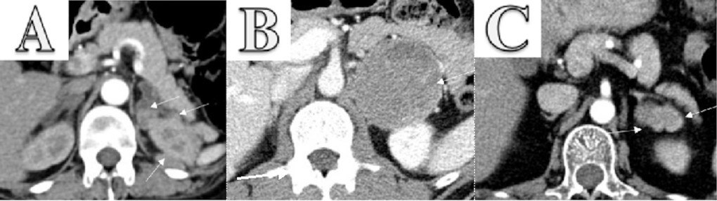
    Hormonal evaluation revealed plasma aldosterone concentration (PAC) <1.0 ng/dL, plasma renin activity (PRA) 0.568 ng/mL/h (0.167–5.380 ng/mL/h), 24-h urine free cortisol (UFC) 357 mg/24h (6–42 mg/24h), ACTH 174 pg/mL, and DHEA-S 353 ug/dL (20.4–186.6 ug/dL). ACTH levels on 2 repeat testings were 229 pg/mL and 342 pg/mL. The rest of the laboratory workup is summarized in Table 1. Considering elevated ACTH and 24-h UFC, a preliminary diagnosis of ACTH-dependent Cushing syndrome was made. An 8-mg dexamethasone suppression test revealed non-suppressed cortisol of 62.99 ug/dL along with dexamethasone 4050 ng/dL (1600–2850 ng/dL). A pituitary MRI was unremarkable for any focal lesion suggesting a diagnosis of ACTH-dependent Cushing’s syndrome secondary to an ectopic source. Imaging studies were then performed to determine the source. A CT scan of the chest and abdomen revealed adenomatous thickening with nodularity of bilateral adrenal glands, and a 1.4-cm nodule in the right middle lobe (Figure 1A, 1B). FDG-PET/CT showed severe bilateral enlargement of the adrenal glands with severe hyper-metabolic uptake (mSUV 9.2 and 9.1 for left and right adrenal glands, respectively) (Figure 2A). The uptake of the right lung nodule on PET/CT was 1.4 mSUV (Figure 2B).

     
    An external file that holds a picture, illustration, etc.
Object name is amjcaserep-22-e934437-g001.jpg

    CT chest, abdomen, and pelvis w/o contrast showed bilateral enlargement of adrenal glands (A, red arrows) and a 1.4-cm nodule in the right middle lobe of the lung (B, blue arrow).

     
    An external file that holds a picture, illustration, etc.
Object name is amjcaserep-22-e934437-g002.jpg

    Whole-body PET/CT following intravenous injection of 40 mCi FDG showed diffuse enlargement of the bilateral adrenal glands with mSUV of 9.2 on the left and 9.1 on the right adrenal gland, respectively (A, red arrows) and low-grade activity with an MSUV of 1.4 in right lung nodule (B, blue arrow).

    Table 1.

    Laboratory on initial presentation.

    Laboratory test Level Reference range
    WBCs 7.8 k/uL 3.7–10.3 k/uL
    RBCs 3.05 M/mL 3.–5.2 M/mL
    Hemoglobin 9.6 g/dL 11.2–15.7 g/dL
    Hematocrit 27.3% 34–45%
    Platelets 98 k/mL 155–369 k/mL
    MCV 89.7 fl 78.2–101.8 fl
    MCH 31.5 pg 26.4–33.3 pg
    MCHC 35.2 g/dL 32.5–35.3 g/dL
    RDW 15.8% 10.1–16.2%
    Glucose 73 mg/dL 74–90 mg/dL
    Sodium 148 mmol/L 136–145 mmol/L
    Potassium 2.4 mmol/L 3.7–4.8 mmol/L
    Bicarbonate 32 mmol/L 22–29 mmol/L
    Chloride 108 mmol/L 97–107 mmol/L
    Calcium 7.0 mg/dL 8.9–10.2 mg/dL
    Magnesium 1.7 mg/dL 1.7–2.4 mg/dL
    Phosphorus 2.3 mg/dL 2.5–4.9 mg/dL
    Albumin 2.4 g/dL 3.3–4.6 g/dL
    Blood urea nitrogen 41 mg/dL 0–30 ng/dL
    Creatinine 1.60 mg/dL 0.60–1.10 mg/dL
    Estimated GFR 31.5 mL/min/1.73m2 >60 mL/min/1.73 m2
    Aspartate transaminase 42 U/L 9–36 U/L
    Alanine transaminase 67 U/L 8–33 U/L
    Alkaline phosphatase 90 U/L 46–142 U/L
    Total protein 4.8 g/dL 6.3–7.9 g/dL
    Arterial blood gas analysis    
    PaCO2 48.9 mmHg 35.0–45.0 mmHg
    PaO2 63.1 mmHg 85.0–100.0 mmHg
    %SAT 92.8% 93.0–97.0
    HCO3 47.8 mm/L 20.0–26.0 mm/L
    Base excess 26.3 mm/L <2.0 mm/L
    pH 7.599 7.350–7.450
    Adrenocorticotropic hormone (ACTH) 174, 229 and 342 pg/mL 15–65 pg/mL
    Urine free cortisol, 24 h 357 ug/24 hr 6–42 mg/24 hr
    8: 00 AM cortisol following 8 mg dexamethasone (4×2 mg doses) previous day 62.99 mg/dL  
    8: 00 AM dexamethasone following 8 mg dexamethasone (4×2 mg doses) previous day 4050 ng/dL 1600–2850 ng/dL

    Based on unsuppressed cortisol following an 8-mg dexamethasone suppression test, negative pituitary MRI, and 1.4-cm lung nodule, we diagnosed ACTH-dependent CS secondary to an ectopic source, most likely from the 1.4-cm lung nodule. While awaiting localization studies, within 3 months of initial presentation, she had 2 hospitalizations, one in May 2021 for acute anemia secondary to bleeding peptic ulcer disease (PUD) requiring endoscopic clipping of the bleeding ulcer, and another in June 2021 for acute on chronic congestive heart failure. The patient’s overall condition continued to deteriorate, and she became progressively weak and wheelchair-bound. A 68-Ga-DOTATATE was planned to establish the source of ectopic ACTH definitively; however, she developed a left hip fracture in July 2021 and could not present for follow-up care. Therefore, she was started on Mifepristone until curative surgery. However, considering the patient’s advanced comorbid conditions, the increased burden of the patient’s health care needs on her elderly husband, and the inability of other family members to provide necessary healthcare-related support, palliative care was pursued. In August 2021, she developed a sacral decubitus ulcer and community-acquired pneumonia. However, she was still alive while receiving palliative care in a nursing home until September 2021.

    Discussion

    Ectopic ACTH syndrome (EAS) is defined as secretion of ACTH from an extra-pituitary source and is the cause of Cushing’s syndrome (CS) in approximately 4–5% of cases [,]. Clinical features of EAS depend on the rate and amount of ACTH production []. Among all forms of Cushing’s (excluding adrenal cortical carcinoma), EAS has the worst outcome, with one of the most extensive combined UK & Athens study demonstrating a 5-year survival rate of 77.6%. Compared to Cushing’s disease (CD), patients with EAS have severe and excessive production of ACTH, resulting in highly elevated cortisol levels. This leads to hypokalemia, metabolic alkalosis, worsening glycemia, hypertension, psychosis, and infections. Metabolic alkalosis and hypokalemia are the 2 most common acid-base and electrolyte abnormalities associated with glucocorticoid excess among these patients. Studies have shown that hypokalemia is seen in up to 90% of patients with EAS. Although hypertension and hypokalemia are often attributed to primary hyperaldosteronism, other causes should be sought. Under normal circumstances, the mineralocorticoid effect of cortisol is insignificant due to local conversion to cortisone by the action of 11 beta-hydroxysteroid dehydrogenase. Excessive cortisol in patients with EAS saturates the action of 11 beta-hydroxysteroid dehydrogenase and leads to the appearance of mineralocorticoid action of cortisol []. In our patient, the initial treatment of hypokalemia was unsatisfactory, so additional endocrine workup was pursued. Elevated urinary cortisol excretion, plasma ACTH levels, unsuppressed cortisol following 8 mg dexamethasone, and lung mass on CT scan strongly suggested that the clinical symptoms were due to EAS. Unfortunately, despite diagnosing the underlying condition contributing to the patient’s symptoms, her clinical condition rapidly deteriorated without surgical treatment.

    Various factors resulted in delayed diagnosis in our patient. First, the patient sought medical care only 3 months after symptom onset. Second, furosemide, a medication commonly used to treat patients with HFrEF, is a frequent culprit of hypokalemia and often is treated with adequate potassium supplementation. Third, multiple hospitalizations resulted in delays in the proper endocrine workup necessary for establishing hypercortisolism. Fourth, localization of the ectopic source requires advanced imaging studies, which are only available in a few tertiary care centers. Fifth, even after tumor localization with PET/CT scan, there is still a need for a more definitive localization study using Ga-DOTATATE scan, which has a higher specificity. However, it was unavailable in our institution and was only available in a few tertiary care centers, with the nearest center being 2.5 h away. Sixth, the impact of the COVID-19 pandemic also played a critical role in promptly providing critical care necessary to the patient. In addition to those, the social situation of our patient also played an essential role in contributing to delays in diagnosis.

    It is well recognized that EAS is associated with various malignancies, mostly of neuroendocrine origin. The most common location of these tumors was found to be the lung (55.3%), followed by the pancreas (8.5%), mediastinum-thymus (7.9%), adrenal glands (6.4%), and gastrointestinal tract (5.4%) []. Prompt surgical removal of ectopic ACTH-secreting tumors is the mainstay of therapy in patients with EAS []. However, localization of such tumors with conventional therapy is often challenging as the sensitivity to localize the tumor is 50–60% for conventional imaging such as CT, MRI, and FDG-PET []. In a study by Isidori et al, nuclear imaging improved the sensitivity of conventional radiological imaging []. Moreover, newer imaging technologies using somatostatin receptor (SSTR) analogs such as 68Ga-DOTATATE PET/CT further improve the ability to localize the tumor. 68Ga-DOTATATE PET/CT, approved in 2016 by the Federal Drug Administration (FDA) for imaging well-differentiated NETs, has a high sensitivity (88–93%) and specificity (88–95%) to diagnose carcinoid tumor []; however, a systematic review reported a significantly lower sensitivity (76.1%) of 68Ga-DOTATATE PET/CT to diagnose EAS [].

    Once localized, the optimal management of EAS is surgical re-section of the causative tumor, which is often curative. However, until curative surgery is done, patients should be medically managed. Drugs used to reduce cortisol levels include ketoconazole, mitotane, and metyrapone [, ]. These are oral medications and decrease cortisol synthesis by inhibiting adrenal enzymes []. Etomidate is the only intravenous drug that immediately reduces adrenal steroid production and can be used when acute reduction in cortisol production is desired [].

    Medical management requires frequent monitoring of cortisol levels and titration of dose to achieve low serum and urine cortisol levels. Mifepristone, an anti-progesterone at a higher dose, works as a glucocorticoid receptor antagonist and can be used to block the action of cortisol. Its use results in variable levels of ACTH and cortisol levels in patients with EAS. Hence, hormonal measurement cannot be used to judge therapeutic response, and clinical improvement is the goal of treatment []. Drugs inhibiting ACTH secretion by NETs such as kinase inhibitors (vandetanib, sorafenib, or sunitinib) are effective in treating EAS secondary to medullary thyroid cancer []. Somatostatin analogs such as octreotide and lanreotide have demonstrated short- and medium-term efficacy in a few EAS patients; however, a few patients failed to improve, necessitating the use of more effective treatment options [,]. Hence, they are not considered a first-line drug as monotherapy and should be used in combination with other agents, or as anti-tumoral therapy in non-excisable metastatic well-differentiated NETs [,]. Cabergoline, a dopamine agonist, has been used with variable therapeutic effects in a few patients []. In 1 patient, the use of combination therapy using Mifepristone and a long-acting octreotide significantly improved EAS []. In our patient, we initiated Mifepristone to reduce the burden associated with frequent biochemical monitoring and planned 68Ga-DOTATATE PET/CT to localize the tumor; however, further diagnostic and therapeutic approaches could not be further undertaken per family wishes.

    Conclusions

    EAS can present with refractory hypokalemia, especially in patients who are already at risk of developing hypokalemia. Diagnosis of EAS is often challenging and requires a multidisciplinary approach. Localization of source of EAS should be done using nuclear imaging, preferably using SSTR analogs, when available. Urgent surgical evaluation remains the mainstay of treatment following tumor localization and can result in a cure. EAS is a rapidly progressive and life-threatening situation that can be fatal if diagnosis or timely intervention is delayed.

    Abbreviations

    ACTH adrenocorticotropic hormone;
    CS Cushing’s syndrome;
    CT computed tomography;
    EAS ec-topic ACTH syndrome;
    MRI magnetic resonance imaging;
    FDG/PET 18-F-fluorodeoxyglucose positron emission tomography;
    NET neuroendocrine tumors;
    SSTR somatostatin receptor;
    EF ejection fraction;
    PAC plasma aldosterone concentration;
    PRA plasma renin activity;
    UFC urine free cortisol;
    DHEA-S dehydroepiandrosterone sulfate;
    68-Ga-DOTATATE Gallium 68 (68Ga) 1,4,7,10-tetraazacyclododecane-1,4,7,10-tet-raacetic acid (DOTA)-octreotate;
    PUD peptic ulcer disease

    Footnotes


    Financial support: None declared


    References:

    1. Pluta RM, Burke AE, Golub RM. JAMA patient page. Cushing syndrome and Cushing disease. JAMA. 2011;306:2742. [PubMed] []
    2. Melmed SKR, Rosen C, Auchus R, Goldfine A. Williams textbook of endocrinology. Elsevier; 2020. []
    3. Rubinstein G, Osswald A, Hoster E, et al. Time to diagnosis in Cushing’s syndrome: A meta-analysis based on 5367 patients. J Clin Endocrinol Metab. 2020;105:dgz136. [PubMed] []
    4. Rosset A, Greenman Y, Osher E, et al. Revisiting Cushing syndrome, milder forms are now a common occurrence: A single-center cohort of 76 subjects. Endocr Pract. 2021;27:859–65. [PubMed] []
    5. Fan L, Zhuang Y, Wang Y, et al. Association of hypokalemia with cortisol and ACTH levels in Cushing’s disease. Ann NY Acad Sci. 2020;1463:60–66. [PubMed] []
    6. Jain SH, Sadow PM, Nose V, Dluhy RG. A patient with ectopic cortisol production derived from malignant testicular masses. Nat Clin Pract Endocrinol Metab. 2008;4:695–700. [PubMed] []
    7. Sarlis NJ, Chanock SJ, Nieman LK. Cortisolemic indices predict severe infections in Cushing syndrome due to ectopic production of adrenocorticotropin. J Clin Endocrinol Metab. 2000;85:42–47. [PubMed] []
    8. Isidori AM, Kaltsas GA, Pozza C, et al. The ectopic adrenocorticotropin syndrome: Clinical features, diagnosis, management, and long-term follow-up. J Clin Endocrinol Metab. 2006;91:371–77. [PubMed] []
    9. Isidori AM, Sbardella E, Zatelli MC, et al. Group ABCS. Conventional and nuclear medicine imaging in ectopic Cushing’s syndrome: A systematic review. J Clin Endocrinol Metab. 2015;100:3231–44. [PMC free article] [PubMed] []
    10. Righi L, Volante M, Tavaglione V, et al. Somatostatin receptor tissue distribution in lung neuroendocrine tumours: A clinicopathologic and immunohistochemical study of 218 ‘clinically aggressive’ cases. Ann Oncol. 2010;21:548–55. [PubMed] []
    11. Ozkan ZG, Kuyumcu S, Balkose D, et al. The value of somatostatin receptor imaging with In-111 Octreotide and/or Ga-68 DOTATATE in localizing Ectopic ACTH producing tumors. Mol Imaging Radionucl Ther. 2013;22:49–55. [PMC free article] [PubMed] []
    12. Paun DL, Vija L, Stan E, et al. Cushing syndrome secondary to ectopic adrenocorticotropic hormone secretion from a Meckel diverticulum neuroendocrine tumor: Aase report. BMC Endocr Disord. 2015;15:72. [PMC free article] [PubMed] []
    13. Grigoryan S, Avram AM, Turcu AF. Functional imaging in ectopic Cushing syndrome. Curr Opin Endocrinol Diabetes Obes. 2020;27:146–54. [PMC free article] [PubMed] []
    14. Poeppel TD, Binse I, Petersenn S, et al. 68Ga-DOTATOC versus 68Ga-DOTATATE PET/CT in functional imaging of neuroendocrine tumors. J Nucl Med. 2011;52:1864–70. [PubMed] []
    15. Varlamov E, Hinojosa-Amaya JM, Stack M, Fleseriu M. Diagnostic utility of Gallium-68-somatostatin receptor PET/CT in ectopic ACTH-secreting tumors: A systematic literature review and single-center clinical experience. Pituitary. 2019;22:445–55. [PubMed] []
    16. Findling JW, Raff H. Cushing’s syndrome: Important issues in diagnosis and management. J Clin Endocrinol Metab. 2006;91:3746–53. [PubMed] []
    17. Diez JJ, Iglesias P. Pharmacological therapy of Cushing’s syndrome: Drugs and indications. Mini Rev Med Chem. 2007;7:467–80. [PubMed] []
    18. Wannachalee T, Turcu AF, Auchus RJ. Mifepristone in the treatment of the ectopic adrenocorticotropic hormone syndrome. Clin Endocrinol (Oxf) 2018;89:570–76. [PubMed] []
    19. Young J, Haissaguerre M, Viera-Pinto O, et al. Cushing’s syndrome due to ectopic ACTH secretion: An expert operational opinion. Eur J Endocrinol. 2020;182:R29–58. [PubMed] []
    20. Pedroncelli AM. Medical treatment of Cushing’s disease: Somatostatin analogues and pasireotide. Neuroendocrinology. 2010;92(Suppl. 1):120–24. [PubMed] []
    21. Moraitis AG, Auchus RJ. Mifepristone improves octreotide efficacy in resistant ectopic Cushing’s syndrome. Case Rep Endocrinol. 2016;2016:8453801. [PMC free article] [PubMed] []

    Articles from The American Journal of Case Reports are provided here courtesy of International Scientific Information, Inc
    • Like 1
  23.  


    The pituitary gland works hard to keep you healthy, doing everything from ensuring proper bone and muscle growth to helping nursing mothers produce milk for their babies. Its functionality is even more remarkable when you consider the gland is the size of a pea.

    “The pituitary is commonly referred to as the ‘master’ gland because it does so many important jobs in the body,” says Karen Frankwich, MD, a board-certified endocrinologist at Mission Hospital. “Not only does the pituitary make its own hormones, but it also triggers hormone production in other glands. The pituitary is aided in its job by the hypothalamus. This part of the brain is situated above the pituitary, and sends messages to the gland on when to release or stimulate production of necessary hormones.”

    These hormones include:

    • Growth hormone, for healthy bone and muscle mass
    • Thyroid-stimulating hormone, which signals the thyroid to produce its hormones that govern metabolism and the body’s nervous system, among others
    • Follicle-stimulating and luteinizing hormones for healthy reproductive systems (including ovarian egg development in women and sperm formation in men, as well as estrogen and testosterone production)
    • Prolactin, for breast milk production in nursing mothers
    • Adrenocorticotropin (ACTH), which prompts the adrenal glands to produce the stress hormone cortisol. The proper amount of cortisol helps the body adapt to stressful situations by affecting the immune and nervous systems, blood sugar levels, blood pressure and metabolism.
    • Antidiuretic (ADH), which helps the kidneys control urine levels
    • Oxytocin, which can stimulate labor in pregnant women

    The work of the pituitary gland can be affected by non-cancerous tumors called adenomas. “These tumors can affect hormone production, so you have too little or too much of a certain hormone,” Dr. Frankwich says. “Larger tumors that are more than 1 centimeter, called macroadenomas, can also put pressure on the area surrounding the gland, which can lead to vision problems and headaches. Because symptoms can vary depending on the hormone that is affected by a tumor, or sometimes there are no symptoms, adenomas can be difficult to pinpoint. General symptoms can include nausea, weight loss or gain, sluggishness or weakness, and changes in menstruation for women and sex drive for men.”

    If there’s a suspected tumor, a doctor will usually run tests on a patient’s blood and urine, and possibly order a brain-imaging scan. An endocrinologist can help guide a patient on the best course of treatment, which could consist of surgery, medication, radiation therapy or careful monitoring of the tumor if it hasn’t caused major disruption.

    “The pituitary gland is integral to a healthy, well-functioning body in so many ways,” Dr. Frankwich says. “It may not be a major organ you think about much, but it’s important to know how it works, and how it touches on so many aspects of your health.”
     

    Adapted from http://www.stjhs.org/HealthCalling/2016/December/The-Pituitary-Gland-Small-but-Mighty.aspx

    • Like 1
  24. Best friends Charly Clive and Ellen Robertson thought carefully about what to call the tumour that was growing in Charly’s brain.

    The doctors had their own name for the golf-ball-sized growth sitting right behind Charly’s left eye — a pituitary adenoma — but the friends decided they needed something less scary. They flirted with calling it Terry Wogan (‘as in Pitui-Terry Wogan,’ says Ellen), but that didn’t seem quite right.

    So Britney Spears fan Charly, then 23, suggested Britney. Bingo! Not only was she ‘iconic and fabulous’, but Britney was also one of life’s survivors. From then on, they were a threesome — Charly, Ellen and Britney the brain tumour — although Ellen is at pains to point out that this Britney was never a friend.

    What a thing to have to deal with, so young. The pair, who met at school in rural Oxfordshire, are now actresses. Charly’s biggest role to date has been in the critically acclaimed 2019 Channel 4 series Pure, while Ellen starred in the Agatha Christie mini-series The Pale Horse.

    But this week they appeared together in Britney, a BBC comedy based on the story of Charly’s brain tumour. The TV pilot (and yes, they are hoping for a full series) is an expansion of a sell-out stage show they performed at the Edinburgh Fringe in 2016.

    The production is admittedly surreal. Viewers are led inside Charly’s brain and the show includes a scene where Charly dons an inflatable sumo-wrestler suit on the day of her diagnosis. Poetic licence? No, it really happened.

    ‘My dad’s mate had given him a sumo suit as a silly Christmas present and so, on Doomsday, we took photos of me in it.’

    The tone was set for how these friends would deal with the biggest challenge of their lives: they would laugh through it, somehow.

    As the women, now 28, point out, what was the alternative?

    Charly says: ‘It was that thing of laughing at the monster so you are not scared of it. If you cry when do you stop? It was easier to make light of it.’

    Their show is not really about a brain tumour. It’s a celebration of friendship. Ellen pretty much moved in with Charly’s family during this time (‘To be in place when I exploded, so she could pick up the debris,’ says Charly).

    The pair live together today, finishing each other’s sentences as we speak on Zoom — and at one point both miming Charly’s brain surgery (with gruesome sound effects).

    This sort of silliness rooted their friendship, which started at the age of 14 when they wrote their own plays (Finding Emo, anyone?) while at school together in Abingdon. Charly later moved to New York to study dramatic arts, and Ellen studied at Cambridge.

    In 2015, Charly came home for a visit, and went to see her GP (played in the drama by Omid Djalili) about her lack of periods and a blind spot in her peripheral vision. An MRI scan showed a mass on her brain. ‘They said it had eroded the bone in my nose and was pressing on the optic nerve, and it was lucky we had caught it,’ she says. ‘The next step would have been discovering it because I’d gone blind.’

    Even worse, the tumour was so close to her carotid artery that removal might kill her — and they still had no idea if it was cancerous. Into the breach stepped Ellen. ‘I saw it as my job to make her laugh, which is what I’d always done anyway,’ she says. They both talk of toppling into limbo, ‘almost like a fantasy world’, says Charly. ‘As I was going through the tests, we’d do impressions of the doctors and create our own scenarios.’

    The friends talk about sitting up into the night, watching TV. There is a touching moment when Charly admits she was afraid to sleep, and Ellen knew it. ‘It’s hard when you are thinking “What if the tumour grows another inch in the night and I don’t wake up?” ’

    Charly was operated on in March 2016, and Ellen remembers the anaesthetist confiding that Charly’s heart had stopped on the operating table.

    ‘He wasn’t the most tactful person we’ve ever met. He said “Oh my God, guys, she died”.’ Charly makes a jazz hands gesture. ‘And guess who is alive again?’ Even at that darkest moment, there were flashes of humour. Ellen laughs at the memory of the surgeon in his scrubs, with wellies on. ‘They had blood on them. I was transfixed. I wanted to ask “Is that Charly’s . . . brain blood?” ’

    In the stage version of the show, the anaesthetist gets two full scenes. ‘He’s the heartthrob of the piece,’ says Charly. ‘A sexy rugger bloke who is crap at talking to people.’

    The days that followed the surgery were hideous — and yet they, too, have been mined for comedy. Charly’s face was bandaged, ‘as if I’d had a Beverly Hills facelift’, and she was warned that she could not sneeze. ‘If I did, bits of my brain would come out my nose,’ she says.

    Ellen read her extracts from Harry Potter but ‘made them smutty’, which confused the already confused Charly further. ‘I was drug-addled and not myself, and in the most bizarre pain, concentrated in my face’.

    ‘That week after the surgery was the worst part of all,’ says Ellen, suddenly serious. ‘She was behaving oddly and there was this unacknowledged fear: was this Charly for ever?’ Oh, the relief when the old Charly eventually re-emerged — albeit a more fragile, often tearful version.

    It was Ellen who persuaded Charly to take their stage show about her illness public — and it went on to win much critical acclaim. ‘I wanted Charly to see it as something other than just this rubbish chapter that needed to be forgotten about,’ says Ellen.

    For her part, Charly credits her best friend as her saviour: ‘I don’t know how I would have got through it all without Ellen.’

    The good news is that Britney was not cancerous, although surgery did not obliterate her entirely. ‘She’s still there, but tiny — just a sludge. I’ve been told that she won’t grow though. If I ever do get another brain tumour, it won’t be Britney.’

    Off they go again, imagining what is happening now inside Charly’s brain. ‘Britney is still in there, trying on outfits for a comeback tour, but it won’t happen,’ says Charly. Ellen nods. ‘It’s over,’ she says. ‘But she’s just left a pair of shoes behind.’

    Britney is available to watch on BBC Three and BBC iPlayer

    Adapted from https://www.dailymail.co.uk/femail/article-10264203/I-laughed-brain-tumour-Id-never-stop-crying-Actress-Charly-Clive.html

×
×
  • Create New...