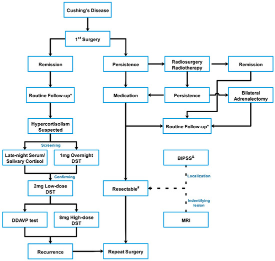
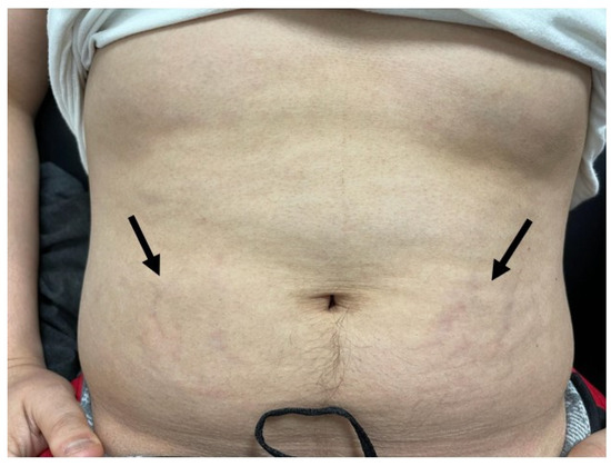
Jump to content

MaryO

~Chief Cushie~
  • Posts

    8,082
  • Joined

  • Last visited

  • Days Won

    555

Posts posted by MaryO

  1. The study covered in this summary was published on Research Square as a preprint and has not yet been peer reviewed.

    Key Takeaways

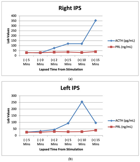
    • Adding a corticotropin-releasing hormone (CRH) stimulation test immediately following a 2-day low-dose dexamethasone suppression test (LDDST) ― what's known as a Dex-CRH test and was first introduced in 1993 ― identified Cushing disease in 5 of 65 people (7.7%) with a confirmed diagnosis but who had previously shown normal cortisol levels on a conventional LDDST.

    • However, the Dex-CRH test also resulted in one (2.5%) false positive case compared with an LDDST alone.

    • Measuring serum dexamethasone levels further improved the diagnostic accuracy of the Dex-CRH test.

    Why This Matters

    • It can be challenging to diagnose Cushing syndrome and to differentiate Cushing disease from nonneoplastic physiologic hypercortisolism caused by conditions that can present with Cushing syndrome–like clinical features, such as diabetes and obesity.

    • The Dex-CRH test, first described in 1993, initially appeared superior to an LDDST alone for ruling out nonneoplastic hypercortisolism, with a report of 100% sensitivity, specificity, and diagnostic accuracy. However, subsequent studies that used different protocols and in which dexamethasone was not measured had results that called into question the accuracy, sensitivity, and specificity of the Dex-CRH test.

    • This study reports the accuracy, sensitivity, and specificity of the Dex-CRH test for diagnosing Cushing disease, performed as first described, in 107 patients, including 74 for whom dexamethasone was also measured.


    Study Design

    • The researchers analyzed data from 107 patients with suspected Cushing disease who underwent a Dex-CRH test during 2002–2014 at the Cleveland Clinic.


    Key Results

    • Sixty-five people received a confirmed diagnosis of Cushing disease and underwent follow-up for a median of 66 months. Cushing disease was not confirmed in 42 patients who were followed for a median of 52 months.

    • The median age of the 107 patients was 40 years, and 82% to 88% were women. The median body mass index for these patients was 34–37 kg/m2.

    • Among the 65 patients with confirmed Cushing disease, five patients (7.7%) had a suppressed cortisol level no greater than 1.4 μg/dL after the LDDST but were appropriately classified as having Cushing disease with a cortisol level that surpassed 1.4 μg/dL by 15 minutes after CRH stimulation.

    • In contrast, 3 of 42 patients (7.1%) in the group without confirmed Cushing disease had an abnormal Dex-CRH test result. For one of these three patients, the LDDST result was borderline normal, with a cortisol level post-DEX of 1.4 μg/dL that increased to 3.1 μg/dL by 15 minutes after CRH stimulation, which resulted in this patient receiving a false positive diagnosis.

    • A cortisol threshold value of more than 1.4 μg/dL during the Dex-CRH test was diagnostic of Cushing disease with sensitivity of 100%, specificity of 93%, and diagnostic accuracy of 97%.

    • Among the 74 patients with dexamethasone measurements, the sensitivity of the Dex-CRH test was unchanged, but the specificity and diagnostic accuracy increased to 97% and 99%, respectively.

    Limitations

    • The study was retrospective.

    • Not all patients underwent measurement of dexamethasone level.

    • No uniform protocol existed for the diagnostic work-up and follow-up of patients suspected of having Cushing disease.

    Disclosures

    • The study did not receive commercial funding.

    • The authors had no financial disclosures.


    This is a summary of a preprint research study , "The Addition of Corticotropin-Releasing Hormone to 2-Day Low Dose Dexamethasone," written by researchers primarily from the Cleveland Clinic and Johns Hopkins University School of Medicine, published on Research Square, and provided to you by Medscape. This study has not yet been peer reviewed. The full text of the study can be found on research square.com.

    From https://www.medscape.com/viewarticle/985984

    • Like 1
  2. Abstract

    Introduction

    Hypertension is one of the most common clinical features of patients with overt and subclinical hypercortisolism. Although previous studies have shown the coexistence of autonomous cortisol and aldosterone secretion, it is unclear whether aldosterone plays a role in hypertension among patients with hypercortisolism. Therefore, we examined the associations of plasma aldosterone concentrations (PACs) with hypertension among patients with overt and subclinical hypercortisolism.

    Methods

    This single-center retrospective cohort study included patients with adrenal tumor and serum cortisol levels after 1-mg dexamethasone suppression test >1.8 µg/dL (50 nmol/L). Using multivariable regression models adjusting for baseline characteristics, we investigated the association of PACs with systolic blood pressure and postoperative improvement of hypertension after the adrenalectomy.

    Results

    Among 89 patients enrolled in this study (median age, 51 years), 21 showed clinical signs of Cushing syndrome (overt hypercortisolism) and 68 did not show clinical presentations (subclinical hypercortisolism). We found that higher PACs were significantly associated with elevated systolic blood pressure among patients with subclinical hypercortisolism (adjusted difference [95% CI] = +0.59 [0.19-0.99], P = 0.008) but not among those with overt hypercortisolism. Among 33 patients with subclinical hypercortisolism and hypertension who underwent adrenalectomy, the postoperative improvement of hypertension was significantly associated with higher PACs at baseline (adjusted risk difference [95% CI] = +1.45% [0.35-2.55], P = 0.01).

    Conclusion

    These findings indicate that aldosterone may contribute to hypertension among patients with subclinical hypercortisolism. Further multi-institutional and population-based studies are required to validate our findings and examine the clinical effectiveness of the intervention targeting aldosterone for such patients.

    Cortisol production in the adrenal gland is regulated by the hypothalamus-pituitary-adrenal (HPA) axis. Subclinical hypercortisolism is a status characterized by the alteration of HPA axis secretion without typical signs or symptoms of overt hypercortisolism (eg, moon face, truncal obesity, easy bruising, thin extremities, proximal myopathy, cutaneous purple striae) [12]. Although overt hypercortisolism can be detected by its clinical presentations or severe complications, it is sometimes challenging for clinicians to appropriately diagnose subclinical hypercortisolism because of the absence of such clinical presentations [2]. The 1-mg overnight dexamethasone suppression test (1-mg DST) measures the response of the adrenal glands to ACTH through the HPA axis and therefore has been widely used for screening and diagnosis of subclinical hypercortisolism [13]. The European Society of Endocrinology Guideline has defined a partial suppression of the HPA axis (ie, serum cortisol levels after 1-mg DST [F-DST] > 1.8 µg/dL [50 nmol/L]) without clinical signs of overt cortisol hypersecretion as “possible autonomous cortisol secretion” and recommended screening these patients for metabolic disorders including hypertension and type 2 diabetes mellitus to offer appropriate treatment of these comorbidities [4].

    Hypertension is one of the most common and distinguishing clinical features in patients with subclinical hypercortisolism [2] as well as overt hypercortisolism [5]. Although hypertension can be triggered by excess cortisol levels [56], it is still unclear whether even slightly elevated cortisol levels among individuals with subclinical hypercortisolism contribute to the occurrence of hypertension. This raises another potential mechanism to cause hypertension such as the coexistence of hyperaldosteronism (ie, excess aldosterone that is an essential steroid hormone for sodium reabsorption, water retention, and blood pressure control) [7]. Previous studies have reported that 10% to 20% of primary aldosteronism is accompanied by cortisol-producing adenoma [8-10], and autonomous cortisol secretion was decreased after the resection of the aldosterone-producing adenoma (a subtype of primary aldosteronism) [11]. Furthermore, a previous mass spectrometry-based analysis revealed that cortisol secretion was frequently found in patients with primary aldosteronism [12]. Although these studies have examined cortisol biosynthesis in primary aldosteronism [13], evidence about whether aldosterone plays a role in the occurrence of hypertension among people with subclinical hypercortisolism is limited.

    To address this knowledge gap, we performed a cohort study examining the association between aldosterone and hypertension among patients with adrenal tumor and F-DST >1.8 µg/dL, stratified by whether patients had clinical signs of Cushing syndrome or not. We first analyzed the cross-sectional association between aldosterone and blood pressure at baseline. Then, we analyzed the longitudinal association between aldosterone at baseline and the improvement rate of hypertension after the adrenalectomy. Last, to further clarify the role of aldosterone in the regulation of blood pressure in subclinical hypercortisolism, we described the difference in aldosterone response to ACTH after the adrenalectomy according to the postoperative improvement of hypertension.

    Materials and Methods

    Data Sources and Study Participants

    A retrospective cohort study was designed to assess the clinical characteristics (focusing on aldosterone) among patients with hypercortisolism at the Yokohama Rosai Hospital from 2008 to 2017. We enrolled 89 patients with adrenal tumor and F-DST > 1.8 µg/dL (50 nmol/L) [3414]. We then categorized them into 2 groups: (1) overt hypercortisolism (F-DST > 5.0 µg/dL [138 nmol/L]) and having clinical signs of Cushing syndrome (moon face, central obesity, dorsocervical fat pad [buffalo hump], purple striae, thin skin, easy bruising, and proximal myopathy] [15]) and (2) subclinical hypercortisolism (not having such clinical signs). All patients with overt hypercortisolism in this study showed F-DST > 5.0 µg/dL (138 nmol/L). The study was approved by the research ethics committee of the Yokohama Rosai Hospital, and all participants provided written informed consent.

    Measurements

    Demographic characteristics were self-reported, and body mass index (BMI) was calculated using measured weight and height. Systolic blood pressure was measured in the sitting position using a standard upper arm blood pressure monitor after a 5-minute rest in a quiet place [16]. The mean of 2 measurements was recorded. If the measurement was done only once on a given occasion, the level obtained was recorded. When the patients were already taking antihypertensives at enrollment, they were asked to report their blood pressure levels at the diagnosis of hypertension (ie, systolic blood pressure before starting antihypertensives). Blood samples were collected at 8:00 AM after the patient had rested in the supine position for 30 minutes. We measured F (µg/dL, × 27.6 for nmol/L) and ACTH (pg/mL, × 0.220 for pmol/L) using chemiluminescent enzyme immunoassay and electrochemiluminescent immunoassay, respectively. Plasma aldosterone concentrations (PACs; ng/dL, × 27.7 for pmol/L) and plasma renin activities (PRAs; ng/mL/h) were measured using radioimmunoassay. Any antihypertensive drugs were replaced with calcium channel antagonists (including dihydropyridine calcium channel antagonists) and/or α blocker several weeks before the measurement of PACs and PRAs according to the clinical guideline of the Japan Endocrine Society [17]. We also measured urine aldosterone (µg/day × 2.77 for nmol/d) and urine cortisol (µg/day, × 2.76 for nmol/d) using radioimmunoassay. The tumor size was estimated using contrast-enhanced thin-section computed tomography scans of the adrenal glands.

    To evaluate whether the patients had autonomous cortisol secretion, we performed 1-mg DST, in which dexamethasone (1 mg) was administered at 11:00 PM, and blood samples were drawn at 8:00 AM the following morning. F and ACTH were measured in 1-mg DST.

    The total or partial adrenalectomy was performed in all cases with overt hypercortisolism. For patients with subclinical hypercortisolism, the adrenalectomy was recommended to those who showed F-DST > 5.0 µg/dL (138 nmol/L) accompanying metabolic disorders [3]. It was also recommended to those who were expected to improve their clinical symptoms and/or metabolic disorders by the tumor resection, which included patients with hypertension possibly resulting from autonomous aldosterone secretion as well as autonomous cortisol secretion from the adrenal gland. The adrenalectomy was conducted when patients agreed with the treatment plan through informed consent. To evaluate whether patients had autonomous aldosterone secretion, we used the screening criterion of primary aldosteronism (ie, PAC/PRA ratio; aldosterone-to-renin ratio [ARR] > 20), followed by the confirmatory tests of primary aldosteronism that included the saline infusion test, captopril challenge, and/or furosemide stimulation test [17].

    For patients who were considered to receive a benefit by the adrenalectomy and who agreed with the examination, we performed the segment-selective adrenal venous sampling to assess the laterality of hyperaldosteronism [18-20]. First, blood samples were collected from the bilateral central adrenal veins before ACTH stimulation. Then, we collected samples from the superior, lateral, and inferior tributaries of the right central adrenal vein and the superior and lateral tributaries of the left central adrenal vein after ACTH stimulation. Aldosterone excess (ie, hyperaldosteronism) was considered when the effluent aldosterone concentrations were > 250 ng/dL before ACTH stimulation and 1400 ng/dL after ACTH stimulation, respectively [18-20]. We used the absolute value instead of the lateralization index because individuals included in our study had elevated cortisol concentrations given the inclusion criteria (ie, F-DST >1.8 µg/dL [50 nmol/L]). For 9 patients with subclinical hypercortisolism who showed bilateral adrenal nodules, the side of adrenalectomy was determined by the nodule size and the results of adrenal venous sampling (ie, laterality of hyperaldosteronism). The adrenalectomy was conducted when patients agreed with the treatment plan through informed consent. Immunohistochemical evaluation of aldosterone synthase cytochrome P450 (CYP11B2) was conducted for some resected nodules.

    To evaluate the postoperative cortisol responsiveness to ACTH, we performed an ACTH stimulation test a year after the adrenalectomy, in which blood samples were collected and PAC and F were measured 30 and 60 minutes after ACTH administration. Postoperative improvement of hypertension was defined as blood pressure <140/90 mmHg without antihypertensives or the reduction of the number of antihypertensives to maintain blood pressure <140/90 mmHg after the adrenalectomy.

    Statistical Analyses

    We describe the demographic characteristics and endocrine parameters at baseline comparing patients with overt hypercortisolism and those with subclinical hypercortisolism using the Fisher exact test for categorical variables and Mann-Whitney U test for continuous variables. Second, for each group, we investigated the association between the baseline characteristics and systolic blood pressure using ordinary least-squares regression models. The model included age, sex, BMI, serum potassium levels, estimated glomerular filtration rate, tumor size, and F and PAC at 8:00 AM. Third, we estimated the risk difference and 95% CI of the improvement rate of hypertension after the adrenalectomy according to these baseline characteristics (including systolic blood pressure) using a modified least-squares regression model with a Huber-White robust standard error [21]. Last, to evaluate whether the improvement of hypertension is related to postoperative cortisol and aldosterone secretion, we compared PAC and F responsiveness to ACTH from peripheral blood samples between patients who improved hypertension and those who did not using the Mann-Whitney U test. The longitudinal and postoperative analyses were performed among patients with subclinical hypercortisolism because only 2 cases with overt hypercortisolism failed to show the improvement of hypertension after the adrenalectomy.

    To assess the robustness of our findings, we conducted the following 2 sensitivity analyses. First, we replaced F at 8:00 AM with F after DST in our regression models. Second, we estimated the risk difference of the improvement rate of hypertension after the adrenalectomy according to the postoperative F and PAC levels after ACTH stimulation, adjusting for the baseline characteristics included in our main model.

    We also conducted several additional analyses. First, to investigate the relationship of change in PAC after adrenalectomy with the improvement rate of hypertension, we included decrease in PAC between before and after adrenalectomy instead of PAC at baseline in the model. Second, to assess the relationship between aldosterone and hypertension among patients with subclinical hypercortisolism without primary aldosteronism, we reran the analyses excluding patients who met the diagnostic criteria of primary aldosteronism. Third, to understand the overall association, we reran the analyses using all samples as a single group to assess the relationship among people with overall (ie, overt and subclinical) hypercortisolism. Last, we compared PAC and F responsiveness with ACTH during adrenal venous sampling between patients with and without postoperative improvement of hypertension. All statistical analyses were performed using Stata, version 15.

    Results

    Among the 89 enrolled patients, 21 showed clinical signs of overt Cushing syndrome and 68 did not. The flow of the study population is shown in Fig. 1. Among 21 patients with overt hypercortisolism, 19 patients had hypertension. All patients underwent adrenalectomy, and 16 patients showed improved hypertension levels after the surgery (1 patient was referred to another hospital; therefore, no information is available). Among 68 patients with subclinical hypercortisolism, 63 had hypertension. After the evaluation of autonomous aldosterone secretion as well as autonomous cortisol secretion, of 33 patients who underwent adrenalectomy, 23 (70%) showed improved hypertension levels after the adrenalectomy (10 patients in the surgery group decided not to undergo adrenalectomy). Patients with subclinical hypercortisolism who underwent adrenalectomy showed lower PRA and higher ARR than those without adrenalectomy (Supplementary Table S1) [22].


    Figure 1.

    Enrollment and follow-up of the study population after the adrenalectomy. aThe prevalence of patients with overt hypercortisolism and hypertension among this study population may be higher than in the general population and therefore needs to be carefully interpreted given that the study institute is one of the largest centers for adrenal diseases in Japan. bAll patients in this category showed autonomous cortisol secretion (ie, serum cortisol levels >5.0 µg/dL [138 nmol/L] after a 1-mg dexamethasone suppression test). cOne case underwent adrenalectomy at another hospital and therefore no information was available after the adrenalectomy. dThe adrenalectomy was performed for 33 patients who were expected to improve their clinical symptoms and/or metabolic disorders, including hypertension. This assessment was mainly based on autonomous cortisol secretion evaluated by a 1-mg dexamethasone suppression test, complicated metabolic disorders, and autonomous aldosterone secretion evaluated by adrenal venous sampling for patients who were positive for the screening and confirmatory tests of primary aldosteronism. Details in the assessment can be found in the Methods section or elsewhere [18-20].

    Demographic Characteristics and Endocrine Parameters Among Patients With Overt and Subclinical Hypercortisolism

    The median age (interquartile range) was 51 years (46, 62 years), and 72% were female. Patients with overt hypercortisolism were relatively younger and showed a higher estimated glomerular filtration rate and larger tumor size compared with patients with subclinical hypercortisolism (Table 1). Other demographic characteristics were similar between these groups. Patients with overt hypercortisolism showed higher F with undetected low ACTH, higher F after DST, and higher urine cortisol levels compared with those with subclinical hypercortisolism who instead showed higher PAC and ARR. Among patients with subclinical hypercortisolism, 9/68 (13.2%) showed undetectable ACTH levels and 25/68 (36%) were positive for PA screening criterion (ie, ARR > 20) followed by at least 1 positive confirmatory test. Based on the results of adrenal venous sampling of these cases, 9 showed aldosterone excess in the right nodules, 6 showed aldosterone excess in the left nodules, and 7 showed aldosterone excess on both sides, respectively (3 cases did not show aldosterone excess on both sides). Immunohistochemical evaluation of CYP11B2 was examined for 6 resected adrenal glands, and all of them showed positive expression.


    Patients’ characteristicsa Patients with overt hypercortisolism (N = 21) Patients with subclinical hypercortisolism (N = 68) P
    Age, y  46 [38-52]  54 [47-63]  0.002 
    Female, n (%)  18 (85.7)  46 (67.7)  0.11 
    Body mass index, kg/m2  23.4 [20.6-26.2]  23.1 [21.7-25.1]  0.94 
    Systolic blood pressure, mm Hg  156 [140-182]  162 [151-191]  0.29 
    Diastolic blood pressure, mm Hg  98 [92-110]  100 [90-110]  0.73 
    Serum potassium, mEq/Lb  3.9 [3.5-4.0]  3.8 [3.6-4.0]  0.98 
    eGFR, mL/min/1.73 m2  86.7 [77.3-123.0]  82.1 [69.8-87.7]  0.02 
    Tumor size by CT scan, mm  28 [25-30]  22 [17-26]  0.001 
    ACTH, 8:00 AM  − c  6.6 [2.4-11.8]  — 
    F, 8:00 AM  16.6 [12.5-18.8]  9.5 [7.7-12.0]  <0.001 
    PRA, 8:00 AM  0.7 [0.4-1.3]  0.5 [0.2-1.0]  0.10 
    PAC, 8:00 AM  8.3 [7.2-9.8]  9.2 [7.2-16.2]  0.09 
    ARR, 8:00 AM  10.0 [6.4-16.7]  21.0 [9.8-46.5]  0.02 
    F after DST  16.5 [14.4-18.7]  5.1 [3.2-7.5]  <0.001 
    Urine cortisol  220.0 [105.0-368.0]  49.5 [37.4-78.5]  <0.001 
    Urine aldosterone  5.7 [3.9-10.1]  7.2 [4.8-13.1]  0.16 

    Conversion to SI units: ACTH, pg/mL × 0.220 for pmol; F, µg/dL × 27.6 for nmol/L; PAC, ng/dL × 27.7 for pmol/L; urine aldosterone, μg/day × 2.77 for nmol/d; Urine cortisol, μg/day × 2.76 for nmol/d.

    Abbreviations: ARR, aldosterone-to-renin ratio; CRH, corticotropin-releasing hormone; CT, thin-section computed tomography; DST, 1-mg dexamethasone suppression test; eGFR, estimated glomerular filtration rate; F, serum cortisol; PRA, plasma renin activity; PAC, plasma aldosterone concentration.

    a

    Data are presented as median (interquartile range) or count (proportions) unless otherwise indicated.

    b

    Serum potassium levels were controlled using potassium supplement/tablets at enrollment.

    c

    Undetected in all cases.

    Association of Demographic Characteristics and Endocrine Parameters With Systolic Blood Pressure

    Among patients with overt hypercortisolism, we did not find a significant association of demographic characteristics and endocrine parameters with systolic blood pressure (Table 2). However, among patients with subclinical hypercortisolism, we found that higher PACs at 8:00 AM were significantly associated with systolic blood pressure (adjusted coefficient [95% CI] = +0.59 [0.19-0.99], P = 0.008). The results did not change when we used F after DST instead of F at 8:00 AM (Supplementary Table S2) [22].

     
    Table 2.

    Cross-sectional association of demographic characteristics and endocrine parameters with systolic blood pressure among patients with overt and subclinical hypercortisolism

    Outcome Systolic blood pressure at baseline
    Groups Patients with overt hypercortisolism Patients with subclinical hypercortisolism
    Parameters  Adjusted coefficient (95% CI)  P  Adjusted coefficient (95% CI)  P 
    Age, y  +1.73 (0.17-3.30)  0.03  +0.49 (−0.13 to 1.10)  0.12 
    Female  −7.48 (−76.75 to 61.79)  0.81  +15.38 (−0.83 to 31.59)  0.06 
    Body mass index  +5.47 (−2.4 to 13.33)  0.15  +1.07 (−0.49 to 2.63)  0.17 
    Serum potassium  +11.29 (−23.42 to 45.99)  0.48  −9.61 (−26.38 to 7.15)  0.26 
    eGFR  −0.12 (−1.00 to 0.77)  0.77  −0.44 (−0.89 to 0.01)  0.06 
    Tumor size  −2.39 (−6.92 to 2.14)  0.26  +0.40 (−0.46 to 1.26)  0.35 
    F, 8:00 AMa,b  +1.96 (−1.27 to 5.18)  0.20  +1.26 (−1.00 to 3.52)  0.27 
    PAC, 8:00 AMa  −2.86 (−7.38 to 1.66)  0.18  +0.59 (0.19-0.99)  0.008 

    Abbreviations: DST, 1-mg dexamethasone suppression test; eGFR, estimated glomerular filtration rate; F, serum cortisol; PRA, plasma renin activity; PAC, plasma aldosterone concentration.

    a

    ACTH and PRA were not included in the main model because they have strong correlation with F and PAC, respectively (ie, multicollinearity). The results did not change when additionally adjusting for ACTH and PRA.

    b

    The results did not change when we replaced F at 8:00 AM with F after DST (Supplementary Table S2).

    Association of Demographic Characteristics and Endocrine Parameters With Hypertension Improvement After the Adrenalectomy Among Patients With Subclinical Hypercortisolism

    Among 33 patients with subclinical hypercortisolism and hypertension who underwent the adrenalectomy, we found that age and higher PAC were significantly associated with a higher improvement rate of hypertension after the adrenalectomy (age, adjusted risk difference [95% CI] = +2.36% [1.08-3.64], P = 0.001; PAC, adjusted risk difference [95% CI] = +1.45% [0.35-2.55], P = 0.01; Table 3). The results did not change when we used F after DST instead of F at 8:00 AM (Supplementary Table S3) [22]. Patients with improved hypertension after the surgery showed significantly lower PACs 60 minutes after a postoperative ACTH stimulation test than those without the improvement of hypertension (P = 0.05), although F and PAC/F ratio were not significantly different between these 2 groups (Table 4). The association between lower PACs after postoperative ACTH stimulation and higher improvement rate of hypertension was also found in the multivariable regression analysis adjusting for baseline characteristics (adjusted risk difference [95% CI] = −1.08% [−1.92 to −0.25], P = 0.01; Supplementary Table S4) [22].


    Table 3.

    Longitudinal association of demographic characteristics and endocrine parameters with hypertension improvement after the adrenalectomy among patients with subclinical hypercortisolisma

    Outcome Hypertension improvement after the adrenalectomy
    Parameters Adjusted risk difference (95% CI) P
    Age  +2.36% (1.08-3.64)  0.001 
    Sex (female)  −11.32% (−61.37 to 38.73)  0.64 
    Body mass index  −5.08% (−10.29 to 0.13)  0.06 
    Systolic blood pressure  −0.67% (−1.77 to 0.43)  0.22 
    Serum potassium  −0.06% (−31.84 to 31.71)  1.00 
    eGFR  +0.53% (−0.36 to 1.42)  0.23 
    Tumor size  +0.79% (−1.35 to 2.93)  0.45 
    F, 8:00 AMb,c  −2.81% (−7.43 to 1.81)  0.22 
    PAC, 8:00 AMb  +1.45% (0.35-2.55)  0.01 

    Abbreviations: eGFR, estimated glomerular filtration rate; F, serum cortisol; PRA, plasma renin activity; PAC, plasma aldosterone concentration.

    a

    Analysis was not performed for patients with overt hypercortisolism because only 2/18 cases failed to show improved hypertension after the adrenalectomy.

    b

    ACTH and PRA were not included in the main model because they have strong correlation with F and PAC, respectively (ie, multicollinearity). The results did not change when additionally adjusting for ACTH and PRA.

    c

    The results did not change when we replaced F at 8:00 AM with F after DST (Supplementary Table S3).


    Table 4.

    Aldosterone and cortisol response to ACTH a year after the adrenalectomy according to hypertension improvement status among patients with subclinical hypercortisolisma

    Outcome: hypertension improvement status after the adrenalectomy Improvement (+) (N = 23) Improvement (−) (N = 10)  
    Parameters Median [IQR] Median [IQR] P
    PAC 60 min after ACTH stimulation  13.6 [10.0-16.7]  15.5 [13.7-43.1]  0.05b 
    F 60 min after ACTH stimulation  16.9 [13.7-20.6]  18.5 [13.5-24.7]  0.61 
    PAC/F ratio 60 min after ACTH stimulation  0.70 [0.52-1.39]  1.27 [0.50-5.44]  0.26 

    Conversion to SI units: F, µg/dL × 27.6 for nmol/L; PAC, ng/dL × 27.7 for pmol/L.

    Abbreviations: F, serum cortisol; PAC, plasma aldosterone concentration.

    a

    Analysis was not performed for patients with overt hypercortisolism because only 2/18 cases failed to show improved hypertension after the adrenalectomy.

    b

    The association was also observed after adjusting for baseline characteristics (eg, age, sex, body mass index, systolic blood pressure, serum potassium, estimated glomerular filtration rate, tumor size) and F 60 min after ACTH stimulation a year after the adrenalectomy (Supplementary Table S4).

    Additional Analyses

    Decreased PAC between before and after adrenalectomy was significantly associated with hypertension improvement (Supplementary Table S5) [22]. When we restricted samples to those without primary aldosteronism, PACs at baseline tended to be associated with systolic blood pressure but the 95% CI included the null (Supplementary Table S6) [22]. Decreased PAC after adrenalectomy was associated with hypertension improvement after the adrenalectomy, whereas PAC at baseline was not associated with that outcome (Supplementary Table S7) [22]. When we analyzed the entire sample (ie, both overt and subclinical hypercortisolism), PAC at baseline was associated with systolic blood pressure at baseline (Supplementary Table S8) [22] and hypertension improvement after the adrenalectomy (Supplementary Table S9) [22]. We also found the higher median value of PAC response to ACTH during adrenal venous sampling at the remained (ie, not resected by the adrenalectomy) side of adrenal gland among patients whose hypertension did not improve compared with those whose hypertension improved after the surgery, but the difference was not statistically significant (Supplementary Table S10) [22].

    Discussion

    In this retrospective cohort study, we found that higher aldosterone levels were associated with higher systolic blood pressure among patients with possible autonomous cortisol secretion and without clinical signs of overt Cushing syndrome (ie, subclinical hypercortisolism). In this group, higher aldosterone before the adrenalectomy was associated with the postoperative improvement of hypertension. Moreover, we found that patients with postoperative improvement of hypertension showed lower aldosterone response to ACTH after the adrenalectomy compared with those without the improvement of hypertension. Decrease in PACs after the adrenalectomy was associated with improved hypertension even among patients with subclinical hypercortisolism who did not have primary aldosteronism at baseline, whereas baseline PAC was not associated with that outcome. We found no evidence that aldosterone is associated with systolic blood pressure among patients with overt hypercortisolism. These findings indicate that elevated aldosterone may contribute to the presence of hypertension and its improvement rate after the adrenalectomy for patients with subclinical hypercortisolism.

    To the best of our knowledge, this is one of the first studies to assess the potential role of aldosterone in hypertension among patients with overt and subclinical hypercortisolism, during both pre- and postoperative phases. Since aldosterone- and cortisol-producing adenoma was reported in 1979 [2324], several studies have assessed the cortisol production in aldosterone-producing adenoma clinically and histologically [8-1025] and showed the correlation between the degree of glucocorticoid excess levels and metabolic markers including BMI, waist circumference, blood pressure, insulin resistance, and high-density lipoprotein [12]. Prior research suggested that aldosterone-producing adenoma might produce cortisol as well as aldosterone even when serum cortisol levels after DST is less than 1.8 µg/dL (50 nmol/L) [11]. Although these studies have focused on cortisol synthesis among patients with aldosterone-producing adenoma, little is known about aldosterone synthesis among patients with cortisol-producing adenoma. Given that patients with hypercortisolism tend to have therapy-resistant hypertension and electrolyte disorders [8], our findings may generate the hypothesis that aldosterone contributes to the incidence and severity of hypertension in patients with possible autonomous cortisol secretion; this warrants further investigation.

    There are several mechanisms by which cortisol excess leads to hypertension, such as regulating endothelial nitric oxide synthase expression modulated by 11β-hydroxysteroid dehydrogenases [26], activating the mineralocorticoid receptor [27] and upregulating vascular endothelin-1 [28]. Moreover, hypercortisolism impairs the production of endothelial vasodilators, including prostacyclin, prostaglandins, and kallikreins [29]. Despite these potential mechanisms, the direct effect of cortisol may not be sufficient to explain hypertension in patients with hypercortisolism, particularly subclinical hypercortisolism, and the presence of cortisol and aldosterone coproducing adenoma indicates another potential pathway to induce hypertension through aldosterone excess. Aldosterone is a steroid hormone not only promoting sodium reabsorption and volume expansion but also activating the mineralocorticoid receptor in the kidney and nonepithelial tissues (eg, adipose tissue, heart, endothelial cells, and vascular smooth muscle cells) [30]. It also induces oxidative stress, inflammation, fibrosis, vascular tone, and endothelial dysfunction [31]; therefore, aldosterone excess could induce hypertension even when it is slightly elevated [32]. A recent multiethnic study showed that aldosterone levels within the reference range were associated with subclinical atherosclerosis partially mediated through elevated blood pressure [33]. These mechanisms support our results indicating the potential contribution of aldosterone to hypertension among patients with subclinical hypercortisolism.

    This study had several limitations. First, we did not have information on the duration of cortisol excess and therefore the estimated effect of cortisol on hypertension in our study might have been underestimated. The duration of exposure to mild hypercortisolism may be one of the important drivers of cardiovascular and metabolic disorders including irreversible vasculature remodeling in patients with subclinical hypercortisolism [2]. Second, we did not have the genetic information of adrenal tumors including aldosterone-producing adenoma. Given the heterogeneity of aldosterone responsiveness to ACTH [34] and postoperative hypertension resolution rate across genetic mutations (eg, KCNJ5, ATP1A1, ATP2B3, CACNA1D, CTNNB1) [35], such information might affect our findings. Third, because of the nature of an observational study, we cannot rule out the unmeasured confounding. Fourth, because aldosterone and cortisol levels were measured at a single point, we may have a risk of mismeasurement. Moreover, when evaluating aldosterone levels, we used dihydropyridine calcium channel blockers to control hypertension based on the clinical guideline of primary aldosteronism in Japan; this might lower serum aldosterone levels. Fifth, because the present study was conducted at a single center, selection bias is inevitable [13]. Given that primary aldosteronism—one of the major causes of secondary hypertension—has still been underdiagnosed, partially because of insufficient recognition of clinical guidelines [36], our findings may indicate the importance of considering aldosterone when evaluating patients with subclinical hypercortisolism accompanied by hypertension. However, we need to carefully interpret the observed “prevalence” in this study because individuals potentially having subclinical hypercortisolism were likely to come to our hospital, which specializes the adrenal disorders, and thus the numbers do not reflect the prevalence in general population. The small number of resected adrenal glands with the evaluation of CYP11B2 expression in this study cohort also limits the prevalence estimation of primary aldosteronism. Finally, as we only followed up 1 year after the adrenalectomy, we could not evaluate the long-term resolution rate of hypertension. To overcome these limitations and generalize our findings, future molecular studies and multicenter longitudinal studies with sufficient individual datasets and longer follow-up are required.

    In conclusion, plasma aldosterone concentrations were associated with systolic blood pressure and improvement rate of hypertension after the adrenalectomy among patients with subclinical hypercortisolism—possible autonomous cortisol secretion without clinical signs of overt Cushing syndrome. Our findings underscore the importance of considering aldosterone when patients have an adrenal tumor with possible autonomous cortisol secretion complicated with hypertension. Future molecular and epidemiological studies are warranted to identify the potential role of aldosterone in hypertension among patients with subclinical hypercortisolism, clarify how often these patients also have primary aldosteronism, and examine the clinical effectiveness of the intervention targeting aldosterone for such patients.

    Funding

    K.I. was supported by the Japan Society for the Promotion of Science (JSPS; 21K20900 and 22K17392) and The Japan Endocrine Society. Study sponsors were not involved in study design, data interpretation, writing, or the decision to submit the article for publication. The funders had no role in the design and conduct of the study; collection, management, analysis, and interpretation of the data; preparation, review, or approval of the manuscript; and decision to submit the manuscript for publication.

    Conflicts of Interest

    All of authors confirm that there is no conflict of interest in relation to this work.

    Data Availability

    Restrictions apply to the availability of some data generated or analyzed during this study to preserve patient confidentiality or because they were used under license. The corresponding author will on request detail the restrictions and any conditions under which access to some data may be provided.


    Abbreviations


    • ARR

      aldosterone-to-renin ratio

    • BMI

      body mass index

    • DST

      dexamethasone suppression test

    • F

      serum cortisol level

    • HPA

      hypothalamus-pituitary-adrenal

    • PAC

      plasma aldosterone concentration

    • PRA

      plasma renin activity

     
    © The Author(s) 2022. Published by Oxford University Press on behalf of the Endocrine Society.
    This is an Open Access article distributed under the terms of the Creative Commons Attribution-NonCommercial-NoDerivs licence (https://creativecommons.org/licenses/by-nc-nd/4.0/), which permits non-commercial reproduction and distribution of the work, in any medium, provided the original work is not altered or transformed in any way, and that the work is properly cited. For commercial re-use, please contact journals.permissions@oup.com
    © The Author(s) 2022. Published by Oxford University Press on behalf of the Endocrine Society.
     
    • Like 1
  3. Abstract

    Purpose: Transsphenoidal surgery is the first-line treatment for Cushing’s disease (CD), even with negative preoperative magnetic resonance imaging (MRI) results. Some patients with persistent or recurring hypercortisolism have negative MRI findings after the initial surgery. We aimed to analyze the efficacy of repeat surgery in two groups of patients and determine if there is an association between positive MRI findings and early remission.
     
    Patients and Methods: Clinical, imaging, and biochemical information of 42 patients who underwent repeat surgery by a single neurosurgeon between 2002 and 2021 was retrospectively analyzed. We compared the endocrinological, histopathological, and surgical outcomes before and after repeat surgery among 14 CD patients with negative MRI findings and 28 patients with positive MRI findings.
     
    Results: Immediate remission was achieved in 29 patients (69.0%) who underwent repeat surgery. Among all patients, 28 (66.7%) had MRI findings consistent with solid lesions. There was no significant difference in remission rates between the recurrence and persistence groups (77.8% vs. 57.1%, odds ratio = 2.625, 95% confidence interval = 0.651 to 10.586). Patients in remission after repeat surgery were not associated with positive MRI findings (odds ratio = 3.667, 95% confidence interval = 0.920 to 14.622).
     
    Conclusions: In terms of recurrence, repeat surgery in patients with either positive or negative MRI findings showed reasonable remission rates. For persistent disease with positive MRI findings, repeat surgery is still an option; however, more solid evidence is needed to determine if negative MRI findings are predictors for failed reoperations for persistent hypercortisolism.


    1. Introduction

    Transsphenoidal pituitary surgery is the primary treatment choice for patients with Cushing’s Disease (CD), which has a reported remission rate of 70% to 90% [1,2]. However, hypercortisolism persists in some of these surgical patients and recurs in 3–29% of patients, even in those who have benefited from remission for more than a decade [3,4].
    In cases in which the primary surgery failed, serval treatments are considered, including reoperation, medication, conventional radiotherapy, radiosurgery, and bilateral adrenalectomy [4]. With remission rates as high as 87% [5], reoperation is a feasible option worth considering. Although some studies have concentrated on the risk factors and long-term outcomes of repeated transsphenoidal surgery [6,7], the necessity of reoperation in patients with varied clinical, imaging, and pathological characteristics has not been adequately discussed. Reoperation is considered when lesions remain visible on magnetic resonance imaging (MRI), given that tumor removal will likely lead to remission, even if it is located in the cavernous sinus [8]. Nevertheless, the incidence of positive MRI findings is typically low in CD patients with either recurrent or persistent disease [5,9,10,11]. Furthermore, MRI has limitations in revealing the accurate structures of the operated area due to distorted anatomy related to the formation of granulation tissue and inflammatory changes after the initial surgery [12]. Unlike the considerable remission rate achieved after the first operation despite negative MRI findings [1], the decision to perform a second operation without visible lesions detected on MRI is challenging for neurosurgeons. These uncertainties emphasize the importance of discussing the risk factors and the necessity of repeat surgery, especially for patients with negative radiological results.
    Our retrospective study aimed to ascertain the treatment preference for reoperation in patients with persistent and recurrent CD and evaluate the significance of MRI findings for selecting patients that are likely to benefit from reoperation. Furthermore, we aimed to provide a reference for surgeons in making decisions on repeat surgical intervention for patients who are most likely to benefit, thereby improving the remission rates associated with reoperation.

    2. Patients and Methods

    We retrospectively identified patients with CD treated with repeated transsphenoidal surgery between 2002 and 2021 at our institution. Patients with three or more pituitary surgeries were excluded from the present study. The preoperative and postoperative evaluations of the first surgeries are shown in Table 1. All patients fulfilled the following inclusion criteria: persistent hypercortisolism after initial surgery or recurrence after remission with a period of normocortisolism or adrenal insufficiency.
    Table 1. Preoperative characteristics of the initial surgery.
    Table
    This study included 42 patients aged 44.4 ± 14.6 years at the time of the repeat operation (Table S1). The median interval between the two operations was 43 months (interquartile range [IQR] = 18–90). The median follow-up duration after the second operation was 15.5 months (IQR = 4–59).

    2.1. Diagnosis

    The diagnostic criteria for recurrence in the present study included new onset or recurrence of symptoms, clinical features, serum cortisol level, 24 h urinary-free cortisol (UFC) level, and biochemical tests (low-dose dexamethasone suppression test and high-dose dexamethasone suppression test (HDDST)), which are frequently used to define CD remission, recurrence, and persistence. An algorithm that is currently used in biochemical assessment and management of recurrent and persistent disease is shown in Figure 1. All tests were performed in a College of American Pathologists-accredited laboratory (No. 7217913). Serum cortisol and UFC were examined using an Access Immunoassay System (Beckman Coulter Inc., Fullerton, CA, USA). The normal ranges were 6.7–22.6 µg/dL and 21–111 µg/24 h, respectively. Plasma adrenocorticotropic hormone (ACTH) levels were measured using an ELSA-ACTH immunoradiometric method (Cisbio Bioassays, Codolet, France). The normal range was 12–78 pg/mL. A serum cortisol value of less than 5 μg/dL was considered to indicate remission. Patients who were not considered to be in remission were discharged and routinely evaluated 6 months after surgery for possible delayed remission. Patients were administered oral cortisone and gradually withdrawn to a physiologic replacement dose after 1 month. The yearly follow-up visit included physical examinations and serum cortisol, UFC, and plasma ACTH assessments. MRI was not performed routinely after surgery unless persistent or recurrent hypercortisolism was confirmed biochemically, as postoperative imaging may not be reliably interpreted for hormone-active pituitary adenoma.
    Jcm 11 06848 g001 550
    Figure 1. Algorithm of the biochemical assessment and treatment of persistent and recurrent Cushing’s disease.
    Contrast-enhanced pituitary MRI at our center was conducted to facilitate diagnosis and surgical planning using a superconducting magnet 1.5/3.0 Tesla scanner (SIGNA; GE Healthcare, Chicago, IL, USA). Before gadolinium injection (0.01 mmol/kg gadopentetate dimeglumine; Magnevist, Berlex Laboratories, Inc., Montville, NJ, USA), T1-weighted spin echo and T2-weighted turbo spin echo images were obtained in the coronal and sagittal planes. Beginning simultaneously with gadolinium injection, coronal and sagittal T1-weighted spin echo images were obtained 2 min after the injection. Imaging studies were independently reviewed by a neuroradiologist, endocrinologist, and the patient’s neurosurgeon. Pituitary imaging prior to the first surgery performed outside of our center was acquired and re-interpreted by the same team. Full agreement was reached on the positive nature of the MRI findings. Otherwise, when MRI findings appeared normal or interpretation was ambiguous, the MRI findings were considered negative.
    Meanwhile, bilateral inferior petrosal sinus sampling (BIPSS) with or without vasopressin (available after 2015) stimulation was performed in nine patients who experienced recurrence but lacked initially positive ACTH staining on the first histological examination to reconfirm whether the Cushing’s syndrome diagnosis was pituitary-dependent. Two patients were evaluated by BIPSS, although the initial pathology was positive. Regarding persistent disease, among eight patients without positive ACTH staining in their first pathological assessment, five were confirmed by positive BIPSS results and five were confirmed by visible radiological lesions. Only one patient with negative ACTH-staining adenoma underwent repeat surgery with either negative BIPSS results or negative imaging findings.

    2.2. Surgical Procedure

    The same surgeon performed surgery on all patients via the mononostril transsphenoidal approach under a microscope or endoscope (available from December 2015). The initial location prior to the first operation did not guide the resection during repeat surgery. For each patient with positive MRI results, the imaging-identified areas for adenoma were biopsied as frozen sections for the initial pathological evaluation. Subsequent resection with a rim of pituitary tissue around the tumor cavity was conducted to confirm neoplasm-free margins. No further exploration was performed before frozen pathology confirmation was available unless the BIPSS result showed an increased ACTH level on the other side.
    For invisible tumors on MRI, the dura mater was opened widely to facilitate exploration of the whole gland, starting from the initial location on MRI before the first surgery or the side with the higher ACTH level in the BIPSS, if available. If no obvious tumor was identified on this side by the neurosurgeon intraoperatively, half of the gland was resected using the guidance of BIPSS lateralization.
    If a tumor was frozen pathologically and identified after half of the gland was removed, the residual gland remained unresected and was only gently explored and sampled in the most suspected area. In some circumstances in which the frozen section was negative, it was subjected to a subtotal adenohypophysectomy of the intermediate lobe and neurohypophysis.
    If invasive adenoma characteristics were also identified, the involved dura and medial wall of the cavernous sinus were resected or coagulated. A sample was collected for postoperative pathological confirmation, if available.

    2.3. Outcome

    Patients were defined as being in remission with an immediate postoperative serum cortisol nadir <5 μg/dL or 24 h UFC at a normal level [13]. Persistent hypercortisolism was defined as an increased postoperative UFC level, while recurrent hypercortisolism was defined as a reappearance of hypercortisolism after a period of normocortisolism or adrenal insufficiency.

    2.4. Statistical Analysis

    Descriptive statistics are presented as means ± standard deviations when normally distributed or medians and ranges when not normally distributed to describe patient outcome measures and incidence of remission among the study population. Statistical significance was set at a p value < 0.05. Fisher’s exact test was used to compare proportions of categorical measures between groups. All analyses were conducted using Instat (GraphPad Software, San Diego, CA, USA).

    3. Results

    3.1. Patient Characteristics

    The basic information and perioperative evaluations of the two operations are shown in Table 1 and Table S1. Among all 27 recurrent cases, the preoperative MRI before the first operation showed a definite pituitary adenoma. The other 12 patients with persistent hypercortisolism had positive MRI findings before the first surgery. The remaining three patients with negative radiographic findings were diagnosed with CD and underwent the first transsphenoidal surgery (TSS) based on their endocrinological results.
    For patients with confirmed persistent or recurrent CD, the imaging findings prior to the second operation of 14 individuals were negative (no solid evidence of tumors), and 28 clearly had positive results for the presence of a solid lesion. All patients who underwent a second surgery for recurrent or persistent hypercortisolism after the initial surgery were endocrinologically re-evaluated before the repeat surgery. There were 38 cases with positive HDDST results among 42 patients. BIPSS was performed in 18 patients with only one that did not reach the criteria of pituitary origin.

    3.2. Outcome

    In our study, 29 of 42 patients (69.0%, 22 recurrent and 7 persistent cases of CD) were in remission after the repeat operation without additional therapy during follow-up (Table S1). At follow-up, compared with patients with persistent disease, the recurrence group had a higher remission rate, although the difference was not significant (77.8% [21/27] vs. 57.1% [8/15]; p > 0.05; odds ratio = 2.625, 95% confidence interval = 0.651 to 10.586). Negative preoperative MRI findings were not associated with lower odds of immediate remission after repeat surgery (p > 0.05; odds ratio = 3.667, 95% confidence interval = 0.920 to 14.622; Table 2).
    Table 2. The remission rate of the recurrent and persistent hypercortisolism patients with or without positive MRI findings.
    Table

    3.3. Association between Outcomes and MRI Findings

    The remission rates of the persistent and recurrent disease groups with positive and negative MRI findings prior to the second procedure are shown in Table 2. Twenty-nine patients whose MRI findings revealed the existence of pituitary adenomas achieved successful outcomes after reoperation (Representative case, #19, Figure 2). The other seven patients who experienced recurrent or persistent hypercortisolism without clear imaging evidence of tumor appearance also benefited from reoperation (Representative case, #11, Figure 3).
    Jcm 11 06848 g002 550
    Figure 2. Preoperative and postoperative MR images of the two operations (AD) demonstrate an in situ relapsed intrasellar mass (yellow arrow). Biochemical results obtained before and after the operations (E) show the tumor-related hormone change. KCZ, ketoconazole; MR, magnetic resonance.
    Jcm 11 06848 g003 550
    Figure 3. MR images (A) demonstrated a pituitary microadenoma on the left side (yellow arrow) before the first operation but not at the subsequent follow-ups (B,C). The biochemical results obtained before the second operation (D) revealed hypercortisolism indicating relapse without obvious MRI confirmation. MR, magnetic resonance; MRI, magnetic resonance imaging.

    3.4. Pathology

    Respectively, 15/27 (55.6%) and 7/15 (46.7%) patients with recurrent and persistent hypercortisolism had ACTH-positive staining in the first pathological findings. Among patients who achieved remission after the second operation, 20 of 29 patients had confirmed adenoma with positive ACTH pathological staining, while 3 patients with adenoma were ACTH-negative. There were five patients that did not achieve remission even though they had positive ACTH-staining adenoma in the second pathological examination. Meanwhile, five patients achieved remission, although no adenomas were found in their pathological specimens. Overall, positive pathology after either the initial or repeated surgery was not a significant predictor for remission after the second surgery.

    3.5. Complications

    Four of forty-two patients experienced major postoperative complications and underwent medical or surgical interventions. Most patients recovered well after the second operation, except in one case with persistent hypercortisolism, where a severe intracranial infection led to death. Another three cases with cerebral spinal fluid leakage related to the second operation were successfully surgically repaired afterwards.
    Hypopituitarism was a common complication in this subgroup of CD. All of the patients in remission after the second TSS underwent glucocorticoid replacement therapy (hydrocortisone or cortisone), adjusted according to the 24 h UFC. A total of 20 patients (20/29, 68.9%) underwent thyroxine replacement therapy. Three patients (3/29, 10.3%) had permanent diabetes insipidus. In the non-remission group, five patients (5/13, 38.5%) experienced hypothyroidism, and two patients (2/13, 15.4%) had permanent diabetes insipidus.

    4. Discussion

    In the present study, we reported outcomes for 42 patients undergoing repeat TSS for recurrent and persistent disease in which an overall remission rate of 69.0% was achieved. Immediate remission rates after reoperation for recurrence have been reported in the literature up to 87% [13,14], which is similar to those of other second-line therapies such as radiation therapy and medical treatment. The CD recurrence rate after the initial TSS is reportedly 10–25% with a follow-up time of 10 years [15,16,17]. Ram et al. reported that surgeons performed a second TSS immediately after the first TSS when the postoperative serum cortisol level did not meet the standard level of remission. With an interval time of 1 to 6 weeks, 71% of patients with persistent disease achieved immediate remission, and 53% (9/17) achieved long-term remission [13]. Another study showed a remission rate of 70% with reoperation performed within 10 days [18]. A second TSS reportedly leads an additional 8% of patients to long-term CD remission [3]. Recurrence groups had slightly higher remission rates, which are insignificant when compared with persistent groups in the present study. Similar findings are demonstrated in the study by Ram et al. implicating that failure of the initial surgery suggested that the patient was more difficult to treat successfully with surgery than most patients with recurrence [13]. Therefore, the selection criteria for potential patients and reoperation strategies require further discussion.

    4.1. Surgical Strategy

    The surgical strategy for the initial CD surgery varies depending on the major concerns of different pituitary surgeons. Some surgeons intend to preserve more normal gland tissue during surgery while others chase higher remission rates. Selective adenectomy is a reasonable choice for visible tumors. Several authors adopted a slightly extended resection with a rim or sometimes 2–3 mm of like-normal tissue around the tumor, which could be considered a partial hypophysectomy [19,20]. A hemi-hypophysectomy is more common in cases in which no tumor was identified during the operation, and the MRI or BIPSS results indicated remarkable lateralization of the tumor origin [21]. Wide exploration of the contralateral side should also be conducted in cases in which BIPSS results are inconsistent with the MRI findings, which may help identify tiny tumors. More extensive procedures, including subtotal or sometimes total pituitary gland resection, have been performed to maximize remission rates up to 75.9–81.8% [20,22], which may be a reasonable recommendation when imaging/intraoperative findings are not definitive, considering the negative impacts on reoperated patients with persistent hypercortisolism rather than hypopituitarism. Interestingly, pathological confirmation rates are fairly low in cases with extended resection even though they show high remission rates. There seems to be a current trend of surgeons performing a partial hypophysectomy, as a total hypophysectomy can lead to hypopituitarism [5,22,23], given that it may not obviously increase remission rates and may decrease quality of life [24].

    4.2. MRI Findings

    Regarding radiological findings, we emphasize that negative MRI findings do not necessarily indicate the inexistence of pituitary adenomas or negative pathological results. A number of cases in the study by Wagenmakers et al. showed that remission achieved after repeated transsphenoidal surgery was not predictable by positive MRI findings before the first or second operation [10]. Preoperative MRI provides a reference for the diagnosis of pituitary adenomas, although it has a limited predictive function for patient prognosis [9], especially for the repeat operation in which the original anatomical structure was more or less destroyed in the initial surgery. A positive MRI finding before the second operation should promote confidence in surgeons. The remission rate after reoperation with positive MRI findings was reportedly as high as 72.7% [10]. According to our study, the two positive-MRI groups with different initial surgical outcomes showed higher remission rates, albeit insignificantly. Positive MRI findings suggest better endocrinological outcomes may be achieved by a second operation in both recurrent and persistent disease groups compared with patients with negative imaging findings. An excellent remission rate (more than 80%) was achieved in the recurrent group with positive MRI findings, thus encouraging a repeat TSS. An acceptable remission rate (over 60%) close to those of alternative treatment options was observed in the recurrent group with negative MRI findings, as well as the persistent group with positive MRI findings. We noted that one patient with persistent CD and negative MRI findings achieved remission after reoperation. Therefore, whether a second surgical treatment is beneficial for these patients should be carefully considered.
    Regarding the recurrent or persistent cases of CD, patients underwent an initial surgery, and we regarded the MRI findings as a possible method to assist in decision making. A second operation is considered when visible lesions remain on MRI under the assumption that removal of the residual tumor leads to remission of the disease. Meanwhile, some recurrent and persistent patients with negative MRI findings also benefited from reoperation. Furthermore, MRI has its limitations in revealing the accurate structures of the originally operated area. The distortion and cicatrization from the previous operation and material packing in the sellar region lead to confusion [12,25]. Unlike the considerable remission rate achieved after the initial operation despite negative MRI findings, reoperation without certain lesion detection on MRI is associated with dissatisfactory remission rates [1], similar to the results of our study. Nevertheless, Knappe and Lüdecke [9] presented a different opinion regarding the significance of MRI findings and reported that it was not usually helpful for determining therapeutic strategies due to its low incidence of detecting existing microadenomas (missed diagnosis in 38–70% of cases). However, the BIPSS results in these cases in which MRI revealed no definitive information on tumors are therefore critical for surgeons to ascertain the pituitary origin of the disease, although another study suggested that MRI and BIPSS do not help locate recurrent tumors [10]. MRI may not help identify tumors in the cavernous sinus or other parasellar regions.

    4.3. Pathology

    We compared the pathological results and remission situations of recurrent patients and persistent patients and failed to find any relationship between pathological results and remission expectations. These findings are supported by the findings of Ram et al. [13], in which no tumors were found in 11 of 17 patients during the second procedure, and 6 of 11 patients achieved remission. In a series by Locatelli et al. [11], no tumors were found in 8 of 12 patients during the second operation, and 5 had surgical remissions. Even in cases of remission, the positive rate of pathological exams was not as high as expected. There was no significant difference in remission rates between patients grouped by pathological results or one-to-one correspondence between histopathological confirmation and surgical outcomes [11]. To date, little evidence supports the prediction of reoperation outcomes by either of the two pathology results.

    4.4. Other Considerations and Factors

    In patients with recurrent and persistent hypercortisolism after their first operation, it was difficult to identify solid lesions on MRI compared with the initial preoperative scans. Notably, BIPSS may provide more information, especially for patients who did not undergo this test before the first operation. Moreover, it may help avoid unnecessary repeat TSS in patients with persistent hypercortisolism by revealing false positives for pituitary ACTH overproduction. BIPSS results have the potential to not only confirm the pituitary origin of the condition (despite the fact that the first histological examination did not show ACTH-positive staining) but also to guide exploration and decision making for a hemi-hypophysectomy or accessing the cavernous sinus, especially for patients without obvious tumors identified intraoperatively. Careful dissection is highly recommended on the side of the obviously lateralized BIPSS results, which sometimes also indicate cavernous sinus invasion not shown on MRI and the necessity of opening the medical wall to achieve extended exploration. The predictive value of BIPSS lateralization in repeated surgery requires further investigation, although it is not optimal in native patients with CD [26].
    According to a study by Lonser et al. [27], over 20% of CD patients had cavernous sinus invasion that was confirmed histologically. The authors advocated for complete resection, including the invaded sella dura and medial cavernous sinus wall by an experienced surgeon’s hands. Notably, endoscopy with magnification and lighting provides a panoramic view to facilitate extended exploration of the sella, including the cavernous sinus, compared with the microscope-based approach. Micko et al. demonstrated that an endoscope allows for a radical inspection of the entire medial wall of the cavernous sinus [28] and increases the lateral angle of visualizations to facilitate differentiation between tumor tissues and other tissues. These advantages over the microscopic transsphenoidal approach are critical for recurrent and unremitted cases; however, further studies with larger sample sizes are needed to verify this conclusion.

    4.5. Other Adjunctive Treatments to Repeat Surgery

    Previous studies have noted that ketoconazole may contribute to enhanced tumor appearance on MRI to facilitate pituitary resection in some circumstances [29]. Castinetti et al. reported that visible lesions may be identified on MRI in one-third of patients who were administered ketoconazole [30].
    In the literature, reoperation for persistent cases without visible lesions on MRI is rarely satisfactory [31], although these patients may benefit from radiosurgery using the entire sellar region as the therapeutic target [32]. The hormonal normalization was achieved after radiosurgery in half of the cases, including those with negative MRI findings [33]. In general, the radiosurgery outcomes and the less commonly used radiotherapy are more favorable, particularly in MRI-negative cases with persistent hypercortisolism compared with repeat surgery, with potentially fewer complications and a shorter length of hospital stay [34,35]. Salvage TSS for refractory CD after radiation therapy has rarely been reported [36] owing to the difficulty of disrupting surgical landmarks, the formation of scar tissue, and the effects of preoperative radiotherapy [34].
    Bilateral adrenalectomy is generally considered the ultima ratio in patients who fail to respond to other treatment options. However, patients who undergo bilateral adrenalectomy will require lifelong surveillance of the corticotroph tumor’s progression, which may lead to Nelson’s syndrome, via MRI and ACTH measurements. Most experts agree that selective transsphenoidal adenomectomy should be recommended as the first-line therapy in patients with Nelson’s syndrome before extrasellar expansion of the tumor occurs [37].

    4.6. Limitations

    Similar to previous studies, our sample size was not large enough to conduct powerful statistical analyses. Some patients lost during follow-up limited the evaluation of long-term outcomes in the current study. We observed a trend in the predictable values of positive preoperative MRI findings, which is not enough evidence to support an apparent relationship. A potential weakness of the present study is that the outcome was only focused on the biochemical benefits of remission after surgical intervention, possibly leading to an underestimation of the risks of hypopituitarism and decreased quality of life. Indeed, larger case series are needed to further investigate the potential predictive factors and best surgical strategy.

    5. Conclusions

    Patients with initial surgical treatment may experience hypercortisolism without positive MRI findings in both recurrent and persistent disease. Our findings suggest that for most patients who experience recurrent or persistent CD, reoperation should be an option even with negative MRI findings. However, further comprehensive investigation on recurrent or persistent CD patients is required. Larger groups of surgically treated CD patients with long follow-up periods should be evaluated to improve reoperation outcomes and determine the appropriate selection criteria for repeat surgery, especially for persistent CD patients.

    Supplementary Materials

    The following supporting information can be downloaded at: https://www.mdpi.com/article/10.3390/jcm11226848/s1, Table S1. Preoperative and postoperative evaluation of the repeated surgery of 42 patients.

    Author Contributions

    B.W. and Y.S. contributed to the study’s conception and design. S.Z. drafted the manuscript. J.R., Z.Z., H.J., Q.S., T.S. and W.W. contributed to data acquisition, analysis, and interpretation. B.W. and Y.S. critically revised the manuscript for important intellectual content. Y.S. and L.B. accept final responsibility for this article. All authors have read and agreed to the published version of the manuscript.

    Funding

    This work was supported in part by the National Natural Science Foundation of China (82000751) and the Shanghai Sailing Program (20YF1438900).

    Institutional Review Board Statement

    This study involving human participants was conducted in accordance with the 1964 Helsinki declaration and its later amendments or comparable ethical standards and was approved by the Ruijin Hospital Ethics Committee of Shanghai Jiao Tong University School of Medicine (approval number 2020-64).

    Informed Consent Statement

    The need for individual consent was waived by the Ethics Committee owing to the retrospective nature of the study.

    Data Availability Statement

    All data generated or analyzed during this study are included in this article. Further enquiries may be directed to the corresponding authors.

    Conflicts of Interest

    The authors have no relevant financial or non-financial interests to disclose.

    References

    1. Sun, Y.; Sun, Q.; Fan, C.; Shen, J.; Zhao, W.; Guo, Y.; Su, T.; Wang, W.; Ning, G.; Bian, L. Diagnosis and therapy for Cushing’s disease with negative dynamic MRI finding: A single-centre experience. Clin. Endocrinol. 2012, 76, 868–876. [Google Scholar] [CrossRef]
    2. Tritos, N.A.; Biller, B.M.K. Current management of Cushing’s disease. J. Intern. Med. 2019, 286, 526–541. [Google Scholar] [CrossRef] [PubMed]
    3. Dimopoulou, C.; Schopohl, J.; Rachinger, W.; Buchfelder, M.; Honegger, J.; Reincke, M.; Stalla, G.K. Long-term remission and recurrence rates after first and second transsphenoidal surgery for Cushing’s disease: Care reality in the Munich Metropolitan Region. Eur. J. Endocrinol. 2014, 170, 283–292. [Google Scholar] [CrossRef] [PubMed]
    4. Rutkowski, M.J.; Flanigan, P.M.; Aghi, M.K. Update on the management of recurrent Cushing’s disease. Neurosurg. Focus 2015, 38, E16. [Google Scholar] [CrossRef] [PubMed]
    5. Burke, W.T.; Penn, D.L.; Repetti, C.S.; Iuliano, S.; Laws, E.R. Outcomes After Repeat Transsphenoidal Surgery for Recurrent Cushing Disease: Updated. Neurosurgery 2019, 85, E1030-e6. [Google Scholar] [CrossRef] [PubMed]
    6. Alexandraki, K.I.; Kaltsas, G.A.; Isidori, A.M.; Storr, H.L.; Afshar, F.; Sabin, I.; Akker, S.A.; Chew, S.L.; Drake, W.M.; Monson, J.P.; et al. Long-term remission and recurrence rates in Cushing’s disease: Predictive factors in a single-centre study. Eur. J. Endocrinol. 2013, 168, 639–648. [Google Scholar] [CrossRef]
    7. Espinosa-de-Los-Monteros, A.L.; Sosa-Eroza, E.; Espinosa, E.; Mendoza, V.; Arreola, R.; Mercado, M. Long-term outcome of the different treatment alternatives for recurrent and persistent cushing disease. Endocr. Pract. 2017, 23, 759–767. [Google Scholar] [CrossRef] [PubMed]
    8. Mastorakos, P.; Taylor, D.G.; Chen, C.-J.; Buell, T.; Donahue, J.H.; Jane, J.A. Prediction of cavernous sinus invasion in patients with Cushing’s disease by magnetic resonance imaging. J. Neurosurg. 2018, 130, 1593–1598. [Google Scholar] [CrossRef]
    9. Knappe, U.J.; Lüdecke, D.K. Persistent and recurrent hypercortisolism after transsphenoidal surgery for Cushing’s disease. Acta Neurochir. Suppl. 1996, 65, 31–34. [Google Scholar] [CrossRef]
    10. Wagenmakers, M.A.; Netea-Maier, R.T.; van Lindert, E.J.; Timmers, H.J.; Grotenhuis, J.A.; Hermus, A.R. Repeated transsphenoidal pituitary surgery (TS) via the endoscopic technique: A good therapeutic option for recurrent or persistent Cushing’s disease (CD). Clin. Endocrinol. 2009, 70, 274–280. [Google Scholar] [CrossRef]
    11. Locatelli, M.; Vance, M.L.; Laws, E.R. Clinical review: The strategy of immediate reoperation for transsphenoidal surgery for Cushing’s disease. J. Clin. Endocrinol. Metab. 2005, 90, 5478–5482. [Google Scholar] [CrossRef] [PubMed]
    12. Wang, F.; Zhou, T.; Wei, S.; Meng, X.; Zhang, J.; Hou, Y.; Sun, G. Endoscopic endonasal transsphenoidal surgery of 1166 pituitary adenomas. Surg. Endosc. 2015, 29, 1270–1280. [Google Scholar] [CrossRef]
    13. Ram, Z.; Nieman, L.K.; Cutler, G.B., Jr.; Chrousos, G.P.; Doppman, J.L.; Oldfield, E.H. Early repeat surgery for persistent Cushing’s disease. J. Neurosurg. 1994, 80, 37–45. [Google Scholar] [CrossRef] [PubMed]
    14. Aranda, G.; Enseñat, J.; Mora, M.; Puig-Domingo, M.; Martínez de Osaba, M.J.; Casals, G.; Verger, E.; Ribalta, M.T.; Hanzu, F.A.; Halperin, I. Long-term remission and recurrence rate in a cohort of Cushing’s disease: The need for long-term follow-up. Pituitary 2015, 18, 142–149. [Google Scholar] [CrossRef] [PubMed]
    15. Swearingen, B.; Biller, B.M.; Barker, F.G., 2nd; Katznelson, L.; Grinspoon, S.; Klibanski, A.; Zervas, N.T. Long-term mortality after transsphenoidal surgery for Cushing disease. Ann. Intern. Med. 1999, 130, 821–824. [Google Scholar] [CrossRef]
    16. Atkinson, A.B.; Kennedy, A.; Wiggam, M.I.; McCance, D.R.; Sheridan, B. Long-term remission rates after pituitary surgery for Cushing’s disease: The need for long-term surveillance. Clin. Endocrinol. 2005, 63, 549–559. [Google Scholar] [CrossRef]
    17. Patil, C.G.; Prevedello, D.M.; Lad, S.P.; Vance, M.L.; Thorner, M.O.; Katznelson, L.; Laws, E.R., Jr. Late recurrences of Cushing’s disease after initial successful transsphenoidal surgery. J. Clin. Endocrinol. Metab. 2008, 93, 358–362. [Google Scholar] [CrossRef]
    18. Trainer, P.J.; Lawrie, H.S.; Verhelst, J.; Howlett, T.A.; Lowe, D.G.; Grossman, A.B.; Savage, M.O.; Afshar, F.; Besser, G.M. Transsphenoidal resection in Cushing’s disease: Undetectable serum cortisol as the definition of successful treatment. Clin. Endocrinol. 1993, 38, 73–78. [Google Scholar] [CrossRef]
    19. Guilhaume, B.; Bertagna, X.; Thomsen, M.; Bricaire, C.; Vila-Porcile, E.; Olivier, L.; Racadot, J.; Derome, P.; Laudat, M.H.; Girard, F. Transsphenoidal pituitary surgery for the treatment of Cushing’s disease: Results in 64 patients and long term follow-up studies. J. Clin. Endocrinol. Metab. 1988, 66, 1056–1064. [Google Scholar] [CrossRef]
    20. Hammer, G.D.; Tyrrell, J.B.; Lamborn, K.R.; Applebury, C.B.; Hannegan, E.T.; Bell, S.; Rahl, R.; Lu, A.; Wilson, C.B. Transsphenoidal microsurgery for Cushing’s disease: Initial outcome and long-term results. J. Clin. Endocrinol. Metab. 2004, 89, 6348–6357. [Google Scholar] [CrossRef]
    21. Bakiri, F.; Tatai, S.; Aouali, R.; Semrouni, M.; Derome, P.; Chitour, F.; Benmiloud, M. Treatment of Cushing’s disease by transsphenoidal, pituitary microsurgery: Prognosis factors and long-term follow-up. J. Endocrinol. Investig. 1996, 19, 572–580. [Google Scholar] [CrossRef] [PubMed]
    22. Carr, S.B.; Kleinschmidt-DeMasters, B.K.; Kerr, J.M.; Kiseljak-Vassiliades, K.; Wierman, M.E.; Lillehei, K.O. Negative surgical exploration in patients with Cushing’s disease: Benefit of two-thirds gland resection on remission rate and a review of the literature. J. Neurosurg. 2018, 129, 1260–1267. [Google Scholar] [CrossRef] [PubMed]
    23. Rees, D.A.; Hanna, F.W.; Davies, J.S.; Mills, R.G.; Vafidis, J.; Scanlon, M.F. Long-term follow-up results of transsphenoidal surgery for Cushing’s disease in a single centre using strict criteria for remission. Clin. Endocrinol. 2002, 56, 541–551. [Google Scholar] [CrossRef] [PubMed]
    24. Santos, A.; Resmini, E.; Gómez-Ansón, B.; Crespo, I.; Granell, E.; Valassi, E.; Pires, P.; Vives-Gilabert, Y.; Martínez-Momblán, M.A.; de Juan, M.; et al. Cardiovascular risk and white matter lesions after endocrine control of Cushing’s syndrome. Eur. J. Endocrinol 2015, 173, 765–775. [Google Scholar] [CrossRef]
    25. Abe, T.; Tanioka, D.; Sugiyama, K.; Kawamo, M.; Murakami, K.; Izumiyama, H. Electromagnetic field system for transsphenoidal surgery on recurrent pituitary lesions—Technical note. Surg. Neurol. 2007, 67, 40–44; discussion 44–45. [Google Scholar] [CrossRef]
    26. Deipolyi, A.; Bailin, A.; Hirsch, J.A.; Walker, T.G.; Oklu, R. Bilateral inferior petrosal sinus sampling: Experience in 327 patients. J. Neurointerventional Surg. 2017, 9, 196–199. [Google Scholar] [CrossRef]
    27. Lonser, R.R.; Ksendzovsky, A.; Wind, J.J.; Vortmeyer, A.O.; Oldfield, E.H. Prospective evaluation of the characteristics and incidence of adenoma-associated dural invasion in Cushing disease. J. Neurosurg. 2012, 116, 272–279. [Google Scholar] [CrossRef]
    28. Micko, A.S.; Wöhrer, A.; Wolfsberger, S.; Knosp, E. Invasion of the cavernous sinus space in pituitary adenomas: Endoscopic verification and its correlation with an MRI-based classification. J. Neurosurg. 2015, 122, 803–811. [Google Scholar] [CrossRef] [PubMed]
    29. Lau, D.; Rutledge, C.; Aghi, M.K. Cushing’s disease: Current medical therapies and molecular insights guiding future therapies. Neurosurg. Focus 2015, 38, E11. [Google Scholar] [CrossRef]
    30. Castinetti, F.; Morange, I.; Jaquet, P.; Conte-Devolx, B.; Brue, T. Ketoconazole revisited: A preoperative or postoperative treatment in Cushing’s disease. Eur. J. Endocrinol. 2008, 158, 91–99. [Google Scholar] [CrossRef]
    31. Valderrábano, P.; Aller, J.; García-Valdecasas, L.; García-Uría, J.; Martín, L.; Palacios, N.; Estrada, J. Results of repeated transsphenoidal surgery in Cushing’s disease. Long-term follow-up. Endocrinol. Nutr. 2014, 61, 176–183. [Google Scholar] [CrossRef] [PubMed]
    32. Sheehan, J.M.; Vance, M.L.; Sheehan, J.P.; Ellegala, D.B.; Laws, E.R., Jr. Radiosurgery for Cushing’s disease after failed transsphenoidal surgery. J. Neurosurg. 2000, 93, 738–742. [Google Scholar] [CrossRef] [PubMed]
    33. Jagannathan, J.; Sheehan, J.P.; Jane, J.A. Evaluation and management of Cushing syndrome in cases of negative sellar magnetic resonance imaging. Neurosurg. Focus 2007, 23, E3. [Google Scholar] [CrossRef] [PubMed]
    34. Benveniste, R.J.; King, W.A.; Walsh, J.; Lee, J.S.; Delman, B.N.; Post, K.D. Repeated transsphenoidal surgery to treat recurrent or residual pituitary adenoma. J. Neurosurg. 2005, 102, 1004–1012. [Google Scholar] [CrossRef]
    35. Jahangiri, A.; Wagner, J.; Han, S.W.; Zygourakis, C.C.; Han, S.J.; Tran, M.T.; Miller, L.M.; Tom, M.W.; Kunwar, S.; Blevins, L.S., Jr.; et al. Morbidity of repeat transsphenoidal surgery assessed in more than 1000 operations. J. Neurosurg. 2014, 121, 67–74. [Google Scholar] [CrossRef]
    36. McCollough, W.M.; Marcus, R.B., Jr.; Rhoton, A.L., Jr.; Ballinger, W.E.; Million, R.R. Long-term follow-up of radiotherapy for pituitary adenoma: The absence of late recurrence after greater than or equal to 4500 cGy. Int. J. Radiat. Oncol. Biol. Phys. 1991, 21, 607–614. [Google Scholar] [CrossRef]
    37. Reincke, M.; Albani, A.; Assie, G.; Bancos, I.; Brue, T.; Buchfelder, M.; Chabre, O.; Ceccato, F.; Daniele, A.; Detomas, M.; et al. Corticotroph tumor progression after bilateral adrenalectomy (Nelson’s syndrome): Systematic review and expert consensus recommendations. Eur. J. Endocrinol. 2021, 184, P1–P16. [Google Scholar] [CrossRef]
     
     
    Publisher’s Note: MDPI stays neutral with regard to jurisdictional claims in published maps and institutional affiliations.

    Share and Cite

    MDPI and ACS Style

    Wang, B.; Zheng, S.; Ren, J.; Zhong, Z.; Jiang, H.; Sun, Q.; Su, T.; Wang, W.; Sun, Y.; Bian, L. Reoperation for Recurrent and Persistent Cushing’s Disease without Visible MRI Findings. J. Clin. Med. 2022, 11, 6848. https://doi.org/10.3390/jcm11226848

    AMA Style

    Wang B, Zheng S, Ren J, Zhong Z, Jiang H, Sun Q, Su T, Wang W, Sun Y, Bian L. Reoperation for Recurrent and Persistent Cushing’s Disease without Visible MRI Findings. Journal of Clinical Medicine. 2022; 11(22):6848. https://doi.org/10.3390/jcm11226848

    Chicago/Turabian Style

    Wang, Baofeng, Shuying Zheng, Jie Ren, Zhihong Zhong, Hong Jiang, Qingfang Sun, Tingwei Su, Weiqing Wang, Yuhao Sun, and Liuguan Bian. 2022. "Reoperation for Recurrent and Persistent Cushing’s Disease without Visible MRI Findings" Journal of Clinical Medicine 11, no. 22: 6848. https://doi.org/10.3390/jcm11226848

    Find Other Styles
     
    Note that from the first issue of 2016, MDPI journals use article numbers instead of page numbers. See further details here.

    Article Metrics

    Citations

    No citations were found for this article, but you may check on Google Scholar

    Article Access Statistics

    Article access statisticsArticle Views20. Nov21. Nov22. Nov23. Nov24. Nov25. Nov26. Nov27. Nov28. Nov29. Nov30. Nov1. Dec2. Dec3. Dec4. Dec5. Dec6. Dec7. Dec8. Dec9. Dec10. Dec0100200300400500
    For more information on the journal statistics, click here.
    Multiple requests from the same IP address are counted as one view.

    From https://www.mdpi.com/2077-0383/11/22/6848


    • Like 1
    • Corcept Therapeutics (NASDAQ:CORT) announced an agreement with Hikma Pharmaceuticals (OTCPK:HKMPF) USA on Thursday to resolve the ongoing patent lawsuit related to Korlym, an oral therapy indicated for patients with Cushing’s syndrome.
       
    • The litigation was filed in the New Jersey district court in 2021, shortly after Hikma (OTCPK:HKMPF) informed Corcept (CORT) about its submission of an Abbreviated New Drug Application (ANDA) seeking FDA approval for a generic version of Korlym.
       
    • Per the terms of the settlement, Corcept (CORT) has allowed Hikma (OTCPK:HKMPF) the rights to market a generic version of Korlym from Oct. 01, 2034, or earlier subject to certain conditions.
       
    • The companies plan to submit the agreement for the review of the U.S. Federal Trade Commission (FTC) and the United States Department of Justice (DOJ).
       
    • A similar patent lawsuit filed by Corcept (CORT) against the U.S. unit of Teva Pharmaceutical (TEVA) remains pending.
       
    • Thanks mainly to higher sales volumes of Korlym, Corcept (CORT) added $366.0M net product revenue in 2021 with ~3% YoY growth.
     
    • Like 1
  4. The study covered in this summary was published on researchsquare.com as a preprint and has not yet been peer reviewed.

    Key Takeaways

    • The hypothalamic-pituitary-adrenal (HPA) axis recovered in nearly three quarters of patients with Cushing disease (CD) within 2 years after successful trans-sphenoidal surgery (TSS), with a median recovery time of 12 months.

    • Preoperative total triiodothyronine (TT3) level appears to be an independent predictor of central adrenal insufficiency (CAI) in CD patients with biochemical remission post surgery.

    Why This Matters

    • Transient CAI typically occurs after successful TSS, requiring physiologic hydrocortisone replacement until HPA recovery.

    • Inadequate replacement may result in glucocorticoid withdrawal symptoms, including adrenal crisis, while overreplacement could lead to glucocorticoid side effects.

    • Findings have been inconsistent regarding recovery time in CD patients and factors predicting HPA axis recovery.

    • The new findings could help clinicians predict HPA axis-function recovery time and adjust cortisone replacement treatment in postoperative CD patients.


    Study Design

    • The retrospective study included 140 patients with biochemical remission following CD surgery at a single institution from 2014–2020.


    Key Results

    • The HPA axis in 103 patients (73.6%) recovered during 2 years' postsurgical follow-up. In 57 patients (55% of this subgroup), it recovered within 12 months.

    • Patients were considered to have recovered if they achieved central adrenal sufficiency (CAS). These patients were significantly younger and had significantly lower midnight levels of adrenocorticotrophic hormone at baseline than those with persistent CAI.

    • The researchers found no significant differences in gender, disease duration, maximal tumor diameter, or history of surgery between the two groups at the time of their diagnosis with CD.

    • Both TT3 and free triiodothyronine levels were significantly lower in patients with persistent CAI vs CAS.

    • There were no significant differences between the two groups in other laboratory parameters, surgical approach, or extended compared with nonextended resection, but more patients in the persistent CAI group underwent partial hypophysectomy.

    • In a multiple logistic regression analysis, TT3 levels at diagnosis independently and significantly predicted HPA recovery at 2-year follow-up post surgery after adjustment for gender, age, duration at diagnosis, maximum tumor diameter, history of surgery, surgical approach (endoscopic or microscopic transsphenoidal surgery), adenomectomy range, and the minimal serum cortisol level within the first 7 postoperative days.

    • Among the 37 patients with persistent CAI at 2 years, 23 (62%) had multiple pituitary axis dysfunctions, including hypothyroidism (19 patients), hypogonadism (19), and central diabetes insipidus (5).

    Limitations

    • This retrospective study could not prove the causality of TT3 level for influencing recovery of the HPA axis. However, the number of enrolled patients was relatively large, and follow-up was regular ― factors that make the conclusion credible and representative, the authors said.

    Disclosures

    • The study received no commercial funding.

    • The authors had no disclosures.

    This is a summary of a preprint research study, "The Recovery Time of Hypothalamic-Pituitary-Adrenal Axis After Curative Surgery in Cushing’s Disease and Its Predictor," by researchers at Huashan Hospital Fudan University, Shanghai, China, published on Research Square and provided to you by Medscape. This study has not yet been peer reviewed. The full text of the study can be found on researchsquare.com.

    Objective

    Patients with Cushing’s disease (CD) experienced transient central adrenal insufficiency (CAI) after successful surgery. However, the reported recovery time of hypothalamic-pituitary-adrenal (HPA) axis varied and the risk factors which could affect recovery time of HPA axis had not been extensively studied. This study aimed to analyze the duration of CAI and explore the risk factors affecting HPA axis recovery in post-operative CD patients with biochemical remission.

    Design and methods

    Medical records of diagnosis with CD in Huashan Hospital were reviewed between 2014 and 2020. 140 patients with biochemical remission and regular follow-up after surgery were enrolled in this retrospective cohort study according to the criteria. Demographic details, clinical and biochemical information at baseline and each follow-up (within 2 years) were collected and analyzed.

    Results

    Overall, 103 patients (73.6%) recovered from transient CAI within 2 years follow-up and the median recovery time was 12 months [95% confidence intervals (CI): 10–14]. The age and midnight ACTH at baseline were significantly lower, while the TT3 and FT3 levels were significantly higher in patients with recovered HPA compared to patients with CAI at 2-year follow-up(p < 0.05). In persistent CAI group, more patients underwent partial hypophysectomy. TT3 at diagnosis was an independent predictor of the recovery of HPA axis, even after adjusting for gender, age, duration, surgical history, maximum tumor diameter, surgical strategy, and postoperative nadir serum cortisol level (p = 0.04, OR: 6.03, 95% CI: 1.085, 22.508). Among patients with unrecovered HPA axis at 2-year follow-up, 23 CAI patients (62%) were accompanied by multiple pituitary axis dysfunction besides HPA axis, including hypothyroidism, hypogonadism, or central diabetes insipidus.

    Conclusion

    HPA axis recovered in 73.6% of CD patients within 2 years after successful surgery, and the median recovery time was 12 months. TT3 level at diagnosis was an independent predictor of postoperative recovery of HPA axis in CD patients. Moreover, patients coexisted with other hypopituitarism at 2-year follow-up had a high probability of unrecovered HPA axis.

    total triiodothyronine

    Cushing’s disease

    central adrenal insufficiency


    Read more at 

    • Like 1
  5. Abstract

    Cushing’s syndrome (CS) is a diagnosis used to describe multiple causes of serum hypercortisolism. Cushing’s disease (CD), the most common endogenous subtype of CS, is characterized by hypercortisolism due to a pituitary tumor secreting adrenocorticotropic hormone (ACTH). A variety of tests are used to diagnose and differentiate between CD and CS. Hypercortisolism has been found to cause many metabolic abnormalities including hypertension, hyperlipidemia, impaired glucose tolerance, and central adiposity. Literature shows that many of the symptoms of hypercortisolism can improve with a low carb (LC) diet, which consists of consuming <30 g of total carbohydrates per day. Here, we describe the case of a patient with CD who presented with obesity, hypertension, striae and bruising, who initially improved some of his symptoms by implementing a LC diet. Ultimately, as his symptoms persisted, a diagnosis of CD was made. It is imperative that practitioners realize that diseases typically associated with poor lifestyle choices, like obesity and hypertension, can often have alternative causes. The goal of this case report is to provide insight on the efficacy of nutrition, specifically a LC diet, on reducing metabolic derangements associated with CD. Additionally, we will discuss the importance of maintaining a high index of suspicion for CD, especially in those with resistant hypertension, obesity and pre-diabetes/diabetes.


    1. Introduction

    Cushing’s syndrome (CS) is a rare disorder of hypercortisolism related to exposure to high levels of cortisol (>20 mcg/dL between 0600–0800 or >10 mcg/dL after 1600) for an extended period [1,2]. CS affects 10 to 15 people per million and is more common among those with diabetes, hypertension, and obesity [3]. The metabolic derangements associated with CS include visceral obesity, elevated blood pressure, dyslipidemia, type II diabetes mellitus (T2DM) and insulin resistance [4]. CS physical exam findings include round face, dorsal fat pad, central obesity, abdominal striae, acne, and ecchymosis [3]. Other symptoms associated with CS include low libido, headache, change in menses, depression and lethargy [2,3,5]. The most common features of CS are weight gain, which is found in 82% of cases, and hypertension, which is found in 50–85% of cases [6]. CS can be caused by exogenous glucocorticoids, known as iatrogenic CS, ectopic ACTH secretion (EAS) from sources like a small cell lung cancer or adrenal adenoma, known as EAS CS, or excess production of ACTH from a pituitary tumor, known as CD [3]. In CD, ACTH subsequently causes increased production of cortisol from the adrenal glands. CD accounts for 80–85% of endogenous cases of CS [3]. Other conditions including alcoholism, depression, severe obesity, bulimia and anorexia nervosa can lead to a Cushing-like state, although are not considered true CS [3]. Many studies have demonstrated that LC diets can ameliorate some of the most common metabolic derangements seen in CD, namely hyperglycemia, weight gain, hypertension and insulin resistance.
     
    A LC diet is a general term for diets which lower the total carbohydrates consumed per day [4]. A ketogenic diet is a subtype of LC that is described as having even fewer carbohydrates, typically less than 30 g/day. By reducing carbohydrate intake and thus limiting insulin production, the body achieves ketosis by producing an elevated number of ketones including β-hydroxybutyric acid, acetoacetic acid, and acetone, in the blood [7]. A carnivore diet, a specific type of a ketogenic diet, is defined as mainly eating animal food such as meat, poultry, eggs and fish. Contrarily, a standard American diet (SAD) is defined as a diet high in processed foods, carbs, added sugars, refined fats, and highly processed dairy products [8]. There are several therapeutic applications for LC diets that are currently supported by strong evidence. These include weight loss, cardiovascular disease, T2DM, and epilepsy. LC diets have clinical utility for acne, cancer, polycystic ovary syndrome (PCOS), and neurologic deficits [9].
    In this case report, the patient endorsed initially starting a LC diet to address weight gain and high blood sugars that he noted on a glucometer. The patient noted a 35 pounds (lbs.) weight loss over the first 1.5 years on his LC diet, as well as improved blood pressure and in his overall health. He then adopted a carnivore diet but found that weight loss was difficult to maintain, although his body composition continued to improveand his clothes fit better. Later, he noted that his blood pressure would at times be poorly controlled despite multiple medications and strict dietary adherence. The patient reported “being in despair” and “not trusting his doctors” because they did not understand how much his diet had helped him. Despite strict adherence, his symptoms of insulin resistance and hypertension persisted. In this report, we will describe how his symptoms of CD were ameliorated by the ketogenic diet. This case report also highlights that when patients are unable to overcome hormonal pathology, clinicians should not blame patients for lack of adherence to a diet, but instead understand the need to evaluate for complex pathology.

    2. Detailed Case Description

    A male patient in his thirties, of Asian descent, had a past medical history of easy bruising, central obesity, headaches, hematuria, and hypertension and past family medical history of hypertension in his father and brother. In 2015, he was at his heaviest weight of 179 lbs. with a body mass index (BMI) of 28 kg/m2, placing him in the overweight category (25.0–29.9 kg/m2). At that time the patient reported he was following a SAD diet and was active throughout the day. The patient stated he ate a diet of vegetables, fruits and carbohydrates, but he was not able to lose weight. The patient stated that he switched to a LC diet, to address weight gain and hyperglycemia, and he reported that he lost approximately 35 lbs. in 1.5 years. The patient described his LC diet as eating green leafy vegetables, low carb fruits, fish, poultry, beef and dairy products. The patient then later switched to a carnivore diet. He noted despite aggressively adhering to his diet, that his weight-loss had plateaued, although his waist circumference continued to decrease. The patient noted his carnivore diet consisted of eating a variety of different meats, poultry, fish and eggs.
     
    The metabolic markers seen in Table 1 were obtained after the patient had started a carnivore diet. The patient’s blood glucose levels decreased overtime despite impaired glucose metabolism being a known side effect of hypercortisolism [4]. The patient’s high-density lipoprotein (HDL) remained in a healthy range (40–59 mg/dL) and his triglycerides stayed in an optimal range (<100 mg/dL), despite dyslipidemia being a complication of CD [4]. When the patient was consuming a SAD diet, he was not under the care of a physician and was unable to provide us with previous biomarkers.
    Table 1. Patient’s metabolic markers on a carnivore diet. Glucose (70 to 99 mg/dL), total cholesterol (desirable <200 mg/dL, borderline high 200–239 mg/dL, high >239 mg/dL), triglycerides (optimal: <100 mg/dL), HDL (low male: <40 mg/dL), low density lipoprotein (LDL) (Optimal: <100 mg/dL).
    Table
    Despite strict adherence to his diet and initial improvement in his weight, his blood pressure and his blood sugar levels, in October of 2021 the patient was admitted to the hospital for hypertensive urgency, with a blood pressure of 216/155. His complaints at the time were unexplained ecchymosis, hematuria and significant headaches that were resistant to Excedrin (acetaminophen-aspirin-caffeine) use. At the hospital, the patient underwent a computed tomography (CT) scan of the head and radiograph of the chest, and both images were negative for acute pathology. During his hospital admission, the patient denied any changes in vision, chest pain or edema of the legs. Ultimately, the patient was told to eat a low-salt diet and to follow-up with a cardiologist. At discharge, the patient was placed on hydrochlorothiazide, labetalol, amlodipine and lisinopril. The patient was then seen by his primary care physician in November of 2021 and his urinalysis at that time showed 30 mg/mL (Negative/Trace) of protein in his urine, without hematuria. The patient’s primary care physician discontinued his hydrochlorothiazide and started the patient on furosemide. Additionally, the primary care physician reinforced cutting out salt and limiting his calories to prevent any further weight gain, which his physician explained would contribute further to his hypertension. He was referred to hematology and oncology in November of 2021 for his symptoms of hematuria and abnormal ecchymosis to his abdomen, thighs and arms. The patient’s coagulation and platelet counts were normal, and his symptoms were noted to be improving. His hematuria and ecchymosis were attributed to his significant Excedrin use from the past 1–2 months, secondary to his headaches, and their anti-platelet effect. It was noted that the patient had significant hemolysis during his hospital admission. However, in his follow up examination, there were no signs of hemolysis, and it was attributed to his hypertensive urgency. Again, a low-salt, calorie-limited diet was recommended. The patient was referred to cardiology where he was evaluated for secondary hypertension, because despite his weight loss and his strict adherence to his diet, his blood pressure was still uncontrolled on multiple medications. He had a normal echocardiogram and renal ultrasound which showed no signs of renal artery stenosis bilaterally. At that time the patient’s serum renin, aldosterone and urine metanephrine levels were all normal. His cardiologist increased his lisinopril, and continued him on amlodipine, furosemide and labetalol and reinforced the recommendations of lowering his salt and preventing weight gain.
     
    The patient first contacted our office in January of 2022. At that time his blood pressure was noted to be 160/120 despite being compliant with current blood pressure medications. The patient reported strict adherence to his carnivore diet by sharing his well-documented meals on his social media accounts. Given the persistent symptoms, despite his significant change in diet and weight loss, we were concerned that a hormonal etiology may be driving his symptoms. The patient was seen in-person, in our office, in March of 2022. At the request of the patient, we again reviewed his social media profile to assess his meal choices and diet. While the patient was eager to show us his carnivore meals, what we incidentally noted in his photos was despite weight loss and strict diet adherence, he had developed moon facies (Figure 1a,b). On the physical exam, we noted his prominent abdominal striae (Figure 2). Several screening tests for Cushing’s syndrome were ordered. A midnight salivary cortisol was ordered, with values of 0.884 ug/dL (<0.122 ug/dL) and 0.986 ug/dL (<0.122 ug/dL) and a urinary free cortisol excretion (UFC) was ordered, with values of 8.8 ug/L (5–64 ug/L). At this point our suspicion was confirmed that the patient had inappropriately elevated cortisol.
    Metabolites 12 01033 g001 550
    Figure 1. The patient’s progression of moon facies, (a) photo from 2019 after initial weight loss (b) photo from office visit in 2022.
    Metabolites 12 01033 g002 550
    Figure 2. The arrows demonstrate early striae visualized on the lower abdomen bilaterally, unclear in image due to poor office lighting.
    Based on screening tests and significant physical exam findings, we referred the patient to endocrinology for a low dose dexamethasone suppression test (DST). They performed a low dose DST revealing a dehydroepiandrosterone (DHEA) of 678 ug/dL (89–427 ug/dL) and ACTH of 23.9 pg/mL (7.2–63.3 pg/mL). The low dose DST and midnight salivary cortisol were both positive indicating hypercortisolism. To begin determining the source of hypercortisolism, the plasma ACTH was evaluated and was 27.2 pg/mL (7.2–63.3 pg/mL). While ACTH was within normal range, a plasma ACTH > 20 pg/mL is suggestive of ACTH-dependent CS, so a magnetic resonance imaging (MRI) of the brain was ordered [2]. The MRI revealed a 4 mm heterogeneous lesion in the central pituitary gland which is suspicious of a cystic microadenoma. To confirm that a pituitary tumor was the cause of the patient’s increased cortisol, the patient was sent for inferior petrosal sinus sampling (IPSS). The results of the IPSS indicated an increase in ACTH in both inferior petrosal sinuses and peripheral after corticotropin-releasing hormone (CRH) stimulation (Figure 3a–c), which was consistent with hypercortisolism.
    Metabolites 12 01033 g003a 550Metabolites 12 01033 g003b 550
    Figure 3. (a) Right IPS venous sampling values for ACTH and prolactin after CRH stimulation over multiple time intervals. (b) Left IPS venous sampling values for ACTH and prolactin after CRH stimulation over multiple time intervals. (c) Peripheral sampling values for ACTH and prolactin after CRH stimulation over multiple time intervals.
    Lab results from the patient’s IPSS venous sampling can be seen above. The graphs depict the lab values of ACTH (7.2–63.3 pg/mL) and prolactin (PRL) (2.1–17.7 ng/mL) before and after CRH stimulation during IPSS. PRL acts as a baseline to indicate successful catheterization in the procedure [10].
     
    Using the ACTH levels from our patient’s IPSS we calculated a ratio of inferior petrosal sinus to peripheral (IPS:P). These results can be seen below (Table 2). The right IPS:P was calculated as 3.60 at 10 min and the left IPS:P as 7.65 at 10 min. These ratios confirmed that the hypercortisolism was due to the pituitary tumor, as it is higher than the 3:1 ratio necessary for diagnosis of CD [11]. The patient is currently scheduled to undergo surgical resection of the pituitary microadenoma.
    Table 2. Right and left petrosal sinus to peripheral serum ACTH ratios.
    Table

    3. Clinical Evaluation for CS

    In this case, the patient presented with uncontrolled hypertension, weight gain despite a strict diet, hyperglycemia, abdominal striae and moon facies. Despite evaluation, both inpatient and outpatient, a diagnosis of CS was not yet explored. When CS is suspected based on clinical findings, the use of exogenous steroids must first be excluded as it is the most common cause of hypercortisolism [3]. If there is still concern for CS, there are three screening tests that can be done which are sensitive but not specific for hypercortisolism. The screening tests include: a 24-h UFC, 2 late night salivary cortisol tests, low dose (1 g) DST [3]. To establish the preliminary diagnosis of hypercortisolism two screening tests must be abnormal [2].
     
    The first step to determine the cause of hypercortisolism is to measure the plasma level of ACTH. Low values of ACTH < 5 pg/mL indicate the cause is likely ACTH-independent CS and imaging of the adrenal glands is warranted as there is a high suspicion of an adrenal adenoma [2,3]. When the serum ACTH is elevated >/20 pg/mL it is likely an ACTH-dependent form of CS [2]. To further evaluate an ACTH-dependent hypercortisolism, an MRI should be obtained as there is high suspicion that the elevated cortisol is coming from a pituitary adenoma. If there is a pituitary mass >6 mm there is a strong indication for the diagnosis of CD [2]. However, pituitary tumors can be quite small and can be missed on MRIs in 20–58% of patients with CD [2]. If there is still a high suspicion of CD with an inconclusive MRI, a high dose DST (8 g) is done. Patients with CD should not respond and their ACTH and DHEA, a steroid precursor, should remain high. Similarly, CRH stimulation test is done and patients with CD should have an increase in ACTH and/or cortisol within 45 min of CRH being given. If the patient has a positive high-dose DST, CRH-stimulation test and an MRI with a pituitary tumor >6 mm no further testing is needed as it is likely the patient has CD [2]. If either of those tests are abnormal, the MRI shows a pituitary tumor < 6 mm, or there is diagnostic ambiguity, the patient should undergo IPSS with ACTH measurements before and after the administration of CRH [4]. IPSS is the gold standard for determining the source of ACTH secretion and confirming CD. In this invasive procedure, ACTH, prolactin, and cortisol levels are sampled prior to CRH stimulation and after CRH stimulation. PRL acts as a baseline to indicate successful catheterization in the procedure [12]. To confirm CD, a ratio of IPS:P is calculated for values prior to and after CRH stimulation. A peak ratio greater than 2.0 before CRH stimulation or a peak ratio greater than 3.0 after CRH stimulation is indicative of CD. In comparing the right and left petrosal sinus sample, an IPS:P ratio greater than 1.4 suggests adenoma lateralization. However, due to high variability, IPSS should not be used for diagnosing lateralization [13].

    4. Discussion

    Surgical intervention remains the primary treatment for CD [4]. However, remission is not guaranteed as symptoms and metabolic diseases have been shown to persist afterwards. In the literature it has been shown that nutrition can have a powerful impact on suppressing, or even reversing metabolic disorders and comorbidities associated with CD. A LC diet has been shown to promote significant weight loss, reduce hypertension, improve dyslipidemia, reverse T2DM and improve cortisol levels (2, 14–15, 18–21).
     
    There are reports of weight loss on a LC diet in the literature. A LC significantly reduced weight and BMI of 30 male subjects [14]. In a group of 120 participants over 24 weeks who followed a LC versus low fat (LF) diet, showed a greater weight loss in the LC group vs. the LF group [15]. Patients diagnosed and treated for CD found that their weight remained largely unchanged even after treatment [6]. In many cases, surgical treatment does not always resolve the associated comorbidity of central adiposity in CD. In such cases, a LC diet can be used before, during and after treatment, as an adjunct, to decrease associated weight gain and comorbidities.
     
    Nutritional intervention can be a powerful adjunct to reduce comorbidities associated with CD. As seen in this case report, the patient’s symptoms of CD, especially hypertension and weight gain, improved with dietary changes despite him having a pituitary microadenoma. Multiple studies showed that a LC diet was able to decrease blood pressure parameters. In a group of 120 participants over 24 weeks who followed a LC versus a LF diet showed a greater decrease in both systolic and diastolic blood pressure in the LC group vs. the LF group [15]. Other literature which studied the effect of a LC diet on hypertension demonstrated the reduction of blood pressure and is thought to be due to ketogenesis. It is thought the production of ketones have a natriuretic effect on the body therefore lowering systemic blood pressure [16].
     
    A LC diet improves lipid profiles and inflammatory markers associated with metabolic syndrome [14]. Literature shows that a LC diet has a greater impact on decreasing triglyceride levels and increasing HDL levels, when compared to a LF diet [15]. Triglyceride levels in patients in CD remission remained high [17]. Therefore, it can be hypothesized that a LC diet would be beneficial, in addition to standard CD treatment, to lower the associated comorbidity of hypertriglyceridemia and metabolic syndrome.
     
    Insulin resistance, a precursor to T2DM, is a common comorbidity of hypercortisolism which can be treated with a LC diet. One study showed that in subjects with T2DM, a decrease in A1c and a reduction in antidiabetic therapy were seen with consumption of a LC diet [18]. Additionally, a cohort of 9 participants following a LC diet were able to collectively lower their A1c on average by 1% while concurrently discontinuing various antidiabetic therapies including insulin [19].
     
    Literature shows that a LC diet can minimize systemic cortisol levels through various mechanisms. Current treatment of CD includes medications which block cortisol production and/or cortisol secretion [2]. LC can imitate similar results seen through medication intervention for CD. Carbohydrate restriction can lower cortisol levels, as carbohydrates stimulate adrenal cortisol secretion and extra-adrenal cortisol regeneration [4]. A ketogenic diet can lower the level of ghrelin, a peptide produced in the stomach that has orexigenic properties [20,21]. Literature shows that ghrelin increases levels of serum cortisol [22]. Therefore, implementing a ketogenic diet would decrease ghrelin, and subsequently minimize the effects of increased ghrelin on serum cortisol. A LC diet decreases visceral fat which itself is an endocrine organ and can increase the synthesis of cortisol [14]. Therefore, decreasing visceral fat also decreases the production of cortisol. A LC was shown to significantly reduced weight, BMI and cortisol levels of 30 obese male subjects [14]. Further, a LC diet excludes foods with a high glycemic index which cause increased stress on the body which subsequently leads to the activation of the hypothalamic-pituitary-axis which causes increased levels of cortisol [14].
     
    This case report illustrated how a LC diet was initially successful at ameliorating the patient’s associated symptoms of hypertension and obesity, making his diagnosis of CD go undetected. Literature shows that while the prevalence of CS on average is a fraction of a percent, it is much higher among patients with poorly controlled diabetes, hypertension and early onset osteoporosis [3]. Two hundred patients with diabetes mellitus were studied and 5.5% were found to have CS [23]. Another study discovered that in subjects with CD, 36.4% were found to have hyperlipidemia, 73.1% with hypertension, and 70.2% with impaired glucose metabolism [17]. It can be concluded that a higher index of suspicion and lower threshold for screening for CS may be necessary in obese and diabetic patient populations. A lower threshold for screening can allow for earlier diagnosis for many patients, and therefore provide better outcomes for those diagnosed with CS.
     
    It is important for clinicians to consider alternative pathology for patients combating metabolic derangements. As depicted in this case, the patient lost 35 lbs. while on a LC diet, despite having hypercortisolism, presumably for months to years prior to the diagnosis of his condition. The patient noted a tendency to gain weight, have elevated blood sugar and blood pressure which prompted him to begin self-treatment with increasingly strict carbohydrate restriction. The patient was able to keep his symptoms of hypercortisolism managed, potentially making the diagnosis difficult for his team of clinicians. From a diagnostic perspective, it’s important to understand that strict dietary adherence can have profound impacts on even the most severe hormonal pathology. Ultimately, this case serves as a reminder of the power of nutrition to address metabolic derangements and simultaneously as a reminder to diagnosticians to never rely on lack of dietary adherence as a reason for persistent metabolic symptoms. The reflexive advice to “not gain weight” and “lower salt intake” in retrospect appears both dogmatic and careless. In this case, the patient had seen several doctors and was even hospitalized and yet his disease state remained unclear and the dietary messaging cursory.

    5. Conclusions

    Many chronic diseases, including diabetes, hypertension and obesity, are generally thought to be caused by dietary and lifestyle choices. However, as exemplified in this report underlying medical problems, such as endocrine disorders, can be the cause of such metabolic derangements. It is critical that practitioners consider other causes of metabolic derangements, as assuming that they are due to poor dietary adherence, can allow them to go undiagnosed. While there is extensive literature on LC diets and their effect on the metabolic derangements associated with hypercortisolism, there needs to be further research on LC as an adjunctive therapy to conventional CD treatment. Ultimately, nutrition can have a powerful impact on suppressing, or even reversing metabolic disorders. As depicted in this case study, a LC diet is powerful enough to temporarily suppress symptoms of CD.

    Author Contributions

    M.K.D., E.-C.P.-M. and T.K. equally contributed to this case report. All authors have read and agreed to the published version of the manuscript.

    Funding

    This research received no external funding.

    Institutional Review Board Statement

    Not applicable.

    Informed Consent Statement

    Written informed consent has been obtained from the patient to publish this paper.

    Data Availability Statement

    The data presented in this study are available in article.

    Acknowledgments

    We would like to thank our patients and the Society of Metabolic Health Practitioners.

    Conflicts of Interest

    T.K. is an unpaid member of the Board of Directors of the Society of Metabolic Health Practitioners and a producer of podcasts on health and nutrition, with all proceeds donated to humanitarian charities; his spouse has ownership interest in a food company. The other author reports no conflicts of interest.

    References

    1. Nieman, L.K. UpToDate. Available online: https://www.uptodate.com/contents/measurement-of-cortisol-in-serum-and-saliva?search=cortisol%20level&source=search_result&selectedTitle=1~150&usage_type=default&display_rank=1 (accessed on 27 September 2022).
    2. Feelders, R.; Sharma, S.; Nieman, L. Cushing’s Syndrome: Epidemiology and Developments in Disease Management. Clin. Epidemiol. 2015, 7, 281. [Google Scholar] [CrossRef] [PubMed]
    3. Guaraldi, F.; Salvatori, R. Cushing Syndrome: Maybe Not so Uncommon of an Endocrine Disease. J. Am. Board Fam. Med. 2012, 25, 199–208. [Google Scholar] [CrossRef] [PubMed]
    4. Guarnotta, V.; Emanuele, F.; Amodei, R.; Giordano, C. Very Low-Calorie Ketogenic Diet: A Potential Application in the Treatment of Hypercortisolism Comorbidities. Nutrients 2022, 14, 2388. [Google Scholar] [CrossRef] [PubMed]
    5. Nieman, L.K. UpToDate. Available online: https://www.uptodate.com/contents/epidemiology-and-clinical-manifestations-of-cushings-syndrome?search=cushings%20diagnosis%20symptoms&source=search_result&selectedTitle=2~150&usage_type=default&display_rank=2 (accessed on 27 September 2022).
    6. Schernthaner-Reiter, M.H.; Siess, C.; Gessl, A.; Scheuba, C.; Wolfsberger, S.; Riss, P.; Knosp, E.; Luger, A.; Vila, G. Factors Predicting Long-Term Comorbidities in Patients with Cushing’s Syndrome in Remission. Endocrine 2018, 64, 157–168. [Google Scholar] [CrossRef] [PubMed]
    7. Giordano, C.; MarchiÃ2, M.; Timofeeva, E.; Biagini, G. Neuroactive Peptides as Putative Mediators of Antiepileptic Ketogenic Diets. Front. Neurol. 2014, 5, 63. [Google Scholar] [CrossRef]
    8. Standard American Diet (SAD). Available online: https://piviohealth.com/knowledge-bank/glossary/standard-american-diet-sad/ (accessed on 2 October 2022).
    9. Paoli, A.; Rubini, A.; Volek, J.S.; Grimaldi, K.A. Beyond Weight Loss: A Review of the Therapeutic Uses of Very-Low-Carbohydrate (Ketogenic) Diets. Eur. J. Clin. Nutr. 2013, 67, 789–796. [Google Scholar] [CrossRef] [PubMed]
    10. Sharma, S.T.; Nieman, L.K. Is Prolactin Measurement of Value during Inferior Petrosal Sinus Sampling in Patients with ACTH-Dependent Cushing’s Syndrome? J. Endocrinol. Investig. 2013, 36, 1112–1116. [Google Scholar] [CrossRef]
    11. Kline, G.; Chin, A.C. Chapter 5—Adrenal disorders. In Endocrine Biomarkers: Clinical Aspects and Laboratory Determination; Elsevier: Amsterdam, The Netherlands, 2017; Available online: https://www.sciencedirect.com/science/article/pii/B9780128034125000057 (accessed on 18 October 2022).
    12. Ghorbani, M.; Akbari, H.; Griessenauer, C.J.; Wipplinger, C.; Dastmalchi, A.; Malek, M.; Heydari, I.; Mollahoseini, R.; Khamseh, M.E. Lateralization of Inferior Petrosal Sinus Sampling in Cushing’s Disease Correlates with Cavernous Sinus Venous Drainage Patterns, but Not Tumor Lateralization. Heliyon 2020, 6, e05299. [Google Scholar] [CrossRef]
    13. Knecht, L. Inferior Petrosal Sinus Sampling in the Diagnosis of Cushing’s Disease. Available online: https://csrf.net/doctors-articles/inferior-petrosal-sinus-sampling-diagnosis-cushings-disease/ (accessed on 18 October 2022).
    14. Polito, R.; Messina, G.; Valenzano, A.; Scarinci, A.; Villano, I.; Monda, M.; Cibelli, G.; Porro, C.; Pisanelli, D.; Monda, V.; et al. The Role of Very Low Calorie Ketogenic Diet in Sympathetic Activation through Cortisol Secretion in Male Obese Population. J. Clin. Med. 2021, 10, 4230. [Google Scholar] [CrossRef] [PubMed]
    15. Yancy, W.S.; Olsen, M.K.; Guyton, J.R.; Bakst, R.P.; Westman, E.C. A Low-Carbohydrate, Ketogenic Diet versus a Low-Fat Diet to Treat Obesity and Hyperlipidemia. Ann. Intern. Med. 2004, 140, 769. [Google Scholar] [CrossRef] [PubMed]
    16. Khan, S.S.; Ning, H.; Wilkins, J.T.; Allen, N.; Carnethon, M.; Berry, J.D.; Sweis, R.N.; Lloyd-Jones, D.M. Association of Body Mass Index with Lifetime Risk of Cardiovascular Disease and Compression of Morbidity. JAMA Cardiol. 2018, 3, 280–287. [Google Scholar] [CrossRef]
    17. Sun, X.; Feng, M.; Lu, L.; Zhao, Z.; Bao, X.; Deng, K.; Yao, Y.; Zhu, H.; Wang, R. Lipid Abnormalities in Patients with Cushing’s Disease and Its Relationship with Impaired Glucose Metabolism. Front. Endocrinol. 2021, 11, 600323. [Google Scholar] [CrossRef] [PubMed]
    18. Bolla, A.; Caretto, A.; Laurenzi, A.; Scavini, M.; Piemonti, L. Low-Carb and Ketogenic Diets in Type 1 and Type 2 Diabetes. Nutrients 2019, 11, 962. [Google Scholar] [CrossRef] [PubMed]
    19. Norwitz, N.G.; Soto-Mota, A.; Kalayjian, T. A Company Is Only as Healthy as Its Workers: A 6-Month Metabolic Health Management Pilot Program Improves Employee Health and Contributes to Cost Savings. Metabolites 2022, 12, 848. [Google Scholar] [CrossRef] [PubMed]
    20. Ebbeling, C.B.; Feldman, H.A.; Klein, G.L.; Wong, J.M.W.; Bielak, L.; Steltz, S.K.; Luoto, P.K.; Wolfe, R.R.; Wong, W.W.; Ludwig, D.S. Effects of a Low Carbohydrate Diet on Energy Expenditure during Weight Loss Maintenance: Randomized Trial. BMJ 2018, 363, k4583. [Google Scholar] [CrossRef] [PubMed]
    21. Marchiò, M.; Roli, L.; Lucchi, C.; Costa, A.M.; Borghi, M.; Iughetti, L.; Trenti, T.; Guerra, A.; Biagini, G. Ghrelin Plasma Levels after 1 Year of Ketogenic Diet in Children with Refractory Epilepsy. Front. Nutr. 2019, 6, 112. [Google Scholar] [CrossRef] [PubMed]
    22. Kärkkäinen, O.; Farokhnia, M.; Klåvus, A.; Auriola, S.; Lehtonen, M.; Deschaine, S.L.; Piacentino, D.; Abshire, K.M.; Jackson, S.N.; Leggio, L. Effect of Intravenous Ghrelin Administration, Combined with Alcohol, on Circulating Metabolome in Heavy Drinking Individuals with Alcohol Use Disorder. Alcohol. Clin. Exp. Res. 2021, 45, 2207–2216. [Google Scholar] [CrossRef] [PubMed]
    23. Catargi, B.; Rigalleau, V.; Poussin, A.; Ronci-Chaix, N.; Bex, V.; Vergnot, V.; Gin, H.; Roger, P.; Tabarin, A. Occult Cushing’s Syndrome in Type-2 Diabetes. Available online: https://academic.oup.com/jcem/article/88/12/5808/2661485 (accessed on 27 September 2022).
     
     
    Publisher’s Note: MDPI stays neutral with regard to jurisdictional claims in published maps and institutional affiliations.
     
    • Like 2
  6. Abstract

    Neuroendocrine carcinomas (NEC) of the cervix are a rare disease entity and account for only 1-2% of cervical carcinomas. The small-cell variant is the most common, with a worse prognosis and a higher rate of lymphatic and hematogenous metastases when compared with other subtypes of NEC. The diagnosis is usually made when the extra-pelvic disease is already apparent. Cushing’s syndrome due to adrenocorticotropic hormone (ACTH)-secreting tumors of the cervix is exceedingly rare. To date, there have been no reported cases in the literature of Cushing’s syndrome induced by the recurrence of metastases years after the initial diagnosis. This is a case of recurrent small-cell neuroendocrine carcinoma of the cervix presenting with Cushing’s syndrome five years after her original diagnosis. We present here the workup, management, and follow-up of this patient, including multisystemic, coordinated medical care.

    Introduction

    Neuroendocrine carcinomas (NECs) are heterogenous groups of tumors derived from neuroendocrine cells. NECs of the cervix are rare and account for 1-2% of all cervical carcinomas, with the small-cell variant being the most common [1,2]. Small-cell NECs have a high rate of lymphatic and hematogenous metastasis even when the carcinoma is limited to the cervix. Patients usually present at a late stage, with the extra-pelvic disease being apparent at the time of diagnosis [2]. Among the different histologic variants of NEC of the cervix, the small-cell variant has the highest rate of recurrence [3]. Adrenocorticotropic hormone (ACTH)-secreting tumors of the cervix are rare [4]. We present a case of recurrent metastatic NEC of the cervix five years after the original diagnosis of NEC of the cervix, now presenting with Cushing’s syndrome [1,2].

    Case Presentation

    A 39-year-old female with a history of recurrent small-cell cervical cancer presented to the emergency department (ED) of our hospital with complaints of weight gain, generalized facial edema, lightheadedness, tingling sensation of her entire face, bilateral leg edema, and abdominal distention.

    Her problems started a month prior to her ED visit, when she started to complain of abdominal distention. She had a computed tomography (CT) abdomen with contrast, which revealed evidence of metastatic disease, including multiple large liver lesions (Figure 1). Subsequently, she had a positron emission tomography (PET) scan, which confirmed the presence of hypermetabolic lesions in the right peritonsillar tissue, liver, right lower quadrant of the abdomen, and bilateral pulmonary nodules with lymphadenopathy in the left hilum (Figure 2). A liver biopsy was done, with the final pathology consistent with recurrent NEC of the cervix. She was started on cisplatin, etoposide, and atezolizumab by gynecologic oncology but started to develop facial swelling and progressive abdominal distention, prompting this ED consult and subsequent admission.

    Abdomial-CT-with-contrast-done-one-month-prior-showed-evidence-of-metastatic-disease-including-multiple-large-liver-lesions.
    Figure 1: Abdomial CT with contrast done one month prior showed evidence of metastatic disease including multiple large liver lesions.
     
     
    PET/CT-demonstrated-the-presence-of-hypermetabolic-lesions-in-the-liver-and-right-lower-quadrant-of-the-abdomen.
    Figure 2: PET/CT demonstrated the presence of hypermetabolic lesions in the liver and right lower quadrant of the abdomen.


    She had a significant medical history of being diagnosed with cervical cancer (FIGO stage 1B2 NEC) five years prior by gynecologic oncology, at which time she underwent concurrent chemo-radiation followed by surgical assessment of her pelvic lymph nodes with robotic pelvic lymph node dissection and bilateral ovarian transposition to avoid premature menopause. She was subsequently treated with cisplatin and pelvic radiation. She had a follow-up cervical biopsy several months after chemotherapy, which showed persistent NEC, but her PET scan showed no evidence of metastatic disease. After undergoing a robotic total laparoscopic hysterectomy, the final pathology showed a persistent microscopic focus of NEC of the cervix with negative margins. She received adjuvant chemotherapy with cisplatin and etoposide for six cycles with regular follow-up pap smears and annual PET scans, with no evidence of recurrence for five years.

    On admission, her vital signs were: blood pressure = 129/79 mm Hg, pulse rate = 85/min, respiratory rate = 18/min, and temperature = 98.5 °F (36.9 °C). Her physical examination was notable for moon facies (a noticeable change from her pictures as recent as two months prior), supraclavicular and dorsocervical fat pads, multiple bruises on her arms, edema of her face and legs, acne of her face and neck, and hair growth of her chin area. No purple striae were seen on the abdomen.

    Laboratory tests revealed leukopenia and thrombocytopenia (which were attributed to her chemotherapy), recently diagnosed diabetes (occasional hyperglycemia and HbA1c 7.7%), and electrolyte imbalances (hypokalemia and hypophosphatemia) (Table 1).

    Sodium 142 mEq/L (135–145 mEq/L)
    Potassium 2.0 mEq/L (3.5–5.0 mEq/L)
    Chloride 98 mEq/L (98–108 mEq/L)
    CO2 35 mEq/L (21–32 mEq/L)
    Anion gap 9 mEq/L (8–16 mEq/L)
    BUN 14 mg/dL (7–13 mEq/L)
    Creatinine 1.13 mg/dL (0.6–1.1 mg/dL)
    Glucose 460 mg/dL (74–100 mg/dL)
    Calcium 7.8 mg/dL (8.5–10.1 mg/dL)
    Phosphorous 1.0 mg/dL (2.5–4.5 mg/dL)
    Albumin 2.5 mg/dL (3.1–4.5 mg/dL)
    AST 43 U/L (15–27 U/L) 
    ALT 76 U/L (12–78 U/L)
    White blood cell count 0.6 k/cmm (4.5–10.0 k/cmm)
    Red blood cell count 3.55 million cells/μL (3.7–5 × 2)
    Hemoglobin 11.9 g/dL (12.0–16.0)
    Hematocrit 34.3% (35.0–47.0)
    Platelet 45 k/cmm (150–440 k/cmm)
    Table 1: Initial laboratory work showed leukopenia, thrombocytopenia, hyperglycemia, hypokalemia, and hypophosphatemia.

    AST: aspartate aminotransferase, CO2: carbon dioxide, BUN: blood urea nitrogen, ALT: alanine aminotransferase.


    Her chest X-ray showed bilateral pleural effusions. Magnetic resonance imaging (MRI) of the brain showed no evidence of pituitary masses, abnormalities, or metastatic disease in the brain. A CT of the chest showed new bilateral non-calcified lung nodules when compared to the previous PET scan, pathologic-sized left hilar adenopathy, and multiple peripherally enhancing hepatic nodules and masses (Figure 3). The adrenal glands were unremarkable. Workup for facial swelling and bilateral leg edema showed no evidence of superior vena cava (SVC) syndrome on both her chest CT and transthoracic echocardiogram.

    Contrast-enhanced-chest-CT-showing-bilateral-noncalcified-lung-nodules.
    Figure 3: Contrast-enhanced chest CT showing bilateral noncalcified lung nodules.


    She was admitted to the intensive care unit (ICU) and started on empiric antibiotics and filgrastim for neutropenia. Replacement therapy for both hypokalemia and hypophosphatemia was given. After both electrolytes were normalized, the patient was started on basal-bolus insulin therapy.

    Based on her clinic presentation of excessive weight gain, new-onset hyperglycemia, hypertension with hypokalemia, and a history of NEC, suspicion of Cushing’s syndrome was high. Further workup showed elevated serum cortisol after 1 mg overnight dexamethasone suppression, elevated 24-hour urine cortisol, and elevated midnight salivary cortisol, which confirmed Cushing’s syndrome (Table 2). ACTH was also elevated, but dehydroepiandrosterone sulfate (DHEAS) was normal. Thyroid function tests showed a slightly low free thyroxine, but this was attributed to an acute illness.

    HgbA1C 7.7% (4.0-6.0%)
    ACTH 1207 pg/mL (7.2–63.3 pg/mL)
    24-hour urine cortisol 7070 μg/24 hr (6–42 μg/24 hr)
    Salivary cortisol >1.000 μg /dL (0.025–0.600 μg/dL)
    Serum cortisol after 1 mg overnight dexamethasone suppression 143.0 μg/dL (3.1–16.7 μg/dL)
    Total testosterone 77 ng/dL (14–76 ng/dL)
    DHEAS 250.0 μg/dL (57.3–279.2 μg/dL)
    Chromogranin A 970.9 ng/mL (0.0–101.8 ng/mL)
    TSH 0.572 mIU/L (0.358–3.74mIU/L)
    Free T4 0.70 ng/dl (0.76–1.46) ng/dl
    Table 2: Work up showed elevated ACTH, elevated 24-hour urine cortisol, elevated salivary cortisol, and elevated serum cortisol after 1 mg overnight dexamethasone suppression test.

    HgbA1C: hemoglobin A1C; ACTH: adrenocorticotropic hormone; DHEAS: dehydroepiandrosterone sulfate; TSH: thyroid stimulating hormone; free T4: free thyroxine.


    A diagnosis of Cushing's syndrome due to metastatic small-cell neuroendocrine carcinoma of the cervix was assumed. A bilateral adrenalectomy, which is the definitive treatment of hypercortisolism when surgical removal of the source of excess ACTH is done, was not done because gynecologic oncology wanted to treat her with chemotherapy urgently due to her metastases and the nature of the disease and felt that surgery and recovery would delay the start of chemotherapy. Ketoconazole was felt to be a poor choice in the setting of liver metastases with worsening liver function tests. The patient was thus started on mifepristone 300 mg daily, as it is indicated for hypercortisolism secondary to endogenous Cushing’s syndrome with diabetes. Nephrology was consulted, and potassium supplementation was transitioned to oral potassium chloride 40 meq tablets four times a day; spironolactone 50 mg twice daily was added for the hypokalemia and hypertension, which occurred after the patient started bevacizumab. Hypokalemia is a common side effect of mifepristone therapy due to the glucocorticoid receptor blockade, which leads to cortisol's spillover effect on unopposed mineralocorticoid receptors. She was discharged home with a basal-bolus insulin regimen.

    Her posthospitalization course was complicated by compression fractures of her lumbar spine one week after discharge with no history of falls. An MRI of the spine showed chronic compression fractures of the T11-L3 vertebral bodies with no evidence of osseous metastatic disease. Dual-energy X-ray absorptiometry (DXA) scan interpretation demonstrated osteoporosis. Vertebral fracture assessment showed morphometric fractures in the lower thoracic and upper lumbar vertebrae. She was subsequently treated with IV administration of 5 mg of zoledronic acid. She was also readmitted multiple times after her initial admission due to the patient's developing neutropenic fever, which was treated with filgrastim and antibiotics.

    After starting mifepristone, her glycemic control improved to the point that insulin therapy could be subsequently discontinued. Her liver enzymes normalized, and ketoconazole was subsequently added for adjunct therapy to treat hypercortisolism, but the dose could not be optimized due to persistently elevated liver function tests. Hypokalemia management and resistant hypertension were additional challenges encountered by this patient.

    At her follow-up visits, she had notably lost weight with the improvement of her leg edema. She continued to follow up with a nephrologist on an outpatient basis, and her normal potassium levels were normal on 40 meq of oral potassium chloride tablets four times a day and spironolactone 150 mg twice a day. She was followed up closely by her gynecologic oncologist and was on bevacizumab, topotecan, and paclitaxel before her unfortunate demise a few months later.

    Discussion

    Cushing’s syndrome due to ectopic ACTH secretion only represents 9-18% of cases. Most primary endocrine tumors responsible for ectopic ACTH secretion are located in the chest [5]. Abdominal and retroperitoneal neuroendocrine tumors are the second- and third-most reported sites [5]. Neuroendocrine tumors of the cervix are incredibly rare [6-9].

    A unique feature of this case is that the patient presented with Cushing’s syndrome due to neuroendocrine tumor metastases found five years after the primary site of the tumor was resected. For this patient, a biopsy of the liver confirmed a metastatic neuroendocrine tumor, but it is unknown if the other sites of metastases are implicated in the production of excess ACTH.

    The management of this disease focuses on controlling hypercortisolism, consequent hyperglycemia, and hypokalemia. Surgical excision of ACTH-secreting neuroendocrine tumors is the most effective, but in cases where that is not possible, bilateral adrenalectomy and medical treatment are the next best treatments for this disease entity [10]. For this patient, bilateral adrenalectomy was not done as gynecologic oncology wanted to treat her with chemotherapy urgently due to the metastases and nature of the disease and felt that surgery and recovery would delay the start of chemotherapy.

    We provided medical management for the patient’s hypercortisolism. Pharmacological therapy for hypercortisolism can be categorized into immediate-acting steroidogenesis inhibitors (metyrapone, ketoconazole, and etomidate), slow-acting cortisol-lowering drugs (mitotane), and glucocorticoid receptor antagonists (mifepristone) [5]. We initially chose mifepristone because it is indicated in patients with type 2 diabetes mellitus and could be given safely despite the patient’s worsening liver function levels [11].

    As demonstrated, the management of recurrent hypokalemia proved challenging in this patient. The phenomenon is well known to be induced by ectopic ACTH. Several mechanisms contribute to this. Activation of renal tubular type 1 (mineralocorticoid) receptors by cortisol is thought to be the mechanism that applies mainly to patients with severe hypercortisolism due to ectopic ACTH secretion. Additionally, there may also be an increase in the production of renin substrate from the liver. The high serum cortisol concentrations may not be completely inactivated by 11β-hydroxysteroid dehydrogenase type 2 in the kidney and overwhelm its ability to convert cortisol to cortisone, resulting in activation of mineralocorticoid receptors resulting in potassium loss in the distal tubules [12]. Hypokalemia may also result from adrenal hypersecretion of mineralocorticoids, such as deoxycorticosterone and corticosterone. This can also be amplified by mifepristone, as it is a glucocorticoid receptor antagonist that increases circulating cortisol levels [12].

    Complications such as hypokalemia, hyperglycemia, acute respiratory distress syndrome, infections, muscle wasting, hypertension, and bone fractures can occur and can arise at any time throughout the course of the disease when urine-free cortisol is fivefold or more above the upper limit of normal [5]. Ketoconazole was initially considered for medical treatment, but due to mildly elevated liver enzymes during the initial presentation, we decided to use mifepristone instead. A small cohort study showed that severe hypercortisolism and increased baseline transaminase levels could be due to cortisol-induced hepatic steatosis [13]. Later in her course, ketoconazole was added to her mifepristone therapy to decrease adrenal cortisol production. Unfortunately, her dose could not be increased due to the patient's persistently elevated liver enzymes.

    Recurrent pancytopenia due to chemotherapy contributed to the protracted nature of this patient’s clinical course. Due to cortisol's immunosuppressive and anti-inflammatory effects, opportunistic infections can arise [14]. Since her initial hospitalization, she has been readmitted several times due to neutropenic fever, which was treated with filgrastim and antibiotics.

    Conclusions

    Ectopic Cushing’s syndrome due to metastatic neuroendocrine small-cell carcinoma is a rare condition with a poor prognosis. The options for treatment are few and not necessarily curative. There needs to be increased awareness of this serious and rare complication. Managing the condition can be a challenge and requires a multidisciplinary team approach to improve outcomes.


    References

    1. Cohen JG, Kapp DS, Shin JY, et al.: Small cell carcinoma of the cervix: treatment and survival outcomes of 188 patients. Am J Obstet Gynecol. 2010, 203:347.e1-6. 10.1016/j.ajog.2010.04.019
    2. Salvo G, Gonzalez Martin A, Gonzales NR, Frumovitz M: Updates and management algorithm for neuroendocrine tumors of the uterine cervix. Int J Gynecol Cancer. 2019, 29:986-95. 10.1136/ijgc-2019-000504
    3. Stecklein SR, Jhingran A, Burzawa J, Ramalingam P, Klopp AH, Eifel PJ, Frumovitz M: Patterns of recurrence and survival in neuroendocrine cervical cancer. Gynecol Oncol. 2016, 143:552-7. 10.1016/j.ygyno.2016.09.011
    4. Chen J, Macdonald OK, Gaffney DK: Incidence, mortality, and prognostic factors of small cell carcinoma of the cervix. Obstet Gynecol. 2008, 111:1394-402. 10.1097/AOG.0b013e318173570b
    5. Young J, Haissaguerre M, Viera-Pinto O, Chabre O, Baudin E, Tabarin A: Management of Endocrine Disease: Cushing's syndrome due to ectopic ACTH secretion: an expert operational opinion. Eur J Endocrinol. 2020, 182:R29-58. 10.1530/EJE-19-0877
    6. Hashi A, Yasumizu T, Yoda I, et al.: A case of small cell carcinoma of the uterine cervix presenting Cushing's syndrome. Gynecol Oncol. 1996, 61:427-31. 10.1006/gyno.1996.0168
    7. Iemura K, Sonoda T, Hayakawa A, et al.: Small cell carcinoma of the uterine cervix showing Cushing's syndrome caused by ectopic adrenocorticotropin hormone production. Jpn J Clin Oncol. 1991, 21:293-8.
    8. Barghouthi N, Perini J, Cheng J: Ectopic adrenocorticotropic hormone production: a case of neuroendocrine cervical small cell carcinoma presenting as Cushing syndrome. AACE Clin Case Rep. 2018, 4:e367-e369. 10.4158/ACCR-2018-0080
    9. Di Filippo L, Vitali G, Taccagni G, Pedica F, Guaschino G, Bosi E, Martinenghi S: Cervix neuroendocrine carcinoma presenting with severe hypokalemia and Cushing's syndrome. Endocrine. 2020, 67:318-20. 10.1007/s12020-020-02202-x
    10. Ilias I, Torpy DJ, Pacak K, Mullen N, Wesley RA, Nieman LK: Cushing's syndrome due to ectopic corticotropin secretion: twenty years' experience at the National Institutes of Health. J Clin Endocrinol Metab. 2005, 90:4955-62. 10.1210/jc.2004-2527
    11. Biller BM, Grossman AB, Stewart PM, et al.: Treatment of adrenocorticotropin-dependent Cushing's syndrome: a consensus statement. J Clin Endocrinol Metab. 2008, 93:2454-62. 10.1210/jc.2007-2734
    12. Fleseriu M, Biller BM, Findling JW, Molitch ME, Schteingart DE, Gross 😄 Mifepristone, a glucocorticoid receptor antagonist, produces clinical and metabolic benefits in patients with Cushing's syndrome. J Clin Endocrinol Metab. 2012, 97:2039-49. 10.1210/jc.2011-3350
    13. Young J, Bertherat J, Vantyghem MC, Chabre O, Senoussi S, Chadarevian R, Castinetti F: Hepatic safety of ketoconazole in Cushing's syndrome: results of a Compassionate Use Programme in France. Eur J Endocrinol. 2018, 178:447-58. 10.1530/EJE-17-0886
    14. Sarlis NJ, Chanock SJ, Nieman LK: Cortisolemic indices predict severe infections in Cushing syndrome due to ectopic production of adrenocorticotropin. J Clin Endocrinol Metab. 2000, 85:42-47. 10.1210/jcem.85.1.6294


    From https://www.cureus.com/articles/111698-recurrent-neuroendocrine-tumor-of-the-cervix-presenting-with-ectopic-cushings-syndrome

    • Like 1
  7. Objective: This extended evaluation (EE) of the SONICS study assessed effects of levoketoconazole for an additional 6 months following open-label, 6-month maintenance treatment in endogenous Cushing’s syndrome.

    Design/Methods: SONICS included dose-titration (150–600 mg BID), 6-month maintenance, and 6-month EE phases. Exploratory efficacy assessments were performed at Months 9 and 12 (relative to start of maintenance). For pituitary MRI in patients with Cushing’s disease, a threshold of ≥2 mm denoted change from baseline in largest tumor diameter.

    Results: Sixty patients entered EE at Month 6; 61% (33/54 with data) exhibited normal mean urinary free cortisol (mUFC). At Months 9 and 12, respectively, 55% (27/49) and 41% (18/44) of patients with data had normal mUFC. Mean fasting glucose, total and LDL-cholesterol, body weight, body mass index, abdominal girth, hirsutism, CushingQoL, and BDI-II scores improved from study baseline at Months 9 and 12. Forty-six patients completed Month 12; 4 (6.7%) discontinued during EE due to adverse events. The most common adverse events in EE were arthralgia, headache, hypokalemia, and QT prolongation (6.7% each). No patient experienced ALT or AST >3× ULN, QTcF interval >460 msec, or adrenal insufficiency during EE. Of 31 patients with tumor measurements at baseline and Month 12 or follow-up, largest tumor diameter was stable in 27 (87%) patients, decreased in 1, and increased in 3 (largest increase 4 mm).

    Conclusion: In the first long-term levoketoconazole study, continued treatment through 12-month maintenance period sustained the early clinical and biochemical benefits in most patients completing EE, without new adverse effects.

    Read the whole article at https://eje.bioscientifica.com/configurable/content/journals$002feje$002faop$002feje-22-0506$002feje-22-0506.xml?t%3Aac=journals%24002feje%24002faop%24002feje-22-0506%24002feje-22-0506.xml&body=pdf-45566

    • Like 2
  8. Abstract

    (1) Background: Cushing’s disease (CD) is a serious endocrine disorder caused by an adrenocorticotropic hormone (ACTH)-secreting pituitary neuroendocrine tumor (PitNET) that stimulates the adrenal glands to overproduce cortisol. Chronic exposure to excess cortisol has detrimental effects on health, including increased stroke rates, diabetes, obesity, cognitive impairment, anxiety, depression, and death. The first-line treatment for CD is pituitary surgery. Current surgical remission rates reported in only 56% of patients depending on several criteria. The lack of specificity, poor tolerability, and low efficacy of the subsequent second-line medical therapies make CD a medical therapeutic challenge. One major limitation that hinders the development of specific medical therapies is the lack of relevant human model systems that recapitulate the cellular composition of PitNET microenvironment. (2) Methods: human pituitary tumor tissue was harvested during transsphenoidal surgery from CD patients to generate organoids (hPITOs). (3) Results: hPITOs generated from corticotroph, lactotroph, gonadotroph, and somatotroph tumors exhibited morphological diversity among the organoid lines between individual patients and amongst subtypes. The similarity in cell lineages between the organoid line and the patient’s tumor was validated by comparing the neuropathology report to the expression pattern of PitNET specific markers, using spectral flow cytometry and exome sequencing. A high-throughput drug screen demonstrated patient-specific drug responses of hPITOs amongst each tumor subtype. Generation of induced pluripotent stem cells (iPSCs) from a CD patient carrying germline mutation CDH23 exhibited dysregulated cell lineage commitment. (4) Conclusions: The human pituitary neuroendocrine tumor organoids represent a novel approach in how we model complex pathologies in CD patients, which will enable effective personalized medicine for these patients.
     

    1. Introduction

    Cushing’s disease (CD) is a serious endocrine disorder caused by an adrenocorticotropic hormone (ACTH)-secreting pituitary neuroendocrine tumor (PitNET) that stimulates the adrenal glands to overproduce cortisol [1,2,3,4]. The WHO renamed pituitary adenomas as PitNETs [5]. While PitNETs have been defined as benign, implying that these tumors cause a disease that is not life threatening or harmful to health, in fact chronic exposure to excess cortisol has wide-ranging and detrimental effects on health. Hypercortisolism causes increased stroke rates, diabetes, obesity, depression, anxiety, and a three-fold increase in the risk of death from cardiovascular disease and cancer [4,6,7,8].
     
    The first-line treatment for CD is pituitary surgery, which is followed by disease recurrence in 50% of patients during the 10-year follow-up period after surgery in the hands of an experienced surgeon [9,10,11]. Studies have demonstrated that surgical failures and recurrences of CD are common, and despite multiple treatments, biochemical control is not achieved in approximately 30% of patients. This suggests that in routine clinical practice, initial and long-term disease remission is not achieved in a substantial number of CD patients [7,12]. Hence, medical therapy is often considered in the following situations: when surgery is contraindicated or fails to achieve remission, or when recurrence occurs after apparent surgical remission. While stereotactic radiosurgery treats incompletely resected or recurrent PitNETs, the main drawbacks include the longer time to remission (12–60 months) and the risk of hypopituitarism [3,13,14]. There is an inverse relationship between disease duration and reversibility of complications associated with the disease, thus emphasizing the importance of identifying an effective medical strategy to rapidly normalize cortisol production by targeting the pituitary adenoma [4,7,12]. Unfortunately, the lack of current standard of care treatments with low efficacy and tolerability makes CD a medical therapeutic challenge.
    The overall goal of medical therapy for CD is to target the signaling mechanisms to lower cortisol levels in the body [15,16]. The drugs offered for treatment of CD vary in the mechanism of action, safety, tolerability, route of administration, and drug–drug interactions [15,16]. In the era of precision medicine [17], where it is imperative to identify effective therapies early, there is an urgent need to accelerate the identification of therapies targeted to the ACTH-secreting pituitary tumor which are tailored for each individual patient. The absence of preclinical models that replicate the complexity of the PitNET microenvironment has prevented us from acquiring the knowledge to advance clinical care by implementing therapies specifically targeting the tumor, which would have a higher efficacy and tolerability for CD patients. In this instance, organoids can replicate much of the complexity of an tumor. An “organoid” is defined as a three-dimensional cell structure, grown from primary cells of dissociated pituitary tumors in Matrigel matrix, which proliferate, and differentiate in three dimensions, eventually replicating key biological properties of the tissue [18]. While pituitary cell lines predominantly represent hormonal lineages, these cultures do not reproduce the primary pituitary tissue because of the tumor transformation and non-physiological 2D culture conditions [19,20,21]. Pituitary tissue-derived organoids have been generated from mouse models [22,23]. While several human and rat pituitary spheroid/aggregate/tumoroid models have been reported, these cultures consist of poorly differentiated cells with high replicative potential which can affect drug response and produce data that poorly translate to the clinic [24,25]. In this study, we developed an organoid model derived from human PitNETs that replicated much of the cellular complexity and function of the patient’s tumor. Organoids derived from corticotroph PitNETs retained the genetic alterations of the patient’s primary tissue.

    2. Materials and Methods

    2.1. Generation and Culture of Human Pituitary Neuroendocrine Tumor (PitNET) Organoids

    Patients with planned transsphenoidal surgery for pituitary tumors were identified in the outpatient neurosurgery clinics. Tissues were collected under the St. Joseph’s Hospital and Barrow Neurological Institute Biobank collection protocol PHXA-05TS038 and collection of outcomes data protocol PHXA-0004-72-29, with the approval of the Institutional Review Board (IRB) and patient consent. Samples were de-identified and shipped to the Zavros laboratory (University of Arizona) for processing.
     
    Pituitary tumor tissue was collected in Serum-Free Defined Medium (SFDM) supplemented with ROCK inhibitor (Y27632, 10 µM), L-glutamine (2 mM), A83-01 (activin receptor-like kinase (Alk) 4/5/7 inhibitor, 0.5 mM), penicillin/streptavidin (1%), kanamycin (1%), amphotericin/gentamycin (0.2%), CHIR-98014 (4 mM), and thiazovivin (TZV, 2.5 mM). Tissues that contained red blood cells were incubated with Red Blood Cell (RBC) Lysis Buffer according to the manufacturer’s protocol (Thermo Fisher Scientific, San Fransisco, CA, USA). Tissues were dissected into small pieces, transferred to digestion buffer (DMEM/F12 supplemented with 0.4% collagenase 2, 0.1% hyaluronic acid, 0.03% trypsin-EDTA) and incubated for 5–10 min at 37 °C with gentle shaking. Tissue was further incubated with Accutase™ (Thermo Fisher Scientific) for 5 min at 37 °C. Enzymatically dissociated cells were pelleted and washed in DPBS supplemented with antibiotics at a 400 relative centrifugal force (RCF) for 5 min. Dissociated adenoma cells were resuspended in Matrigel™, and Matrigel™ domes containing the cells were then plated in culture dishes and overlaid with pituitary growth media (Supplemental Table S1). The culture was maintained at 37 °C at a relative humidity of 95% and 5% CO2. Organoid growth medium was replenished every 3–4 days and passaged after 15 days in culture.

    2.2. Generation of Induced Pluripotent Stem Cells (iPSCs)

    Induced pluripotent stem cell lines (iPSC lines) were generated from control individuals (no reported disease) or CD patients according to published protocols by the University of Arizona iPSC Core [26]. All human iPSC lines were tested and found to be negative for mycoplasma contamination using the Mycoalert Mycoplasma testing kits (LT07-318, Lonza), and no karyotype abnormalities were found (KaryoStat+, Thermo).

    2.3. Pituitary Organoids Generated from iPSCs

    Six well culture plates were coated with 2 mL/well 0.67% Matrigel (diluted in E8 media, UA iPSC core, 151169-01) and incubated at 37 °C at a relative humidity of 95% and 5% CO2 overnight. The iPSC lines were reprogrammed from the blood of either a healthy donor (JCAZ001) or a CD patient (iPSC7 and iPSC1063) at the University of Arizona iPSC Core. Passage 12 iPSCs were plated onto the coated plates and incubated at 37 °C at a relative humidity of 95% and 5% CO2. At 70% confluency, cells were passaged to freshly coated 24 well plates at a ratio of 1:8 and grown to 85–90% confluency before beginning the directed differentiation schedule. From days 0 to 3, cells were cultured in E6 media supplemented with 1% penicillin/streptomycin, 10 μM SB431542, and 5 ng/mL BMP4. BMP4 was withdrawn from the culture at day 3. Starting on day 4, the cells were cultured in E6 media, supplemented with 10 μM SB431542, 30 ng/mL human recombinant SHH, 100 ng/mL FGF8b, 10 ng/mL FGF18, and 50 ng/mL FGF10. Fifteen days after culture, the cells were harvested in cold E6 media by pipetting and resuspended in Matrigel™ (20,000 cells/50 mL Matrigel™). Matrigel™ domes containing the cells were plated in culture dishes and overlaid with differentiation media containing E6 media which was supplemented with 10 μM Y-27632, 30 ng/mL human recombinant SHH, 100 ng/mL FGF8b, 10 ng/mL FGF18, and 50 ng/mL FGF10 (Supplemental Table S2). Organoids were cultured for a further 15 days at 37 °C at a relative humidity of 95% and 5% CO2.

    2.4. Spectral Flow Cytometry (Cytek™ Aurora)

    The multicolor flow cytometry panel was designed using the Cytek® Full Spectrum Viewer online tool to calculate the similarity index (Supplemental Figure S1). The organoids were harvested in cold SFDM media and centrifuged at 400× g for 5 min. Supernatant was discarded and organoids were dissociated to single cells using Accutase® (Thermo Fisher Scientific 00-4555-56). The enzymatic reaction was stopped using prewarmed DPBS, and cells were then centrifuged at 400× g for 5 min and incubated with fluorochrome-conjugated/unconjugated primary surface or cytoplasmic antibodies (Supplemental Figure S1) at 4 °C for 30 min. Cells were then washed with Cell Staining Buffer (BioLegend # 420-201) and incubated with secondary antibodies (Supplemental Figure S1) at 4 °C for 30 min. Cells were fixed using Cytofix/Cytoperm™ Fixation/Permeabilization Solution (BD Biosciences # 554714) at 4 °C for 20 min, followed by washing with Fixation/Permeabilization wash buffer. Cells were labeled with fluorochrome-conjugated/unconjugated intracellular primary antibodies (Supplemental Figure S1) at 4 °C for 30 min, then washed and incubated with secondary antibodies at 4 °C for 30 min. Cells were resuspended in cell staining buffer and fluorescence and measured using the Cytek Aurora 5 Laser Spectral Flow Cytometer. An unstained cell sample was fixed and used as a reference control. UltraComp eBeads™, Compensation Beads (Thermo Fisher Scientific # 01-2222-42) were stained with the individual antibodies and used as single stain controls for compensation and gating. Data were acquired using the Cytek™ Aurora and analyzed using Cytobank software (Beckman Coulter, Indianapolis, IN, USA).

    2.5. Whole Mount Immunofluorescence

    Organoids were immunostained using published protocols by our laboratory [27,28,29]. Proliferation was measured by using 5-ethynyl-2′-deoxyuridine (EdU) incorporation according to the Manufacturer’s protocol (Click-IT EdU Alexa Fluor 555 Imaging Kit, Thermo Fisher Scientific C10338). Co-staining was performed by blocking fixed organoids with 2% donkey serum (Jackson Immuno Research, # 017-000-121) diluted in 0.01% PBST for 1hr at room temperature. Organoids were then incubated overnight at 4 °C with primary antibodies, followed by secondary antibodies and Hoechst (Thermo Fisher Scientific H1399, 1:1000 in 0.01% PBST) for 1 h at room temperature. Human specific primary antibodies used included: rabbit anti-ACTH (Thermo Fisher Scientific 701293, 1:250), rabbit anti-Synaptophysin (Thermo Fisher Scientific PA5-27286, 1:100), species PIT1 (Thermo Fisher Scientific PA5-98650, 1:50), rabbit anti-LH (Thermo Fisher Scientific PA5-102674, 1:100), mouse anti-FSH (Thermo Fisher Scientific MIF2709, 1:100), mouse anti-PRL (Thermo Fisher Scientific CF500720, 1:100), Alexa Flour conjugated GH (NB500-364AF647, 1:100), and mouse anti-CAM5.2 (SIGMA 452M-95, 1:250). The secondary antibodies used included Alexa Fluor 488 Donkey Anti Rabbit IgG (H+L) (Thermo Fisher Scientific A21206, 1:100) or Alexa Fluor 647 Donkey Anti Mouse IgG (H+L) (Thermo Fisher Scientific A31571, 1:100). Organoids were visualized and images were acquired by confocal microscopy using the Nikon CrestV2 Spinning Disk (Nikon, Melville, NY, USA). Fluorescence intensity and percentage of EdU positive cells of total cells, were calculated using Nikon Elements Software (Version 5.21.05, Nikon, Melville, NY, USA).

    2.6. Nuclear Morphometric Analysis (NMA)

    Nuclear Morphometric Analysis (NMA) using treated organoids was performed based on a published protocol that measures cell viability based on the changes in nuclear morphology of the cells, using nuclear stain Hoechst or DAPI [30]. Images of organoid nuclei were analyzed using the ImageJ Nuclear Irregularity Index (NII) plugin for key parameters, which included cell area, radius ratio, area box, aspect, and roundness. Using the published spreadsheet template [30], the NII of each cell was calculated with the following formula: NII = Aspect − Area Box + Radius Ratio + Roundness. The area vs. NII of vehicle-treated cells were plotted as a scatter plot using the template, and was considered as the normal cell nuclei. The same plots were generated for each condition, and the NII and area of treated cells were compared to the normal nuclei, and classified as one of the following NMA populations: Normal (N; similar area and NII), Mitotic (S; similar area, slightly higher NII), Irregular (I; similar area, high NII), Small Regular (SR; apoptotic, low area and NII), Senescent (LR; high area, low NII), Small Irregular (SI; low area, high NII), or Large Irregular (LI; high area, high NII). Cells classified as SR exhibited early stages of apoptosis, and cells classified as either I, SI, or LI exhibited significant nuclear damage. The percentage of cells in each NII classification category were calculated and plotted as a histogram using GraphPad Prism.

    2.7. ELISA

    Concentration of secreted ACTH in conditioned media that was collected from organoid cultures was measured using the Human ACTH ELISA Kit (Novus Biologicals, NBP2-66401), according to the manufacturer’s protocol. The enzyme–substrate reaction was measured spectrophotometrically (BioTek Gen5 Micro Plate Reader Version 3.11, Santa Clara, CA, USA) at a wavelength of 450 nm, and the ACTH concentration (pg/mL) was interpolated by a standard curve with a 4-parameter logistic regression analysis, using GraphPad Prism (Version 9.2.0, San Diego, CA, USA).

    2.8. Drug Assay

    Patient adenoma-derived pituitary organoids were grown in 96-well plates and treated with 147 small molecules taken from the NCI AOD9 compound library for 72 h. (https://dtp.cancer.gov/organization/dscb/obtaining/available_plates.html (accessed on 22 August 2021)). Drugs were diluted from 10 mM DMSO stock plates into 100 M DMSO working stocks with a final concentration of 1μM. All vehicle controls were treated with 0.1% DMSO. Organoid proliferation was measured using a CellTiter 96® AQueous One Solution Cell Proliferation Assay kit (MTS, Promega, G3582, Madison, WI, USA) according to the manufacturer’s instruction. Organoid death was calculated based on the absorbance readings at 490 nm, collected from the MTS assay relative to the vehicle controls. Drug screens were performed with biological replicates in the same screen. Drugs were selected based on their ability to target key signaling pathways as well as clinical relevance to the treatment. Drug sensitivity is represented by cell viability, and is significant at <0.5 suppressive effect of the drugs. The percent of cell viability relative to the vehicle control was calculated. Correlation coefficients across each organoid were calculated using the Pearson method to assess confidence in replication. The variance component was detected for each drug across all organoids. A random effect model was run with a single random factor for each drug, and estimated variance was calculated by rejecting the null hypothesis that variation was not present among samples. The drug responses were grouped by variance factor, into large (vc > 100), median (100 > vc > 50), and small (vc < 50). A heatmap was used to display the differential responses in cell viability for the drugs.
    Drugs that clustered together and showed response within corticotrophs were investigated further based on their mode of action. Pathways (Kegg and Reactome) and gene ontology mapping were conducted for the genes that were being targeted by the drugs, in order to evaluate the key responses in cellular processes. A network was constructed in Cytoscape v 3.8.2 (San Diego, CA, USA) for the purpose of association between the drugs and genes.

    2.9. Drug Dose Responses

    Organoids were grown in Matrigel™ domes within 96-well round-bottom culture plates. Recombinant human SHH was removed from the pituitary organoid growth media, 24 h prior to drug treatment. Organoids were treated with either vehicle (DMSO), cabergoline (Selleckchem S5842), ketoconazole (Selleckchem S1353), roscovitine (Selleckchem S1153), GANT61 (Stemcell Technologies 73692), pasireotide (TargetMol TP2207), mifeprostone (Selleckchem S2606), etomidate (Selleckchem S1329), mitotane (Selleckchem S1732), metyropane (Selleckchem S5416), or osilodrostat (Selleckchem S7456) at concentrations of 0, 1, 10, 100, 1000, and 10,000 nM, for 72 h. The percentage of cell viability was measured using an MTS assay (Promega G3580). Absorbance was measured at 490 nm and normalized to the vehicle. Concentrations were plotted in a logarithmic scale, and a nonlinear dose response curve regression was calculated using GraphPad Prism. An IC50 value for each drug treatment was determined based on the dose response curve, using GraphPad Prism analysis software.

    2.10. Calculation of Area under the Curve (AUC)

    AUC (area under the curve) was determined by plotting the normalized % cell viability versus transformed concentration of the drugs, using a trapezoidal approximation for the area [31]. The formula was based on splitting the curve into trapezoids with bases equal to the % viability (V) and height equal to the interval length (difference in concentrations (C), and then summing the areas of each trapezoid:
    n0(Vn+Vn1)2(CnCn1)
     

    2.11. Quantitative RT PCR (qRT-PCR)

    RNA was collected from patient-derived organoid cultures using the RNeasy Mini Kit (Qiagen). cDNA was generated from the extracted RNA, and then pre-amplified using TaqMan PreAmp Master Mix (Thermo Fisher Scientific 391128). The primers used were human-specific GAPDH (Thermo Fisher Scientific, Applied Biosystems Hs02786624_g1), NR5A1 (SF1) (Thermo Fisher Scientific, Hs00610436_m1), PIT1 (Thermo Fisher Scientific, Hs00230821_m1), TPit (Thermo Fisher Scientific, Hs00193027), and POMC (Thermo Fisher Scientific, Hs01596743_m1). Each PCR reaction was performed using a final volume of 20 µL, composed of 20X TaqMan Expression Assay primers, 2X TaqMan Universal Master Mix (Applied Biosystems, TaqMan® Gene Expression Systems), and a cDNA template. Amplification of each PCR reaction was conducted in a StepOne™ Real-Time PCR System (Applied Biosystems, Foster City, CA, USA), using the following PCR conditions: 2 min at 50 °C, 10 min at 95 °C, denaturing for 15 s at 95 °C, and annealing/extending for 1 min at 60 °C, for a total of 40 cycles. Relative fold change was calculated using the 2 − ∆∆Ct method [32], where CT = threshold cycle. Results were analyzed as the average fold change in gene expression compared to the control, and GAPDH served as an internal control.

    2.12. Whole Exome Sequencing

    WES was performed by the University of Arizona Center for Applied Genetics and Genomic Medicine. Isolated DNA from patient adenoma tissue will be quantified using the Qubit quantitation system with standard curve, as per the supplier protocol (Thermo Fisher Scientific). All samples were further tested for quality using the Fragment Analyzer (Advanced Analytical), following the manufacturer-recommended protocols. Whole exome sequencing (WES) was performed by array capture and approximately 60 Mb of exome target sequence, using the SureSelectXT Human All Exon V6 enrichment (Agilent) or equivalent (which one was used). All exome library builds were quantified via qPCR and subsequently sequenced to a minimum 20X coverage, using paired-end chemistry on the Illumina NovaSeq platform. Whole exome sequencing (WES) was performed by hybridization capture of approx. 35 Mb of the exome target sequence, using the Swift Exome Hyb Panel (Swift Biosciences 83216). All exome library builds were quantified via qPCR and subsequently sequenced to a minimum 20X coverage, using paired-end chemistry on the Illumina NextSeq500 or NovaSeq platform (Illumina). DNA reads were trimmed, filtered by quality scores and aligned to the human genome (hg38) with Burrows–Wheeler Aligner with default parameters. Picard (http://broadinstitute.github.io/picard (accessed on 22 December 2021)) was used to mark duplicates. Germline single nucleotide variants (SNV) were called using the Genome Analysis Tool Kit (GATK), using the given guidelines. Mutations were annotated using ANNOVAR for coding sequences. Variants that passed the quality filter were further investigated for similarity. Concordance between tissue and organoids was calculated using Jaccard similarity index (Jij = Mij/(Mi + Mj − Mij) where Mi is the number of variants in tissues, Mj is the number of variants in organoids, and Mij is the number of identical variants in both tissue and organoid.

    2.13. Single Cell RNA Sequencing (scRNA-Seq)

    Cultures were collected on day 15 of the pituitary directed differentiation schedule, and cells were dissociated into a single-cell suspension using Cell Dissociation Buffer (Thermo Fisher Scientific 13151014). Cells (15,000 cells/sample) were resuspended in the sample buffer (BD Biosciences 65000062), filtered using cell strainer (40 microns), and loaded into a BD Rhapsody cartridge (BD Biosciences 400000847) for single-cell transcriptome isolation. Based on the BD Rhapsody system whole-transcriptome analysis for single-cell whole-transcriptome analysis, microbead-captured single-cell transcriptomes were used to prepare a cDNA library. Briefly, double-stranded cDNA was first generated from the microbead-captured single-cell transcriptome in several steps, including reverse transcription, second-strand synthesis, end preparation, adapter ligation, and whole-transcriptome amplification (WTA). Then, the final cDNA library was generated from double-stranded full-length cDNA by random priming amplification using a BD Rhapsody cDNA Kit (BD Biosciences, 633773), as well as the BD Rhapsody Targeted mRNA and WTA Amplification Kit (BD Biosciences, 633801). The library was sequenced in PE150 mode (paired-end with 150-bp reads) on NovaSeq6000 System (Illumina). A total of 80,000 reads were demultiplexed, trimmed, mapped to the GRCh38 annotation, and quantified using the whole transcriptome analysis pipeline (BD Rhapsody™ WTA Analysis Pipeline v1.10 rev6, San Jose, CA, USA) on the Seven Bridges Genomics platform (https://igor.sbgenomics.com (accessed on 4 April 2022)), prior to clustering analysis in Seurat. For QC and filtration, read counting and unique molecular identifier (UMI) counting were the principal gene expression quantification schemes used in this single-cell RNA-sequencing (scRNA-seq) analysis. The low-quality cells, empty droplets, cell doublets, or multiplets were excluded based on unique feature count (less than 200 or larger than 2500), as they may often exhibit either an aberrantly high gene count or very few genes. Additionally, the mitochondrial QC metrics were calculated, and the cells with >5% mitochondrial counts were filtered out, as the percentage of counts originating from a set of low-quality or dying cells often exhibit extensive mitochondrial contamination. After the removal of unwanted cells from the single cell dataset, the global-scaling normalization method LogNormalize was employed. This method normalizes the feature expression measurements for each cell by the total expression, multiplies this by a scale factor (10,000), and log-transforms the result. The molecules per gene per cell, based on RSEC error correction (RSEC_MolsPerCell file) matrix files from iPSCctrl and iPSCCDH23 samples, were imported into Seurat v4, merged, and processed (as stated above) for UMAP reduction, cluster identification, and differential marker assessment using the FindAllMarkers function within Seurat.

    2.14. Statistical Analyses

    Sample size was based on assessment of power analysis using SigmaStat software. Data collected from each study from at least 4 in vitro technical replicates were analyzed by obtaining the mean ± standard error of the mean (SEM), unless otherwise stated. The significance of the results was then tested using commercially available software (GraphPad Prism, GraphPad software, San Diego, CA, USA).

    3. Results

    3.1. Generation and Validation of Human PitNET Tissue Derived Organoids

    Human PitNET tissue was harvested during endoscopic transsphenoidal pituitary surgery from 35 patients in order to generate organoids. These cultures are referred to as human PitNET tissue derived organoids (hPITOs). Supplementary Table S3 summarizes the neuropathology reports and clinical diagnosis from these cases. In summary, 12 corticotroph (functional, CD), and 3 silent corticotroph tumors (nonfunctional tumors), 9 gonadotroph tumors, 8 lactotroph tumors, and 3 somatotroph tumors (acromegaly) were used to generate hPITOs (Supplementary Table S3).
    Bright-field microscopy images of hPITOs that were generated from corticotroph adenomas from patients diagnosed with CD (Figure 1a–e). Silent/nonfunctioning tumors (Figure 1f,g) revealed morphological diversity among the organoid lines between individual patients and amongst subtypes. Confocal microscopy was used to capture a z-stack through the hPITO38, immunofluorescently stained for CAM5.2 (red), ACTH (green), and Hoechst (nuclear staining, blue) and emphasizes the 3D cellular structure of the hPITOs (Supplemental Video S1). Lactotroph, gonadotroph, and somatotroph adenomas were used to generate hPITOs, and showed the same morphological divergence amongst subtypes and between each patient line (Supplemental Figure S2). Proliferation was measured within the cultures using 5-ethynyl-2′-deoxyuridine (EdU) uptake and showed that the percentage of EdU+ve cells/total Hoechst+ve nuclei directly correlated with the pathology MIB-1 (Ki67) score (red, R2 = 0.9256) (Figure 1a–g, Supplemental Figure S2). ACTH concentration, which was measured by ELISA using organoid conditioned culture media collected from each hPITO line, showed the highest expression in the corticotroph adenoma organoids generated from CD patients (Figure 1h).
    Cells 11 03344 g001 550
    Figure 1. Morphology and function of corticotroph hPITOs. (ag) Brightfield images, immunofluorescence staining using antibodies specific for CAM5.2 (red), ACTH (green), and EdU (magenta, inset) of organoid cultures generated from patients with Cushing’s disease (hPITOs 1, 7, 10, 33, 35) or nonfunctional corticotroph adenomas (hPITO8, 12). Quantification of %EdU positive cells/total cell number is shown and compared to the Ki67 score given in the pathology report (Supplemental Table S3). An ELISA was performed using conditioned media collected from (h) corticotroph hPITO cultures and (i) lactotroph, somatotroph, and gonadotroph hPITO cultures for the measurement of ACTH secretion (pg/mL).

    3.2. Characterization of Cell Lineages in Pituitary Adenoma-Derived Organoids by Spectral Cytek™ Aurora Analysis

    In order to validate the similarity in cell lineages identified between the organoid line and the patient’s tumor, we compared the immunohistochemistry from the neuropathology report (Supplemental Table S3) to the expression pattern of pituitary adenoma-specific markers, which were measured using Cytek™ Aurora spectral flow cytometry (Figure 2). The location of cells that are found in each cluster based on the highly expressed antigens are shown in the representative tSNE (viSNE) maps (Figure 2a). Compared to nonfunctional adenoma-derived hPITOs, organoids derived from corticotroph adenomas of CD patients highly expressed proliferating (Ki67+) T-Pit+ ACTH cells (Figure 2a). Interestingly, there was an increase in SOX2+ cells within the total cell population, associated with Crooke’s cell adenoma hPITOs (Figure 2a). Within the total cell population, cell clusters expressing CD45 and vimentin were also measured (Figure 2a). Data for the analysis of corticotroph hPITOs, derived from CD patients and individuals with nonfunctional adenomas, were summarized in a heatmap for each subtype organoid line based on quantified cell abundance (percent of total cells) using spectral flow cytometry (Figure 2b).
    Cells 11 03344 g002 550
    Figure 2. Cell heterogeneity of corticotroph hPITOs. (a) viSNE maps define spatially distinct cell populations using pituitary specific cell lineage, stem cell, and transcription factor markers. Cell populations were quantified in organoids generated from CD patients with corticotroph adenomas (sparsely granulated and Crooke’s cell adenoma) or patients with nonfunctional corticotroph adenomas. (b) Quantification of the abundance of cells expressing pituitary specific markers as a percent total. viSNE maps define spatially distinct cell populations in organoid cultures generated from CD patient with (c) corticotroph adenoma (hPITO37, Crooke’s cell adenoma) and adjacent normal tissue (hPITO37N), or (d) sparsely granulated corticotroph adenomas (hPITO38) and adjacent normal tissue (hPITO38N).
    Organoid cultures derived from pituitary adenomas (hPITO37 and hPITO38) were compared to organoids derived from adjacent normal pituitary tissue (hPITO37N and hPITO38N) (Figure 2c,d). While Pit1 lineages including cells expressing GH and PRL, as well as SF1 lineages expressing FSH and LH, were detected in the hPITO37N and hPITO38N organoid cultures, these cell populations were significantly reduced within the patient’s matched adenoma tissue (Figure 2c,d). Overall, hPITOs derived from CD patients expressed increased stem and progenitor cell markers, including CXCR4, SOX2, and CD133 (Figure 2). Collectively, our findings of the characterization of the hPITO cultures support our prediction that this in vitro model recapitulates much of the patient’s adenoma pathophysiology.

    3.3. Inherent Patient Differences to Drug Response Is Reflected in the Organoid Culture

    Tumor recurrence can occur in as many as 30–50% of CD patients after successful surgical treatment [10,33,34]. Unfortunately, bilateral adrenalectomy is the chosen surgical treatment for patients with persistent CD [35]. Bilateral adrenalectomy leads to the increased risk for development of Nelson’s syndrome (progressive hyperpigmentation due to ACTH secretion and expansion of the residual pituitary tumor). Although the risk of developing Nelson’s syndrome following adrenalectomy can be reduced by 50% with stereotactic radiotherapy [35], there is a need to develop medical therapies that directly target the pituitary adenoma. Thus, we established a high-throughput drug screening assay using patient-derived PitNET organoids. After 72 h of treatment, cell viability was measured using an MTS assay, and data were represented as a heatmap whereby blue indicated higher cell death, and red suggested higher cell viability. The replicates behaved consistently with the drug response, with correlation scores of >0.8 for these samples (Figure 3a). We estimated the variance component for each drug across all organoids. Variation among samples was found to be significant (p ≤ 0.05) for each of the 83 drugs. The drug responses were grouped by variance factor into large, median, and small. The larger the variance, the more variable the drug response was across the organoids. We noted a set of drugs that showed a significant differential response across the functional corticotroph organoids. Unsupervised clustering of drug responses across organoids shows a pattern that relates to our statistically calculated results (Figure 3a,c), and the replicates for each independent organoid cluster together. The drugs with higher variance components across all the functional corticotrophs cluster together as a group (Figure 3a). These drugs show cell viability of 10% to 60% across different organoids. Analyzing the pattern more closely, we observe that, within a pathologically defined group, there was a differential organoid response to drugs as well as inherent patient differences to drugs within this group. Figure 3 demonstrates a variation in drug responsiveness amongst the organoid lines generated from individual patients. Importantly, there was further divergence in drug responsiveness amongst the individual organoid lines within each pathologically defined corticotroph subtype. These data clearly demonstrate that the inherent patient difference to drug response which is often observed among CD patients is reflected in the organoid culture.
    Cells 11 03344 g003 550
    Figure 3. Drug screen using hPITOs generated from CD patients. (a) High-throughput drug screening of hPITOs reveals sensitivities to a range of therapeutic agents. Cell viability with high values (indicating resistance) are depicted in red, and low values (indicating sensitivity) are in blue in the clustered heatmap. (b,c) Clusters showing response to therapeutic agents with the most variance across the organoids. (d) Network of drugs from the clusters b and c and their gene targets, showing their participation in signaling pathways and cellular processes.
    Drugs that clustered together and showed correlated responses were investigated further for their mode of action based on target genes (Figure 3d). The genes were analyzed for their associations in cellular pathways and gene ontology functional processes. Identified drug–gene pairs were interconnected by cellular pathways that are known to regulate cell cycle, WNT signaling, hedgehog signaling, and neuroactive ligand-receptor interaction signaling pathways (Figure 3d). These identified genes are also known to be influenced by multiple cellular functions, such as cytokine–cytokine receptor interactions and Notch signaling. Proteosome 20S subunit genes PSMAs/PSMBs and the HDAC gene family are involved in many cellular functions. The ephrin receptors (EPHs), adrenoceptor alpha receptors (ADRs), dopamine receptors (DRDs), and the 5-hydroxytryptamine serotonin receptors (HTRs) gene families influence neuronal functions and are targeted by multiple drugs in our focused cluster. These data reveal potential therapeutic pathways for CD patients.
    Divergent half maximal inhibitory concentration (IC50) values, as documented by an MTS cell viability assay, were observed in response to drug treatment among hPITOs lines 28, 33, 34, 35, and 37. Note that a shift of the curve to the right indicates a higher IC50 (i.e., more resistant to that drug). Cell viability assays were normalized to vehicle-treated controls in order to ensure that toxicity was specific to the drug effects (Figure 4). Dose response curves for organoid 33 and organoid 34 showed better responses at lower doses for cabergoline compared to Metyrapone and osilodrostat, but different for organoid 35, where Metyrapone and osilodrostat gave better responses than Cabergoline (Figure 4a–h). For the drugs mifepristone and GANT61, 33 and 34 had the same level of response to both the drugs. However, when the two organoid responses were compared, 34 had a better response than 33 (Figure 4a–h). Similar divergent drug responses were observed in hPITO lines 37 and 38 (Figure 4i,k). However, organoids generated from adjacent normal pituitary tissue from patients 37 and 38 were nonresponsive to the same standard of care of investigational drugs for CD (Figure 4j,l). These data were consistent with observation made in the drug screen (Figure 3a–c), and demonstrate that there was an inherent difference to drug response within the organoid cultures of the same corticotroph subtype.
    Cells 11 03344 g004 550
    Figure 4. Drug dose responses by hPITOs generated from CD patients. Dose responses to mifepristone, GANT61, cabergoline, and osilodrostat. (a,e) hPITO28, (b,f) hPITO33, (c,g) hPITO34, and (d,h) hPITO35. Dose responses to cabergoline, ketoconazole, roscovitine, GANT61, pasireotide, mifepristone, etomidate, mitotane, metyrapone, and osilodrostat in (i) hPITO37, (j) organoids generated from adjacent normal pituitary tissue (hPITO37N), (k) hPITO38, (l) hPITO38N, and (m) hPITO39. (n) IC50 and integrated area under the curve in response to mifepristone, ketoconazole, and pasireotide using hPITO39 cultures. Nuclear morphometric analysis of hPITO39 cultures in response to (o,p) vehicle, (q,r) mifepristone, (s,t) pasireotide, and (u,v) ketoconazole. Morphometric classification of NII was based on the normal (N), small (S), small regular (SR), short irregular (SI), large regular (LR), large irregular (LI), and irregular (I) nuclear morphology. Representative Hoechst staining of organoids in response to drug treatments for the calculation of the nuclear irregularity index (NII) are shown in the insets in (p,r,t,v).
    In addition to cell viability, Nuclear Morphometric Analysis (NMA) using treated organoids was performed based on a published protocol that measures cell viability according to the changes in nuclear morphology of the cells, using nuclear stain Hoechst or DAPI [30]. Nuclear Irregularity Index (NII) was measured based on the quantification of the morphometric changes in the nuclei in response to the standard-of-care drugs mifepristone, pasireotide, and ketoconazole in hPITO39 (Figure 4o–v). The area vs. NII of vehicle-treated cells were plotted as a scatter plot using the template, and considered as the normal cell nuclei (Figure 4o). The same plots were generated for mifepristone (Figure 4q), pasireotide (Figure 4s), and ketoconazole (Figure 4u). The NII and area of treated cells were compared to those of the normal nuclei, and classified as one of the following NMA populations: Normal (N; similar area and NII), Mitotic (S; similar area, slightly higher NII), Irregular (I; similar area, high NII), Small Regular (SR; apoptotic, low area and NII), Senescent (LR; high area, low NII), Small Irregular (SI; low area, high NII), or Large Irregular (LI; high area, high NII) (Figure 4p,r,t,v). Cells classified as SR exhibited early stages of apoptosis, and cells classified as either I, SI, or LI exhibited significant nuclear damage. Data showed that mifepristone induced significant apoptosis in hPITO39 cultures (Figure 4r), compared to responses to pasireotide (Figure 4t) and ketoconazole (Figure 4v). These responses were consistent with the IC50 and the total area under the curve in response to drugs (Figure 4m,n). Measurement of NII is an approach which may be used to confirm potential drug targets identified from the drug screen.

    3.4. Organoid Responsiveness to Pasireotide Correlates with SSTR2 and SSTR5 Expression

    Organoid lines hPITO28, 31, 33, 34, and 35 exhibited divergent IC50 values in response to SSTR agonist pasireotide (Figure 5a). hPITO34 was the most responsive to pasireotide, with a low IC50 value of 6.1 nM (Figure 5a). Organoid lines hPITO33 and hPITO35 were the least responsive, with IC50 values of 1.2 µM and 1 µM, respectively, in response to pasireotide (Figure 5a). The expression of SSTR subtypes 1–5 among the different organoid lines were measured by qRT-PCR and IHC (Figure 5b). One of the least responsive organoid lines, hPITO28, exhibited lower differential expression in SSTR2 and SSTR5 compared to the highly responsive hPITO34 line (Figure 5a,b). Gene expression levels of SSTR2 and SSTR5 within hPITO28 and 34 correlated with protein levels within the patient’s tumor tissue (Figure 5c–f). Given the greater binding affinity for SSTR5 compared to SSTR2 by pasireotide, these data were consistent with greater responsiveness to the drug by hPITO34 in comparison to hPITO28 (Figure 5a,c–f). The expression of SSTR subtypes 2 and 5 within the organoid cultures correlated with the expression patterns of the patient’s tumor tissues (Figure 5a,c–f).
    Cells 11 03344 g005 550
    Figure 5. SSTR1-5 expression in hPITOs and patient’s PitNET tissue. (a) Dose response of hPITO28, 31, 33, 34, and 35 lines to pasireotide. (b) Differential expression of SSTR subtypes 1–5 (SSTR1, SSTR2, SSTR3, SSTR4, SSTR5) in hPITO28, hPITO31, hPITO33, hPITO34, and hPITO35. Immunohistochemistry of (c,e) SSTR2 and (d,f) SSTR5 expression in patient PitNET tissue (Pt28 and Pt34), from which hPITO28 and 34 were generated.

    3.5. Organoids Derived from Pituitary Corticotroph Adenomas Retain the Genetic Alterations of the Patient’s Primary Tumor

    In order to identify the genetic features of the organoids derived from pituitary adenomas of CD patients, we performed whole-exome sequencing (WES) of hPITOs and the corresponding primary adenoma tissues. We performed WES analysis of each hPITO line, and compared the results with those for the corresponding primary adenoma tissues. We showed the concordance rate of exonic variants between the primary tumor tissues obtained from CD patients and the corresponding organoid line. We identified, on average, approximately 5000 mutations across each of the 14 paired samples of organoids and tissues. For the variants detected, all seven pairs showed a Jaccard index ranging from 0.5 to 0.8. Out of seven pairs, five (hPITO24, 25, 28 and 35) pairs had a Jaccard score of 0.8, while hPITO33 and 34 pairs had 0.7, and hPITO1 had 0.5. In order to investigate the similarity across the SNV (single nucleotide variation) sites, we calculated the Jaccard index of exon sites for synonymous and non-synonymous events, and found scores for all pairs ranging from 0.8 to 0.9. Furthermore, for only non-synonymous events, Jaccard scores also ranged from 0.8 to 0.9, except for hPITO1, which showed overall lower concordance, and had a score of 0.4 to 0.5. Figure 6 shows non-synonymous mutations found in organoid and tissue pairs for some of the key genes that are known to be involved in pituitary adenoma disease. Concordance indices between organoids and the matched patient’s adenoma tissues is reported in Figure 6. Therefore, WES data demonstrated that organoids derived from pituitary corticotroph adenomas retained the genetic alterations of the patient’s primary tumor tissue.
    Cells 11 03344 g006 550
    Figure 6. Genomic landscape of hPITOs recapitulates genetic alterations commonly found PitNETs. Overview of single nucleotide variation events detected in hPITOs in genes commonly altered in PitNETs. The mutation frequency across the organoid population is depicted on the right. Color coding of the figure shows that organoid lines are derived from the same patient tumor tissue. ORG: organoid line, TIS: matched patient’s PitNET tissue.

    3.6. IPSC Pituitary Organoids Generated from a CD Patients Expressing Familial Mutations Reveal Corticotroph Adenoma Pathology In Vitro

    Extensive research has revealed the role of somatic and germline mutations in the development of CD adenomas [36,37]. Pituitary organoids were developed from iPSCs generated from the PBMCs of CD patients and carrying germline mutations that were identified by WES (Supplemental Figure S4). Chromosomal aberrations were not found when comparing against the reference dataset in the iPSCs generated from the CD patients (Supplemental Figure S3a,b). PBMCs isolated from patients diagnosed with CD were analyzed by WES in order to determine the expression of germline mutations. WES revealed the expression of a more recently identified gene predisposing patients to CD, namely cadherin-related 23 [38] (Supplemental Figure S5).
    Pituitary organoids were then developed from iPSCs which were generated from the PBMCs of patients with CD (iPSCCDH23 and iPSCMEN1) and a healthy individual (iPSCctrl). Expression of PIT1 (pituitary-specific positive transcription factor 1), ACTH (adrenocorticotropic hormone), GH (growth hormone), FSH (follicle-stimulating hormone), LH (luteinizing hormone), PRL (prolactin), and synaptophysin (synaptophysin) with co-stain Hoechst (nuclei, blue) was measured by immunofluorescence, using chamber slides collected at 15 of the differentiation schedules (Supplemental Figure S6). While pituitary tissue that was differentiated from iPSCctrl expressed all major hormone-producing cell lineages (Supplemental Figure S6a), there was a significant increase in the expression of ACTH and synaptophysin, with a concomitant loss of PIT1, GH, FSH, LH, and PRL in iPSCsMEN1 (Supplemental Figure S6b,c). Interestingly, iPSCCDH23 cultures exhibited a significant increase in the expression of ACTH, GH, LH, and synaptophysin, with a concomitant loss of PIT1, FSH, and PRL (Supplemental Figure S6b,c). Immunofluorescence of iPSCs collected on the fourth day of the differentiation schedule revealed no expression of PIT1, ACTH, GH, FSH, LH, or PRL in (data not shown). Compared to control lines, iPSC lines expressing mutated CDH23 secreted significantly greater concentrations of ACTH earlier in the differentiation schedule (Supplemental Figure S7a). The upregulated expression of pituitary corticotroph adenoma-specific markers in iPSCCDH23 and iPSCMEN1 demonstrates that the iPSC-derived organoids represented the pathology of corticotroph adenomas in vitro.

    3.7. ScRNA-seq Reveals the Existence of Unique Proliferative Cell Populations in iPSCCDH23 Cultures When Compared to iPSCsctrl

    Using Seurat to identify cell clusters, as well as Uniform Manifold Approximation and Projection 9UMAP, clustering analysis identified 16 distinct cell populations/clusters consisting of known marker genes. Clusters 1, 5, and 7 of the iPSCsCDH23 were distinct from the iPSCctrl cultures (Figure 7a,b). Pituitary stem cells were characterized in iPSCctrl and iPSCCDH23 cultures (Figure 7b). Clusters 1 and 5 expressed markers consistent with the corticotroph subtype cell lineage (Figure 5c). Markers of dysregulated cell cycles and increased proliferation were identified in cell cluster 7 (Figure 7c). Expression of the E2 factor (E2F) family of transcription factors, which are downstream effectors of the retinoblastoma (RB) protein pathway and play a crucial role in cell division control, were identified in distinct cell cluster 7, which was identified within the iPSCCDH23 cultures (Figure 7c). Stem cell markers were also upregulated in cell cluster 7, and identified within the iPSCCDH23 cultures (Figure 7c). Using Cytobank software to analyze organoids collected 30 days post-differentiation, cells were gated on live CK20 positive singlets, and 9000 events per sample were analyzed by the viSNE algorithm. ViSNE plots are shown in two dimensions with axes identified by tSNE- 1 and tSNE-2, and each dot representing a single cell positioned in the multidimensional space (Figure 7d). Individual flow cytometry standard files were concatenated into single flow cytometry standard files, from which 12 spatially distinct populations were identified (Figure 7e). Overlaying cell populations identified by traditional gating strategies onto viSNE plots identified unique cell populations within the iPSCCDH23 cultures (Figure 7e). There were distinct cell populations between the iPSCctrl and iPSCCDH23 organoids, in addition to expression of hormone and cell lineage markers such as ACTH, TPit, PRL, and PIT1 (Figure 7e). The cell populations that exhibited high expression of Ki67 within the iPSCctrl organoid cultures included SOX2+ and PIT1+ populations (Figure 7f). The highly proliferating cell populations within the iPSCCDH23 organoid cultures included those that expressed CD90+/VIM+/CXCR4+ (mesenchymal stem cells), CXCR4+/SOX2+ (stem cells), TPit+ (corticotroph cell lineage), CD133+/CD31+ (endothelial progenitor cells), and CK20+/VIM+/CXCR4+ (hybrid epithelial-mesenchymal stem cells) (Figure 7f). Overall, the iPSCCDH23 organoids were significantly more proliferative compared to the iPSCctrl cultures (Figure 7f). Immunofluorescence staining of iPSCCDH23 organoids revealed increased mRNA expression of TPit and POMC, which correlated with increased ACTH protein compared to iPSCsctrl (Supplemental Figure S6). As shown in Supplemental Figure S6b,c, iPSCCDH23 cultures also exhibited a significant increase in the expression of GH and LH (Supplemental Figure S6b,c).
    Cells 11 03344 g007 550
    Figure 7. Single cell analysis of iPSCctrl and iPSCCDH23 cultures 15 and 30 days post-directed differentiation. (a) UMAP plots showing identified cell clusters 0–16 in iPSCctrl and iPSCCDH23 cultures 15 days post-directed differentiation. (b) Violin plots of representative identified markers of the corticotroph cell lineage, where 2 subpopulations were observed among iPSCctrl and iPSCCDH23 cultures. Arrows highlight clusters 1, 5, and 7. (c) Violin plots showing expression of genes representative of stem cells, Wnt, NOTCH, Hh and SST signaling, anterior pituitary (corticotroph) cell lineage, and cell cycle in clusters 1, 5, and 7 of iPSCCDH23 cultures. Plot width: cell number, plot height: gene expression. (d) viSNE maps showing concatenated flow cytometry standard files for both samples and iPSCctrl and iPSCCDH23 organoids 30 days post-directed differentiation. (e) Overlay of manually gated cell populations onto viSNE plots. (f) Fluorescent intensity of Ki67 of viSNE maps for both samples and iPSCctrl and iPSCCDH23 organoids. iPSCctrl = 22518 events; iPSCCDH23 = 17542 events.
    Collectively, Figure 7 demonstrates that the development of pituitary organoids generated from iPSCs of CD patients may reveal the existence of cell populations which, potentially, contribute to the support of adenoma growth and progression, as well as an expansion of stem and progenitor cells that may be the targets for tumor recurrence.

    4. Discussion

    Our studies demonstrate the development of organoids generated from human PitNETs (hPITOs) can potentially be used to screen for the sensitivity and efficacy of responses to targeted therapies for CD patients that either fail to achieve remission or exhibit recurrence of disease after surgery. In addition, we have documented that induced pluripotent stem cells (iPSCs) generated from a CD patient expressing germline mutation CDH23 (iPSCCDH23) reveals the disease pathogenesis under directed differentiation. Many early in vitro experiments have used pituitary cell lines, spheroids, aggregates, and/or tumoroids that do not replicate the primary PitNET microenvironment [19,20,21], and lack a multicellular identity [39,40]. The development of PitNET tissue-generated organoids is limited to the use of transgenic mouse models as the source [22,23,41]. The recent organoid cultures reported by Nys et al. [42] have been generated from single stem cells isolated from PitNET tissue, and are claimed to be true organoids due to their clonality. However, multicellular complexity was not validated by the protein expression or hormone secretion from pituitary cell lineages in these cultures [42]. According to the National Cancer Institute (NCI, NIH), an ‘organoid’ is defined as “a tiny, 3-dimensional mass of tissue that is made by growing stem cells (cells from which other types of cells develop) in the laboratory” [43]. The hPITOs reported here begin from single and/or 3–4 cell clusters dissociated from the PitNET tissue that harbors the stem cells. Supplemental Video S2 demonstrates a process of ‘budding,’ as well as lumen formation as organoids grow and differentiate. We document differentiation and function by comprehensive spectral flow cytometry, ELISA, and response to standard of care drugs. The growth of PitNET organoids reported in the current study is consistent with that of gastrointestinal tissue derived cultures that begin from cell clusters, crypts, or glands [27,44,45].
    Our studies report a PitNET tissue organoid culture with a multicellular identity consisting of differentiated cell lineages, stem/progenitor cells, and immune and stromal cell compartments, which replicates much of the patient’s own adenoma pathology, functionality, and complexity. We have also demonstrated that iPSCs, derived from the blood of a CD patient, can be directly differentiated into pituitary organoids that resemble similar characteristics to the tumor tissue. Many investigators have proposed the use of organoids in personalized medicine, but have focused these efforts on targeted treatment of cancers [27,46,47,48]. The findings reported in these studies are the first to implement this approach for the potential treatment of PitNETs. Collectively, we have developed a relevant human in vitro approach to potentially advance our knowledge as well as our approach to studies in the field of pituitary tumor research. Both the hPITOs and the iPSCCDH23 may be implemented in studies that strive to (1) define the molecular and cellular events that are crucial for the development of PitNETs leading to CD, and (2) accelerate the identification of effective targeted therapies for patients with CD.
    While published studies have advanced our understanding of the molecular mechanisms of the pathogenesis of corticotroph adenomas and elucidated candidate therapeutic targets for CD, these reports fall short of directly informing clinical decisions for patient treatment. Using organoids to screen potential drugs and compounds can potentially improve therapeutic accuracy. Figure 3 demonstrated a variation in drug responsiveness amongst the organoid lines generated from individual patients. Importantly, there was further divergence in drug responsiveness amongst the individual organoid lines within each pathologically defined corticotroph subtype. For example, hPITOs generated from patients with sparsely granulated corticotroph adenomas (hPIT0s 10, 25, 34, 35) and Crooke’s cell adenomas (hPITOs 7, 33) showed variable responses regardless of similar pathologically defined subtypes. In addition, the response of the tumor cells within the organoids to the standard of care drugs that directly target the pituitary in the body, including mifepristone and cabergoline, was only 50% in hPITO34 and hPITO35, and almost 0% in the other lines, including hPITO7, 10, and 25. These data clearly demonstrate that the inherent patient difference to drug response that is often observed among CD patients is reflected in the organoid culture. This culture system may be an approach that will provide functional data revealing actionable treatment options for each patient. Patient-derived organoids from several tumors have served as a platform for testing the efficacy of anticancer drugs and predicting responses to targeted therapies in individual patients [27,46,48,49,50]. An example of the use of organoids in identifying drug responsiveness within an endocrine gland is that of papillary thyroid cancer [51]. Organoids developed from PTC patients were used as a preclinical model for studying responsiveness to anticancer drugs in a personalized approach [51]. However, our study is the first report of the use of hPITOs for drug screening. Connecting genetic and drug sensitivity data will further categorize corticotroph subtypes associated with CD. WES analysis of each hPITO line was compared to the results for the corresponding primary adenoma tissues. We showed the concordance rate of exonic variants between the primary tumor tissues obtained from CD patients and the corresponding organoid line. On average, approximately 80% of the variants observed in the CD patients’ adenoma tissues were retained in the corresponding hPITOs.
    Pituitary organoids were also developed from iPSCs generated from PBMCs of a CD patient expressing a germline genetic alteration in cadherin-related 23 CDH23 (iPSCCDH23), a CD patient expressing an MEN1 mutation (iPSCMEN1), and a healthy individual (iPSCctrl). Foundational studies performed by investigators at the genome level have revealed significant knowledge regarding the pathophysiology of CD [36,37,52,53]. In some instances, CD is a manifestation of genetic mutation syndromes that include multiple endocrine neoplasia type 1 (MEN1), familial isolated pituitary adenoma (FIPA), and Carney complex [54,55]. CDH23 syndrome is clinically associated with the development of Usher syndrome, deafness, and vestibular dysfunction [56]. Several mutations in CDH23 are associated with inherited hearing loss and blindness [57]. However, none of the variants found in this study were linked to any symptoms of deafness or blindness. A possible explanation is that deafness-related CDH23 mutations are caused by either homozygous or compound heterozygous mutations [57]. In a study that linked mutations in CDH23 with familial and sporadic pituitary adenomas, it was suggested that these genetic alterations could play important roles in the pathogenesis of CD [38]. Genomic screening in a total of 12 families with familial PitNETs, 125 individuals with sporadic pituitary tumors, and 260 control individuals showed that 33% of the families with familial pituitary tumors and 12% of individuals with sporadic pituitary tumors expressed functional or pathogenic CDH23 variants [38]. Consistent with the expected pathology and function of a PitNET from a patient with CD, iPSCCDH23 organoids exhibited hypersecretion of ACTH, and expression of transcription factors and cell markers were reported in the pathology report for corticotroph PitNETs. Collectively, these findings warrant further investigation to determine whether carriers of CDH23 mutations are at a high risk of developing CD and/or hearing loss. Specifically, clinical investigation is required to determine whether pituitary MRI scans should be adopted in the screening of CDH23-related diseases, including Usher syndrome and age-related hearing loss.
    Pituitary organoids generated from iPSCs of a CD patient revealed the existence of cell populations that potentially contribute to the support of PitNET growth and disease progression, as well as an expansion of stem and progenitor cells that may be the targets for tumor recurrence. Organoids derived from both pituitary adenomas and iPSCs exhibited increased expression of stem cell and progenitor markers at both the protein and transcriptomic levels. Unique clusters that were proliferative in the iPSCCDH23 organoids expressed a hybrid pituitary cell population which was in an epithelial/mesenchymal state (CK20+/VIM+/CXCR4+/Ki67+). In support of our findings, a similar report of a hybrid epithelial/mesenchymal pituitary cell has been made as part of the normal developmental stages of the human fetal pituitary [58]. Previous studies have suggested that pituitary stem cells undergo an EMT-like process during cell migration and differentiation [59,60,61]. Consistent with our findings are extensive studies using single cells isolated from human pituitary adenomas to show increased expression of stem cell markers SOX2 and CXCR4 [22,23,41,62,63]. Within the clusters identified in the iPSCCDH23 culture were cell populations expressing stem cell markers, including SOX2, NESTIN, CXCR4, KLF4, and CD34. The same iPSCCDH23 cell clusters, 4, 8, 9, and 11, co-expressed upregulated genes of NOTCH, Hedgehog, WNT, and TGFβ signaling, which are pivotal not only in pituitary tumorigenesis and pituitary embryonic development, but also in ‘tumor stemness’ [22,23,41,62,63,64]. We also noted that clusters of cell populations 5 and 14 unique within the iPSCCDH23 cultures expressed upregulated genes which were indicative of high proliferation. We observed upregulated expression of the E2F family of transcription factors (E2Fs) E2F1 and E2F7. These findings are of significance, given that there is evidence to show that upregulation of E2Fs is fundamental for tumorigenesis, metastasis, drug resistance, and recurrence [65]. Within the pituitary adenoma microenvironment, whether these stem cells directly differentiate into pituitary tumors or support the growth of the adenoma is largely unknown. In addition, whether pituitary stem cell populations become activated in response to injury is also understudied. Although the role of stem cells has been identified using a mouse model through implantation of the cells within the right forebrain [66], the identification of pituitary tumor-initiating stem cells using in vivo orthotopic transplantation models is impossible in mice. Pituitary tumors harboring the stem cells may require engraftment within the environment from which the cells are derived in order to enable growth and differentiation of the tumor. However, it is technically impossible to implant cells orthotopically in the murine pituitary. The pituitary tumor organoid cultures presented in these studies may offer an approach by which isolation, identification, and characterization of this stem cell population is possible. Therefore, we would gain knowledge on the mechanisms of pituitary tumor pathogenesis and reveal potential novel targets for therapeutic interventions by using the iPSC generated pituitary organoid culture.
    PitNETs associated with the development of CD cause serious morbidity due to chronic cortisol exposure that dysregulates almost every organ system in the body. Overall, existing medical therapies remain suboptimal, with negative impact on health and quality of life, including considerable risk of therapy resistance and tumor recurrence. To date, little is known about the pathogenesis of PitNETs. Here, we present a human organoid-based approach that will allow us to acquire knowledge of the mechanisms underlying pituitary tumorigenesis. Such an approach is essential to identify targeted treatments and improve clinical management of patients with CD.

    5. Conclusions

    Cushing’s disease (CD) is a serious endocrine disorder caused by an adrenocorticotropic hormone (ACTH)-secreting pituitary neuroendocrine tumor (PitNET), which stimulates the adrenal glands to overproduce cortisol. The absence of preclinical models that replicate the PitNET microenvironment has prevented us from acquiring the knowledge to identify therapies that can be targeted to the tumor with a higher efficacy and tolerability for patients. Our studies demonstrate the development of organoids generated from human PitNETs or induced pluripotent stem cells as an essential approach to identifying targeted therapy methods for CD patients.

    Supplementary Materials

    The following supporting information can be downloaded at: https://www.mdpi.com/article/10.3390/cells11213344/s1, Figure S1: Antibodies used and Cytek® Full Spectrum Viewer showing calculated similarity indices; Figure S2: Morphology and proliferation of lactotroph, somatotroph, and gonadotroph hPITOs; Table S1: Pituitary Growth Media; Table S2: Components used for pituitary organoids generated from iPSCs; Table S3: clinical characteristics of pituitary adenoma samples used for the generation of organoids; Table S4: Average correlation of replicates reported in Figure 3; Table S5: pituitary cell lineage or stem cell markers used in the scRNA-seq analysis; Video S1: hPITO38 EdU ACTH 3.

    Author Contributions

    Conceptualization, Y.Z.; methodology, J.C., Y.Z., J.M.C., B.N.S., S.M. and K.W.P.; software, J.C., Y.Z., J.M.C., S.M., Y.C., P.M. and R.P.; validation, Y.Z., J.C., J.M.C., A.S.L., K.C.J.Y. and R.P.; formal analysis, J.C., Y.Z., J.M.C., R.P., Y.C., S.M. and P.M.; investigation, Y.Z.; resources, Y.Z., J.C., J.E., C.A.T., B.H. and A.S.L.; data curation, J.C., Y.Z., J.M.C., R.P. and S.M.; writing—original draft preparation, Y.Z., J.C, S.M., J.M.C., Y.C., B.H. and R.P.; writing—review and editing, Y.Z., J.C., J.M.C., A.S.L., K.C.J.Y., S.M., J.E., C.A.T., K.W.P., B.H., Y.C., P.M., B.N.S. and R.P.; visualization, Y.Z., J.C., J.M.C., A.S.L., K.C.J.Y. and R.P.; supervision, Y.Z.; project administration, Y.Z.; funding acquisition, Y.Z. All authors have read and agreed to the published version of the manuscript.

    Funding

    This research was supported by the Department of Cellular and Molecular Medicine (University of Arizona College of Medicine) startup funds (Zavros). This research study was also partly supported by the National Cancer Institute of the National Institutes of Health under award number P30 CA023074 (Sweasy).

    Institutional Review Board Statement

    The study was conducted in accordance with the Declaration of Helsinki and approved by the Institutional Review Board of St. Joseph’s Hospital and Barrow Neurological Institute Biobank collection protocol PHXA-05TS038, and collection of outcomes data protocol PHXA-0004-72-29, and patient consent (protocol date of approval).

    Informed Consent Statement

    Written informed consent was obtained from all subjects involved in the study.

    Data Availability Statement

    The datasets generated during the analysis of the present study are available in the ReDATA repository, https://doi.org/10.25422/azu.data.19755244.v1. The datasets generated in the current study are also available from the corresponding author on reasonable request. All data generated or analyzed during this study are included in this published article (and its Supplementary Information Files).

    Acknowledgments

    We acknowledge the technical support of Maga Sanchez in the Tissue Acquisition and Cellular/Molecular Analysis Shared Resource (TACMASR University of Arizona Cancer Center) for assistance with embedding and sectioning of organoids. We would also like to acknowledge Patty Jansma (Marley Imaging Core, University Arizona) and, Douglas W Cromey (TACMASR imaging, University of Arizona Cancer Center) for assistance in microscopy. The authors thank the patients who consented to donate pituitary tumor tissues and blood for the development of the organoids. Without their willingness to participate in the study, this work would not be possible.

    Conflicts of Interest

    The authors declare no conflict of interest.

    References

    1. Cushing, H. Posterior Pituitary Activity from an Anatomical Standpoint. Am. J. Pathol. 1933, 9, 539–548.19. [Google Scholar] [PubMed]
    2. Cushing, H. The basophil adenomas of the pituitary body and their clinical manifestations (pituitary basophilism) 1932. Obes. Res. 1994, 2, 486–508. [Google Scholar] [CrossRef] [PubMed]
    3. Ironside, N.; Chen, C.J.; Lee, C.C.; Trifiletti, D.M.; Vance, M.L.; Sheehan, J.P. Outcomes of Pituitary Radiation for Cushing’s Disease. Endocrinol. Metab. Clin. N. Am. 2018, 47, 349–365. [Google Scholar] [CrossRef]
    4. Loriaux, D.L. Diagnosis and Differential Diagnosis of Cushing’s Syndrome. N. Engl. J. Med. 2017, 377, e3. [Google Scholar] [CrossRef]
    5. Asa, S.L.; Mete, O.; Perry, A.; Osamura, R.Y. Overview of the 2022 WHO Classification of Pituitary Tumors. Endocr. Pathol. 2022, 33, 6–26. [Google Scholar] [CrossRef]
    6. Nishioka, H.; Yamada, S. Cushing’s Disease. J. Clin. Med. 2019, 8, 1951. [Google Scholar] [CrossRef] [PubMed]
    7. Feelders, R.A.; Hofland, L.J. Medical treatment of Cushing’s disease. J. Clin. Endocrinol. Metab. 2013, 98, 425–438. [Google Scholar] [CrossRef]
    8. Limumpornpetch, P.; Morgan, A.W.; Tiganescu, A.; Baxter, P.D.; Nyawira Nyaga, V.; Pujades-Rodriguez, M.; Stewart, P.M. The Effect of Endogenous Cushing Syndrome on All-cause and Cause-specific Mortality. J. Clin. Endocrinol. Metab. 2022, 107, 2377–2388. [Google Scholar] [CrossRef]
    9. Ciric, I.; Zhao, J.C.; Du, H.; Findling, J.W.; Molitch, M.E.; Weiss, R.E.; Refetoff, S.; Kerr, W.D.; Meyer, J. Transsphenoidal surgery for Cushing disease: Experience with 136 patients. Neurosurgery 2012, 70, 70–80; discussion 71–80. [Google Scholar] [CrossRef]
    10. Alexandraki, K.I.; Kaltsas, G.A.; Isidori, A.M.; Storr, H.L.; Afshar, F.; Sabin, I.; Akker, S.A.; Chew, S.L.; Drake, W.M.; Monson, J.P.; et al. Long-term remission and recurrence rates in Cushing’s disease: Predictive factors in a single-centre study. Eur. J. Endocrinol. 2013, 168, 639–648. [Google Scholar] [CrossRef]
    11. Sonino, N.; Zielezny, M.; Fava, G.A.; Fallo, F.; Boscaro, M. Risk factors and long-term outcome in pituitary-dependent Cushing’s disease. J. Clin. Endocrinol. Metab. 1996, 81, 2647–2652. [Google Scholar] [CrossRef] [PubMed]
    12. Van der Pas, R.; Feelders, R.A.; Gatto, F.; De Bruin, C.; Pereira, A.M.; Van Koetsveld, P.M.; Sprij-Mooij, D.M.; Waaijers, A.M.; Dogan, F.; Schulz, S.; et al. Preoperative normalization of cortisol levels in Cushing’s disease after medical treatment: Consequences for somatostatin and dopamine receptor subtype expression and in vitro response to somatostatin analogs and dopamine agonists. J. Clin. Endocrinol. Metab. 2013, 98, E1880–E1890. [Google Scholar] [CrossRef]
    13. Kondziolka, D. Cushing’s disease and stereotactic radiosurgery. J. Neurosurg. 2013, 119, 1484–1485; discussion 1485. [Google Scholar] [CrossRef] [PubMed]
    14. Mehta, G.U.; Sheehan, J.P.; Vance, M.L. Effect of stereotactic radiosurgery before bilateral adrenalectomy for Cushing’s disease on the incidence of Nelson’s syndrome. J. Neurosurg. 2013, 119, 1493–1497. [Google Scholar] [CrossRef] [PubMed]
    15. Tritos, N.A. Adrenally Directed Medical Therapies for Cushing Syndrome. J. Clin. Endocrinol. Metab. 2021, 106, 16–25. [Google Scholar] [CrossRef] [PubMed]
    16. Gheorghiu, M.L.; Negreanu, F.; Fleseriu, M. Updates in the Medical Treatment of Pituitary Adenomas. Horm. Metab. Res. 2020, 52, 8–24. [Google Scholar] [CrossRef]
    17. Kaiser, U.B. Cushing’s disease: Towards precision medicine. Cell. Res. 2015, 25, 649–650. [Google Scholar] [CrossRef]
    18. Bissell, M.S.a.M.J. Organoids: A historical perspective of thinking in three dimensions. J. Cell Biol. 2017, 216, 31–40. [Google Scholar] [CrossRef]
    19. Danila, D.C.; Zhang, X.; Zhou, Y.; Dickersin, G.R.; Fletcher, J.A.; Hedley-Whyte, E.T.; Selig, M.K.; Johnson, S.R.; Klibanski, A. A human pituitary tumor-derived folliculostellate cell line. J. Clin. Endocrinol. Metab. 2000, 85, 1180–1187. [Google Scholar] [CrossRef]
    20. Bjoro, T.; Torjesen, P.A.; Ostberg, B.C.; Sand, O.; Iversen, J.G.; Gautvik, K.M.; Haug, E. Bombesin stimulates prolactin secretion from cultured rat pituitary tumour cells (GH4C1) via activation of phospholipase C. Regul. Pept. 1987, 19, 169–182. [Google Scholar] [CrossRef]
    21. Bjoro, T.; Sand, O.; Ostberg, B.C.; Gordeladze, J.O.; Torjesen, P.; Gautvik, K.M.; Haug, E. The mechanisms by which vasoactive intestinal peptide (VIP) and thyrotropin releasing hormone (TRH) stimulate prolactin release from pituitary cells. Biosci. Rep. 1990, 10, 189–199. [Google Scholar] [CrossRef] [PubMed]
    22. Cox, B.; Laporte, E.; Vennekens, A.; Kobayashi, H.; Nys, C.; Van Zundert, I.; Uji, I.H.; Vercauteren Drubbel, A.; Beck, B.; Roose, H.; et al. Organoids from pituitary as a novel research model toward pituitary stem cell exploration. J. Endocrinol. 2019, 240, 287–308. [Google Scholar] [CrossRef] [PubMed]
    23. Vennekens, A.; Laporte, E.; Hermans, F.; Cox, B.; Modave, E.; Janiszewski, A.; Nys, C.; Kobayashi, H.; Malengier-Devlies, B.; Chappell, J.; et al. Interleukin-6 is an activator of pituitary stem cells upon local damage, a competence quenched in the aging gland. Proc. Natl. Acad. Sci. USA 2021, 118, e2100052118. [Google Scholar] [CrossRef] [PubMed]
    24. Zhang, D.; Hugo, W.; Redublo, P.; Miao, H.; Bergsneider, M.; Wang, M.B.; Kim, W.; Yong, W.H.; Heaney, A.P. A human ACTH-secreting corticotroph tumoroid model: Novel Human ACTH-Secreting Tumor Cell in vitro Model. EBioMedicine 2021, 66, 103294. [Google Scholar] [CrossRef] [PubMed]
    25. Tsukada, T.; Kouki, T.; Fujiwara, K.; Ramadhani, D.; Horiguchi, K.; Kikuchi, M.; Yashiro, T. Reassembly of anterior pituitary organization by hanging drop three-dimensional cell culture. Acta. Histochem. Cytochem. 2013, 46, 121–127. [Google Scholar] [CrossRef] [PubMed]
    26. Narsinh, K.H.; Jia, F.; Robbins, R.C.; Kay, M.A.; Longaker, M.T.; Wu, J.C. Generation of adult human induced pluripotent stem cells using nonviral minicircle DNA vectors. Nat. Protoc. 2011, 6, 78–88. [Google Scholar] [CrossRef]
    27. Steele, N.G.; Chakrabarti, J.; Wang, J.; Biesiada, J.; Holokai, L.; Chang, J.; Nowacki, L.M.; Hawkins, J.; Mahe, M.; Sundaram, N.; et al. An Organoid-Based Preclinical Model of Human Gastric Cancer. Cell. Mol. Gastroenterol. Hepatol. 2019, 7, 161–184. [Google Scholar] [CrossRef]
    28. Bertaux-Skeirik, N.; Feng, R.; Schumacher, M.A.; Li, J.; Mahe, M.M.; Engevik, A.C.; Javier, J.E.; Peek, R.M.J.; Ottemann, K.; Orian-Rousseau, V.; et al. CD44 plays a functional role in Helicobacter pylori-induced epithelial cell proliferation. PLoS Pathog. 2015, 11, e1004663. [Google Scholar] [CrossRef]
    29. Feng, R.; Aihara, E.; Kenny, S.; Yang, L.; Li, J.; Varro, A.; Montrose, M.H.; Shroyer, N.F.; Wang, T.C.; Shivdasani, R.A.; et al. Indian Hedgehog mediates gastrin-induced proliferation in stomach of adult mice. Gastroenterology 2014, 147, 655–666.e9. [Google Scholar] [CrossRef]
    30. Filippi-Chiela, E.C.; Oliveira, M.M.; Jurkovski, B.; Callegari-Jacques, S.M.; da Silva, V.D.; Lenz, G. Nuclear morphometric analysis (NMA): Screening of senescence, apoptosis and nuclear irregularities. PLoS ONE 2012, 7, e42522. [Google Scholar] [CrossRef]
    31. Gagnon, R.C.; Peterson, J.J. Estimation of confidence intervals for area under the curve from destructively obtained pharmacokinetic data. J. Pharm. Biopharm. 1998, 26, 87–102. [Google Scholar] [CrossRef] [PubMed]
    32. Livak, K.; Schmittgen, T. Analysis of relative gene expression data using real-time quantitative PCR and the 2(-Delta Delta C(T)) Method. Methods 2001, 25, 402–408. [Google Scholar] [CrossRef] [PubMed]
    33. Hinojosa-Amaya, J.M.; Varlamov, E.V.; McCartney, S.; Fleseriu, M. Hypercortisolemia Recurrence in Cushing’s Disease; a Diagnostic Challenge. Front. Endocrinol. 2019, 10, 740. [Google Scholar] [CrossRef] [PubMed]
    34. Patil, C.G.; Prevedello, D.M.; Lad, S.P.; Vance, M.L.; Thorner, M.O.; Katznelson, L.; Laws, E.R., Jr. Late recurrences of Cushing’s disease after initial successful transsphenoidal surgery. J. Clin. Endocrinol. Metab. 2008, 93, 358–362. [Google Scholar] [CrossRef]
    35. Katznelson, L. Bilateral adrenalectomy for Cushing’s disease. Pituitary 2015, 18, 269–273. [Google Scholar] [CrossRef]
    36. Reincke, M.; Sbiera, S.; Hayakawa, A.; Theodoropoulou, M.; Osswald, A.; Beuschlein, F.; Meitinger, T.; Mizuno-Yamasaki, E.; Kawaguchi, K.; Saeki, Y.; et al. Mutations in the deubiquitinase gene USP8 cause Cushing’s disease. Nat. Genet. 2015, 47, 31–38. [Google Scholar] [CrossRef]
    37. Chen, J.; Jian, X.; Deng, S.; Ma, Z.; Shou, X.; Shen, Y.; Zhang, Q.; Song, Z.; Li, Z.; Peng, H.; et al. Identification of recurrent USP48 and BRAF mutations in Cushing’s disease. Nat. Commun. 2018, 9, 3171. [Google Scholar] [CrossRef]
    38. Zhang, Q.; Peng, C.; Song, J.; Zhang, Y.; Chen, J.; Song, Z.; Shou, X.; Ma, Z.; Peng, H.; Jian, X.; et al. Germline Mutations in CDH23, Encoding Cadherin-Related 23, Are Associated with Both Familial and Sporadic Pituitary Adenomas. Am. J. Hum. Genet. 2017, 100, 817–823. [Google Scholar] [CrossRef]
    39. Ikeda, H.; Mitsuhashi, T.; Kubota, K.; Kuzuya, N.; Uchimura, H. Epidermal growth factor stimulates growth hormone secretion from superfused rat adenohypophyseal fragments. Endocrinology 1984, 115, 556–558. [Google Scholar] [CrossRef]
    40. Baek, N.; Seo, O.W.; Kim, M.; Hulme, J.; An, S.S. Monitoring the effects of doxorubicin on 3D-spheroid tumor cells in real-time. Onco. Targets 2016, 9, 7207–7218. [Google Scholar] [CrossRef]
    41. Laporte, E.; Nys, C.; Vankelecom, H. Development of Organoids from Mouse Pituitary as In Vitro Model to Explore Pituitary Stem Cell Biology. J. Vis. Exp. 2022. [Google Scholar] [CrossRef] [PubMed]
    42. Nys, C.; Lee, Y.L.; Roose, H.; Mertens, F.; De Pauw, E.; Kobayashi, H.; Sciot, R.; Bex, M.; Versyck, G.; De Vleeschouwer, S.; et al. Exploring stem cell biology in pituitary tumors and derived organoids. Endocr. Relat. Cancer 2022, 29, 427–450. [Google Scholar] [CrossRef]
    43. Available online: https://www.cancer.gov/publications/dictionaries/cancer-terms/def/organoid (accessed on 20 September 2022).
    44. Mahe, M.M.; Aihara, E.; Schumacher, M.A.; Zavros, Y.; Montrose, M.H.; Helmrath, M.A.; Sato, T.; Shroyer, N.F. Establishment of Gastrointestinal Epithelial Organoids. Curr. Protoc. Mouse Biol. 2013, 3, 217–240. [Google Scholar] [CrossRef] [PubMed]
    45. Schumacher, M.A.; Aihara, E.; Feng, R.; Engevik, A.; Shroyer, N.F.; Ottemann, K.M.; Worrell, R.T.; Montrose, M.H.; Shivdasani, R.A.; Zavros, Y. The use of murine-derived fundic organoids in studies of gastric physiology. J. Physiol. 2015, 593, 1809–1827. [Google Scholar] [CrossRef] [PubMed]
    46. Holokai, L.; Chakrabarti, J.; Lundy, J.; Croagh, D.; Adhikary, P.; Richards, S.S.; Woodson, C.; Steele, N.; Kuester, R.; Scott, A.; et al. Murine- and Human-Derived Autologous Organoid/Immune Cell Co-Cultures as Pre-Clinical Models of Pancreatic Ductal Adenocarcinoma. Cancers 2020, 12, 3816. [Google Scholar] [CrossRef] [PubMed]
    47. Boj, S.F.; Hwang, C.I.; Baker, L.A.; Chio, I.I.C.; Engle, D.D.; Corbo, V.; Jager, M.; Ponz-Sarvise, M.; Tiriac, H.; Spector, M.S.; et al. Organoid models of human and mouse ductal pancreatic cancer. Cell 2015, 160, 324–338. [Google Scholar] [CrossRef]
    48. Tiriac, H.; Belleau, P.; Engle, D.D.; Plenker, D.; Deschenes, A.; Somerville, T.D.D.; Froeling, F.E.M.; Burkhart, R.A.; Denroche, R.E.; Jang, G.H.; et al. Organoid Profiling Identifies Common Responders to Chemotherapy in Pancreatic Cancer. Cancer Discov. 2018, 8, 1112–1129. [Google Scholar] [CrossRef]
    49. Driehuis, E.; van Hoeck, A.; Moore, K.; Kolders, S.; Francies, H.E.; Gulersonmez, M.C.; Stigter, E.C.A.; Burgering, B.; Geurts, V.; Gracanin, A.; et al. Pancreatic cancer organoids recapitulate disease and allow personalized drug screening. Proc. Natl. Acad. Sci. USA 2019 116, 26580–26590. [CrossRef]
    50. Jung, Y.H.; Choi, D.H.; Park, K.; Lee, S.B.; Kim, J.; Kim, H.; Jeong, H.W.; Yang, J.H.; Kim, J.A.; Chung, S.; et al. Drug screening by uniform patient derived colorectal cancer hydro-organoids. Biomaterials 2021, 276, 121004. [Google Scholar] [CrossRef]
    51. Chen, D.; Tan, Y.; Li, Z.; Li, W.; Yu, L.; Chen, W.; Liu, Y.; Liu, L.; Guo, L.; Huang, W.; et al. Organoid Cultures Derived From Patients With Papillary Thyroid Cancer. J. Clin. Endocrinol. Metab. 2021, 106, 1410–1426. [Google Scholar] [CrossRef]
    52. Reincke, M.; Theodoropoulou, M. Genomics in Cushing’s Disease: The Dawn of a New Era. J. Clin. Endocrinol. Metab. 2021, 106, e2455–e2456. [Google Scholar] [CrossRef] [PubMed]
    53. Ma, Z.Y.; Song, Z.J.; Chen, J.H.; Wang, Y.F.; Li, S.Q.; Zhou, L.F.; Mao, Y.; Li, Y.M.; Hu, R.G.; Zhang, Z.Y.; et al. Recurrent gain-of-function USP8 mutations in Cushing’s disease. Cell. Res. 2015, 25, 306–317. [Google Scholar] [CrossRef] [PubMed]
    54. Melmed, S. Pathogenesis of pituitary tumors. Nat. Rev. Endocrinol. 2011, 7, 257–266. [Google Scholar] [CrossRef] [PubMed]
    55. Stratakis, C.A.; Tichomirowa, M.A.; Boikos, S.; Azevedo, M.F.; Lodish, M.; Martari, M.; Verma, S.; Daly, A.F.; Raygada, M.; Keil, M.F.; et al. The role of germline AIP, MEN1, PRKAR1A, CDKN1B and CDKN2C mutations in causing pituitary adenomas in a large cohort of children, adolescents, and patients with genetic syndromes. Clin. Genet. 2010, 78, 457–463. [Google Scholar] [CrossRef]
    56. Mouchtouris, N.; Smit, R.D.; Piper, K.; Prashant, G.; Evans, J.J.; Karsy, M. A review of multiomics platforms in pituitary adenoma pathogenesis. Front. Biosci. 2022, 27, 77. [Google Scholar] [CrossRef] [PubMed]
    57. Bolz, H.; von Brederlow, B.; Ramirez, A.; Bryda, E.C.; Kutsche, K.; Nothwang, H.G.; Seeliger, M.; del, C.S.C.M.; Vila, M.C.; Molina, O.P.; et al. Mutation of CDH23, encoding a new member of the cadherin gene family, causes Usher syndrome type 1D. Nat. Genet. 2001, 27, 108–112. [Google Scholar] [CrossRef] [PubMed]
    58. Zhang, S.; Cui, Y.; Ma, X.; Yong, J.; Yan, L.; Yang, M.; Ren, J.; Tang, F.; Wen, L.; Qiao, J. Single-cell transcriptomics identifies divergent developmental lineage trajectories during human pituitary development. Nat. Commun. 2020, 11, 5275. [Google Scholar] [CrossRef]
    59. Cheung, L.Y.; Davis, S.W.; Brinkmeier, M.L.; Camper, S.A.; Perez-Millan, M.I. Regulation of pituitary stem cells by epithelial to mesenchymal transition events and signaling pathways. Mol. Cell. Endocrinol. 2017, 445, 14–26. [Google Scholar] [CrossRef]
    60. Shintani, A.; Higuchi, M. Isolation of PRRX1-positive adult pituitary stem/progenitor cells from the marginal cell layer of the mouse anterior lobe. Stem Cell. Res. 2021, 52, 102223. [Google Scholar] [CrossRef]
    61. Yoshida, S.; Nishimura, N.; Ueharu, H.; Kanno, N.; Higuchi, M.; Horiguchi, K.; Kato, T.; Kato, Y. Isolation of adult pituitary stem/progenitor cell clusters located in the parenchyma of the rat anterior lobe. Stem Cell. Res. 2016, 17, 318–329. [Google Scholar] [CrossRef]
    62. Laporte, E.; Vennekens, A.; Vankelecom, H. Pituitary Remodeling Throughout Life: Are Resident Stem Cells Involved? Front. Endocrinol. 2020, 11, 604519. [Google Scholar] [CrossRef] [PubMed]
    63. Vankelecom, H.; Roose, H. The Stem Cell Connection of Pituitary Tumors. Front. Endocrinol. 2017, 8, 339. [Google Scholar] [CrossRef] [PubMed]
    64. Mertens, F.; Gremeaux, L.; Chen, J.; Fu, Q.; Willems, C.; Roose, H.; Govaere, O.; Roskams, T.; Cristina, C.; Becu-Villalobos, D.; et al. Pituitary tumors contain a side population with tumor stem cell-associated characteristics. Endocr. Relat. Cancer 2015, 22, 481–504. [Google Scholar] [CrossRef] [PubMed]
    65. Chen, H.Z.; Tsai, S.Y.; Leone, G. Emerging roles of E2Fs in cancer: An exit from cell cycle control. Nat. Rev. Cancer 2009, 9, 785–797. [Google Scholar] [CrossRef] [PubMed]
    66. Xu, Q.; Yuan, X.; Tunici, P.; Liu, G.; Fan, X.; Xu, M.; Hu, J.; Hwang, J.Y.; Farkas, D.L.; Black, K.L.; et al. Isolation of tumour stem-like cells from benign tumours. Br. J. Cancer 2009, 101, 303–311. [Google Scholar] [CrossRef] [PubMed]
     
     
    Publisher’s Note: MDPI stays neutral with regard to jurisdictional claims in published maps and institutional affiliations.
    • Like 1
  9. Objective: The first-line treatment for Cushing’s disease is transsphenoidal surgery, after which the rates of remission are 60 to 80%, with long-term recurrence of 20 to 30%, even in those with real initial remission. Drug therapies are indicated for patients without initial remission or with surgical contraindications or recurrence, and ketoconazole is one of the main available therapies. The objective of this study was to evaluate the safety profile of and the treatment response to ketoconazole in Cushing’s disease patients followed up at the endocrinology outpatient clinic of a Brazilian university hospital.

    Patients and methods: This was a retrospective cohort of Cushing’s disease patients with active hypercortisolism who used ketoconazole at any stage of follow-up. Patients who were followed up for less than 7 days, who did not adhere to treatment, or who were lost to follow-up were excluded.

    Results: Of the 172 Cushing’s disease patients who were followed up between 2004 and 2020, 38 received ketoconazole. However, complete data was only available for 33 of these patients. Of these, 26 (78%) underwent transsphenoidal surgery prior to using ketoconazole, five of whom (15%) had also undergone radiotherapy; seven used ketoconazole as a primary treatment. Ketoconazole use ranged from 14 days to 14.5 years. A total of 22 patients had a complete response (66%), three patients had a partial response (9%), and eight patients had no response to treatment (24%), including those who underwent radiotherapy while using ketoconazole. Patients whose hypercortisolism was controlled or partially controlled with ketoconazole had lower baseline 24-h urinary free cortisol levels than the uncontrolled group [times above the upper limit of normal: 0.62 (SD, 0.41) vs. 5.3 (SD, 8.21); p < 0.005, respectively] in addition to more frequent previous transsphenoidal surgery (p < 0.04). The prevalence of uncontrolled patients remained stable over time (approximately 30%) despite ketoconazole dose adjustments or association with other drugs, which had no significant effect. One patient received adjuvant cabergoline from the beginning of the follow-up, and it was prescribed to nine others due to clinical non-response to ketoconazole alone. Ten patients (30%) reported mild adverse effects, such as nausea, vomiting, dizziness, and loss of appetite. Only four patients had serious adverse effects that warranted discontinuation. There were 20 confirmed episodes of hypokalemia among 10/33 patients (30%).

    Conclusion: Ketoconazole effectively controlled hypercortisolism in 66% of Cushing’s disease patients, being a relatively safe drug for those without remission after transsphenoidal surgery or whose symptoms must be controlled until a new definitive therapy is carried out. Hypokalemia is a frequent metabolic effect not yet described in other series, which should be monitored during treatment.


    Introduction

    Cushing’s disease (CD) results from a pituitary tumor that secretes adrenocorticotropic hormone (ACTH), which leads to chronic hypercortisolism. It is a potentially fatal disease with high morbidity and a mortality rate of up to 3.7 times than that of the general population (14) associated to several clinical–metabolic disorders caused by excess cortisol and/or loss of circadian rhythm (5). In general, its management is a challenge even in reference centers (6, 7).

    Transsphenoidal surgery (TSS), the treatment of choice for CD, results in short-term remission in 60 to 80% of patients (8). However, recurrence rates of 20 to 30% are found in long-term follow-up, even in those with clear initial remission (9). Drug therapies can help control excess cortisol in patients without initial remission, in cases of recurrence, and in those with contraindications or high initial surgical risk (10).

    Nevertheless, specific drugs that act on the pituitary adenoma, which could directly treat excess ACTH, have a limited effect, and only pasireotide is approved for this purpose in Brazil (11, 12). In this scenario, adrenal steroidogenesis blockers are important. One such off-label medication is the antifungal drug ketoconazole, a synthetic imidazole derivative that inhibits the enzymes CYP11A1, CYP17, CYP11B2, and CYP11B1. Because of its hepatotoxicity and the availability of other drugs, it has been withdrawn from the market in several countries (13). In Europe, it is still approved for use in CD, although in the United States, it is recommended for off-label use almost in CD (1416). Due to the potential benefits for hypercortisolism, ketoconazole has been replaced by levoketoconazole, which the European Union has recently approved for CD with a lower expected hepatotoxicity (17).

    Thus, when adrenal inhibitors are used as an alternative treatment for CD, information about the outcomes of drugs such as ketoconazole are important. Clinical studies on these effects in CD are scarce, mostly retrospective, multicenter, or from developed countries (14, 18). A recent meta-analysis on the therapeutic modalities for CD included only four studies (246 patients) that evaluated urinary cortisol response as a treatment outcome and eight studies (366 patients) describing the prevalence of some side effects: change in transaminase activity, digestive symptoms, skin rash, and adrenal insufficiency. Hypokalemia was not mentioned in this meta-analysis (19).

    The objective of this study was to evaluate the safety profile of and treatment response to ketoconazole in CD patients followed during a long term in the endocrinology outpatient clinic of a Brazilian university hospital.

    Patients and methods

    Patients

    We retrospectively evaluated 38 patients (27 women) diagnosed with CD. These patients, whose treatment included ketoconazole at any time between 2004 and 2020, are part of a prospective cohort series from the Hospital de Clínicas de Porto Alegre neuroendocrinology outpatient clinic.

    The diagnostic criteria for hypercortisolism were based on high 24-h urinary free cortisol levels (24-h UFC) in at least two samples, non-suppression of serum cortisol after low-dose dexamethasone testing (>1.8 µg/dl), and/or loss of cortisol rhythm (midnight serum cortisol >7.5 µg/dl or midnight salivary cortisol >0.208 nmol/L). CD was diagnosed by normal or elevated ACTH levels, evidence of pituitary adenoma >0.6 cm on magnetic resonance image (MRI), and ACTH central/periphery gradient on inferior petrosal sinus catheterization when MRI was normal or showed an adenoma <0.6 cm.

    CD was considered to be in remission after the improvement of hypercortisolism symptoms or clinical signs of adrenal insufficiency, associated with serum cortisol within reference values, normalization of 24-h UFC and/or serum cortisol <1.8 μg/dl at 8 am after 1 mg dexamethasone overnight, and/or normalization of midnight serum or salivary cortisol. In patients with active disease, to evaluate the ketoconazole treatment response, 24-h UFC was used as a laboratory parameter, as recommended in similar publications (14, 16, 20, 21), but in some cases, we considered elevated late night salivary cortisol and/or 1 mg dexamethasone overnight cortisol (even with normal 24-h UFC), given the greater assessment sensitivity seen through these two methods in the detection of early recurrence when compared with 24-h UFC (22).

    Inclusion criteria

    We included patients with CD and active hypercortisolism who used ketoconazole either as primary treatment, after TSS without hypercortisolism remission, or after a recurrence.

    Exclusion criteria

    We excluded patients with CD and active hypercortisolism who used ketoconazole but had <7 days of follow-up, irregular outpatient follow-up, treatment non-adherence, and incomplete medical records or those who were lost to follow-up.

    Evaluated parameters

    Prior to ketoconazole treatment, all patients underwent an assessment of pituitary function and hypercortisolism, including serum cortisol, ACTH, 24-hour UFC, cortisol suppression after 1 mg dexamethasone overnight, midnight serum cortisol, and/or midnight salivary cortisol. The evaluated parameters were sex, age at diagnosis, weight, height, prevalence and severity of hypertension and DM, pituitary tumor characteristics, prior treatment (surgery, radiotherapy, or other medications), symptoms at disease onset, biochemical tests (renal function, hepatic function, and lipid profile), number of medications used to treat associated comorbidities, data on medication tolerance, and reasons for discontinuation, when necessary.

    The clinical parameters observed during treatment were control of blood pressure and hyperglycemia, anthropometric measurements (weight, height, and body mass index), jaundice, and any other symptoms or adverse effects reported by patients.

    The biochemical evaluation included fasting glucose, glycated hemoglobin, lipid profile (total cholesterol, high-density lipoprotein, low-density lipoprotein, and triglycerides), markers of liver damage (transaminases, bilirubin, gamma-glutamyl transferase, and alkaline phosphatase), electrolytes (sodium and potassium), and renal function (creatinine and urea). Hypecortisolism was accessed preferentially by 24-h UFC, however, late-night salivary cortisol and cortisol after 1 mg overnight dexamethasone could also be used.

    Study design

    This retrospective cohort study included patients with CD who were followed up at the Hospital de Clínicas de Porto Alegre Endocrinology Division, with their medical records from the first outpatient visit and throughout clinical follow-up collected. This study was approved by the Hospital de Clínicas de Porto Alegre Research Ethics Committee (number 74555617.0.0000.5327).

    Outcomes

    Hypercortisolism was considered controlled when the 24-h UFC and/or late-night salivary cortisol (LNSC) and/or overnight 1 mg dexamethasone suppression test (DST) levels were normalized in at least two consecutive assessments. Hypercortisolism was considered partially controlled when there was a 50% over-reduction in 24-h UFC and/or LNSC and/or DST levels but still above normal. A reduction lower than 50% in these parameters was considered as non-response.

    We also assessed the ketoconazole doses that resulted in 24-h UFC normalization, maximum dose, medication tolerance, adverse effects, and changes in liver, kidney, and biochemical function. Due to the characteristics of this study, these outcomes were periodically evaluated in all patient consultations, which occurred usually every 2 to 4 months.

    Data collection

    This retrospective cohort evaluated outpatient medical records and any tests indicated by the attending physician as a pragmatic study. Ketoconazole use followed the department’s care protocol, which is based on national and international guidelines (4), and all patients received a similar care routine: the recommended initial prescription was generally taken in two to six doses at 100 to 300 mg/day. It was then increased by 200 mg every 2 to 4 months until hypercortisolism was controlled or side effects developed, especially those related to liver function. The maximum prescription was 1,200 mg/day. Clinical follow-up of these patients was performed 30 days after starting the medication and every 2–4 months thereafter (23).

    Clinical, anthropometric, laboratory, and other exam data were collected through a review of the hospital’s electronic medical records for the entire follow-up period. Data from the first and last consultation were considered in the final analysis of all parameters.

    Statistical analysis

    Baseline population characteristics were described as mean and standard deviation (SD) or median with interquartile ranges (25–75) for continuous variables. The chi-square test was used to compare qualitative variables, and Student’s t-test or ANOVA was used to compare the quantitative variables. The Mann–Whitney U-test was used for unpaired data. P-values <0.05 were considered significant. Statistical analysis was performed in SPSS 18.0 (SPSS Inc., Chicago, IL, USA) and R package geepack 1.3-1.

    Results

    Treatment with ketoconazole was indicated for 41 of the 172 CD patients. In 3/41 patients, ketoconazole was unallowed due to concomitant liver disease, and 38 received ketoconazole during CD treatment between 2004 and 2020. Of these, five were excluded due to insufficient data to determine the response to ketoconazole (short treatment time, irregular follow-up, incomplete medical records, or lost to follow-up). The baseline characteristics of every sample are shown in Table 1. Thus, 33/41 patients were included in the final analysis. The patients were predominantly women (84.2%) and white (89.5%); 11 had microadenoma, 15 had macroadenoma, and 11 had no adenoma visualized. In 12/33 patients, pituitary imaging was not performed immediately before starting ketoconazole. Hypertension was observed in 26 patients (78%) and DM in 12 patients (36%). The mean age at CD diagnosis was 31.7 years.

    Table 1
    www.frontiersin.org

    TABLE 1 Baseline clinical data of Cushing’s disease patients treated with ketoconazole.

    Of the 33 patients with complete data, 26 (78%) underwent TSS prior to starting ketoconazole, five of whom (15%) had also undergone radiotherapy. Thus, seven patients used ketoconazole as primary treatment since performing a surgical procedure was impossible at that time. Of these, four had no response to ketoconazole, one had a partial response, and two had a complete response. At follow-up, four of these patients underwent their first TSS, and three continued the ketoconazole therapy, achieving full UFC control. Among those who used ketoconazole after TSS (n = 26), 20 had a complete response, two had a partial response, and four had no response. Figure 1 shows the study flow chart and patient distribution throughout the treatment.

    Figure 1
    www.frontiersin.org

    FIGURE 1 Flowchart of ketoconazole treatment in Cushing's disease patients.

    Individual patient data are described in Table 2. The duration of ketoconazole use ranged from 14 days (in one patient who used it pre-TSS) to 14.5 years. The total follow-up time of the 22 patients with controlled CD ranged from 3 months to 14.5 years, with a mean of 5.33 years and a median of 4.8 years.

    Table 2
    www.frontiersin.org

    TABLE 2 Individual data.

    Therapeutic response

    Relative therapeutic response data are described in Table 3. Patients whose hypercortisolism was controlled or partially controlled with ketoconazole had lower baseline 24-h UFC than the uncontrolled group [times above the upper limit of normal: 0.62 (SD, 0.41) vs. 5.3 (SD, 8.21); p < 0.005, respectively], in addition to more frequent prior TSS (p < 0.04). In some patients (4/33), 24-h UFC was in the normal range at the beginning of ketoconazole therapy, but they were prescribed with the medication due to the clinical recurrence of CD associated to cortisol non-suppression after 1 mg dexamethasone overnight and/or abnormal midnight salivary or serum cortisol.

    Table 3
    www.frontiersin.org

    TABLE 3 Baseline characteristics of Cushing’s disease patients according to therapeutic response to ketoconazole.

    Figure 2 shows that the prevalence of uncontrolled patients remained stable over time (approximately 30%) despite dose adjustments or association with other drugs, which led to no differences. When analyzing only the results of the last follow-up visit (eliminating fluctuations during follow-up), 22 patients had a complete response (66%), three patients had a partial response (9%), and eight patients had no response to ketoconazole treatment (24%), which includes patients who underwent radiotherapy during ketoconazole treatment.

    Figure 2
    www.frontiersin.org

    FIGURE 2 Prevalence of controlled hypercortisolism during follow-up of Cushing's disease patients treatesd with ketoconazole.

    During follow-up, no significant differences were found in blood pressure control or in dehydroepiandrosterone sulfate, cortisol, ACTH, or glucose levels. Worsening of hypertension control was observed in association with hypokalemia in some cases, as described in side effects. The ketoconazole doses ranged from 100 to 1,200 mg per day, and there were no significant dose or response differences between the groups (Table 4). Figure 3 shows the patients, their dosages, and 24-h UFC control at the first and last consultation, showing a trend toward hypercortisolism reduction in approximately 70% of the cohort (25 of 33). Only four patients used doses lower than 300 mg at the end of follow-up. One of them used before TSS and suspended its use after surgery. One patient, who has already undergone radiotherapy, discontinued ketoconazole due to intolerance, despite adequate control of hypercortisolism. Another one, who had also undergone radiotherapy, was lost to follow-up when it was controlled using 100 mg daily, and one remained controlled using 200 mg, without previous radiotherapy.

    Table 4
    www.frontiersin.org

    TABLE 4 Final dose of ketoconazole used in patients with Cushing’s disease.

    Figure 3
    www.frontiersin.org

    FIGURE 3 First and last consultation 24çhour UFC results vs. ketoconazole dosage in Cushing's disease patients.

    Side effects

    Regarding adverse effects (Table 5), there was no significant difference between the controlled/partially controlled group and the uncontrolled group regarding liver enzyme changes or drug intolerance. Mild adverse effects, including nausea, vomiting, dizziness, and loss of appetite, occurred in 10 patients (30%). Only four patients had serious adverse effects that warranted discontinuing the medication. In two cases, ketoconazole was discontinued due to a significantly acute increase in liver enzymes (drug-induced hepatitis) during the use of 400 and 800 mg of ketoconazole. Non-significant elevation of transaminases (up to three times the normal value) was observed in three cases. A slight increase in gamma-glutamyltransferase occurred in six patients. In these nine patients with elevated liver markers, the daily dose ranged from 400 to 1,200 mg. None of those with mild increases in liver markers needed to discontinue ketoconazole.

    Table 5
    www.frontiersin.org

    TABLE 5 Adverse effects of ketoconazole in Cushing’s disease patients treated with ketoconazole.

    One female patient developed pseudotumor cerebri syndrome, which was treated with acetazolamide. She did not need to discontinue ketoconazole, having used it for more than 10 years without new side effects and achieving complete control of hypercortisolism (24). Another patient became pregnant during follow-up while using the medication, but no maternal or fetal complications occurred (25).

    Hypokalemia was also observed during follow-up. Twenty episodes of reduced potassium levels occurred in 10 patients over the course of treatment. Of these episodes, six occurred in controlled patients, three in partially controlled patients, and 11 in uncontrolled patients (Table 6). The hypokalemia was managed with spironolactone (25 to 100 mg) and oral potassium supplementation.

    Table 6
    www.frontiersin.org

    TABLE 6 Characteristics of Cushing’s disease patients who developed hypokalemia during ketoconazole treatment.

    Ketoconazole and associations

    Of the patients who used an association of cabergoline and ketoconazole, one did so since the beginning of follow-up, while another nine were prescribed cabergoline during follow-up due to non-response to ketoconazole alone. Of these 10 patients, two did not start the medication due to problems in obtaining the drug. Thus, in two of the nine patients on the maximum tolerated dose of ketoconazole or who could not tolerate a higher dose due to hepatic enzymatic changes, 1.5–4.5 mg of cabergoline per week was associated. In patients not controlled with ketoconazole plus cabergoline, mitotane (two patients) or pasireotide (two patients) was added. Only two of nine patients responded to the combination of cabergoline and ketoconazole. Data on these associations are shown in Table 7.

    Table 7
    www.frontiersin.org

    TABLE 7 Effects of associating cabergoline with ketoconazole in Cushing’s disease patients.

    Considering that one of the indications for the treatment of hypercortisolism may be complementary to radiotherapy, we analyzed the eight patients who underwent radiotherapy after transsphenoidal surgery. In these patients, doses of ketoconazole from 200 to 1,200 mg were used, and in six patients there was a normalization of the UFC in 1 to 60 months of treatment. Thus, the association of ketoconazole with radiotherapy was effective in normalizing the 24-h UFC in 75% of cases.

    Clinical follow-up

    New therapeutic approaches were attempted in some patients during follow-up: radiotherapy (eight patients), new TSS (five patients), and bilateral adrenalectomy (four patients). At the end of this analysis, 11 patients remained on ketoconazole, all with controlled hypercortisolism. Among the 11 patients who were not fully controlled by the last visit, five were using ketoconazole as pre-TSS therapy and underwent TSS as soon as possible, while three others underwent radiotherapy and two underwent bilateral adrenalectomy. One patient was lost to follow-up.

    Discussion

    According to the current consensus about CD, drug treatment should be reserved for patients without remission after TSS, those who cannot undergo surgical treatment, or those awaiting the effects of radiotherapy (4, 16). Drugs available in this context may act as adrenal steroidogenesis blockers (ketoconazole, osilodrostat, metyrapone, mitotane, levoketoconazole, and etomidate), in pituitary adenoma (somatostatinergic receptor ligands—pasireotide), dopamine receptor agonists (cabergoline), or glucocorticoid receptor blockers (mifepristone) (16, 26). Among these alternatives, the drug of choice still cannot be determined. Thus, the best option must be established individually, considering aspects such as remission potential, safety profile, availability, cost, etc. (16, 27, 28).

    For over 30 years, ketoconazole has been prescribed off-label for CD patients with varied rates of remission of hypercortisolism, and it can be used in monotherapy or associated with other drugs (29, 30). The Brazilian public health system does not provide drugs for the treatment of CD, and among medications with a better profile for controlling hypercortisolism, such as osilodrostat, levoketoconazole, and pasireotide, only pasireotide has been approved by the national regulatory authority (ANVISA). Due to such pragmatic considerations, ketoconazole is among the most commonly used drugs in our health system, whether recently associated or not with cabergoline (7).

    In this cohort, the most prevalent response type was complete (66%). Since 75% of the CD patients who used ketoconazole had a complete or partial response, there was a clear trend towards improvement in hypercortisolism. When only those who used ketoconazole post-TSS were evaluated, the rate of control increased to 76%. We found that patients with a higher initial 24-h UFC tended to have less control of excess cortisol, a difference that was not observed when analyzing ketoconazole dose or follow-up time.

    In our series and at the prescribed doses, the combination of cabergoline and ketoconazole was not effective in the management of hypercortisolism since only two of nine patients (22%) had their 24-hour UFC normalized. However, it should be observed that this association was used in patients who had more severe CD and, consequently, were less likely to have a favorable response. The effects of cabergoline in CD patients remain controversial, although some studies have shown promising responses (31, 32).

    Previous reviews found that the efficacy of ketoconazole for hypercortisolism control was quite heterogeneous, ranging from 14 to 100% in 99 patients (33, 34). Our cohort’s response rate was lower than that of Sonino et al. (89%) (20) but higher than that of a multicenter cohort by Castinetti et al. (approximately 50%) (14). Regarding other smaller series (3537) our results reinforce some findings that demonstrate a percentage of control greater than 50% of the cases.

    Our analyses showed a trend toward a response that continued, with some oscillations, over time. The rate of uncontrolled patients remained stable over time (approximately 30%), regardless of association with other drugs (cabergoline, mitotane, or pasireotide) or dose adjustments. Speculatively, it would appear that patients who respond to ketoconazole treatment would show some type of response as soon as therapy begins.

    Our cohort has the longest follow-up time of any study on ketoconazole use in CD, nearly 15 years. Our results demonstrate that patients who benefit from ketoconazole (i.e., control of hypercortisolism and associated comorbidities) can safely use it for a long term since those who did not experience liver enzyme changes at the beginning of treatment also had no long-term changes.

    Another relevant information for clinical practice is the result of treatment with ketoconazole associated with radiotherapy, which demonstrated normalizing the 24-h UFC in 75% of cases, a finding that reinforces the use of this therapeutic combination, especially in cases that are more resistant to different treatment modalities.

    As described in the literature, adverse effects, such as nausea, vomiting, dizziness, headache, loss of appetite, and elevated transaminases, are relatively frequent (38). In our cohort, 10 patients (30%) had mild adverse effects, and four (12%) had more serious adverse effects requiring discontinuation. In other studies, up to 20% of patients required discontinuation due to side effects (14). We documented 20 episodes of hypokalemia during ketoconazole treatment, some with worsening blood pressure control. In most cases, hypokalemia has occurred in association with the use of diuretic drugs, which may have potentiated potassium spoliation, reinforcing the need of stringent surveillance in hypertensive Cushing’s disease patients using this combination. It can also result from the enzymatic blockade that could lead to the elevation of adrenal mineralocorticoid precursors (pex. deoxycorticosterone), with consequent sodium retention and worsening hypertension. Although it has not been analyzed in other series with ketoconazole, this side effect has been observed in patients who received other adrenal-blocking drugs, such as osilodrostat and metyrapone (16). This alteration seems to be transient in some patients; in our series, it was managed by suspending drugs that could worsen hypokalemia and introducing spironolactone and/or potassium supplementation. Hypokalemia may also result from continuing intense adrenal stimulation by ACTH and changes in the activity of the 11-beta-hydroxysteroid dehydrogenase enzyme, which increase the mineralocorticoid activity of cortisol, as observed in patients with severe hypercortisolism in uncontrolled CD (39). Hypogonadism occurred in one male patient. In two adolescent patients (one female and one male), hypercortisolism was effectively controlled without altering the progression of puberty. As described in other cohorts, this effect was expected due to the high doses, which block adrenal and testicular androgen production (20).

    Thus, our findings confirm previous reports in the literature and add important information about the side effects and safety of long-term ketoconazole use in CD treatment. Our data reinforce the current recommendations about ketoconazole for recurrent cases or those refractory to surgery, including proper follow-up by an experienced team specializing in evaluating clinical and biochemical responses and potential adverse effects (7, 18, 40). Despite the severity of many of our CD patients, no ketoconazole-related death occurred during follow-up, including long-term observation. On the other hand, no patient progressed to definitive remission of hypercortisolism, even after many years of treatment with ketoconazole.

    Conclusions

    In our cohort of patients, ketoconazole proved to be an effective and safe alternative for CD treatment, although it can produce side effects that require proper identification and management, allowing effective long-term treatment. We found side effects that have been rarely described in the literature, including hypokalemia and worsening hypertension, which require specific care and management. Thus, ketoconazole is an effective alternative for CD patients who cannot undergo surgery, who do not achieve remission after pituitary surgery, or who have recurrent hypercortisolism.

    Data availability statement

    The raw data supporting the conclusions of this article will be made available by the authors without undue reservation.

    Ethics statement

    The studies involving human participants were reviewed and approved by the Hospital de Clínicas de Porto Alegre Research Ethics Committee. Written informed consent for participation was not required for this study in accordance with the national legislation and the institutional requirements.

    Author contributions

    CV and MAC created the research format. CV, RBM, and MCBC realized the search on medical records. CV performed the statistical analysis. MAC, ACVM, and TCR participated in the final data review and discussion. ACVM participated in the final data review and discussion as volunteer collaborator. All authors contributed to the article and approved the submitted version.

    Funding

    This work was supported by the “Coordenação de Aperfeiçoamento de Pessoal de Nı́vel Superior” (CAPES), Ministry of Health - Brazil, through a PhD scholarship; and the Research Incentive Fund (FIPE) of Hospital de Clı́nicas de Porto Alegre.

    Acknowledgments

    The authors would like to thank the HCPA Research and Graduate Studies Group (GPPG) for the statistical technical support provided by Rogério Borges. We also thank the Research Incentive Fund of Hospital de Clínicas de Porto Alegre and Coordenação de Aperfeiçoamento de Pessoal de Nível Superior (CAPES), by funds applied. We also thank the Graduate Program in Endocrinology and Metabolism (PPGEndo UFRGS) for all the support in the preparation of this research.

    Conflict of interest

    The authors declare that the research was conducted in the absence of any commercial or financial relationships that could be construed as a potential conflict of interest.

    Publisher’s note

    All claims expressed in this article are solely those of the authors and do not necessarily represent those of their affiliated organizations, or those of the publisher, the editors and the reviewers. Any product that may be evaluated in this article, or claim that may be made by its manufacturer, is not guaranteed or endorsed by the publisher.

    References

    1. Fleseriu M, Castinetti F. Updates on the role of adrenal steroidogenesis inhibitors in cushing’s syndrome: a focus on novel therapies. Pituitary (2016) 19(6):643–53. doi: 10.1007/s11102-016-0742-1

    PubMed Abstract | CrossRef Full Text | Google Scholar

    2. Pivonello R, De Leo M, Cozzolino A, Colao A. The treatment of cushing’s disease. Endocr Rev (2015) 36(4):385–486. doi: 10.1210/er.2013-1048

    PubMed Abstract | CrossRef Full Text | Google Scholar

    3. Nieman LK, Biller BMK, Findling JW, Newell-Price J, Savage MO, Stewart PM, et al. The diagnosis of cushing’s syndrome: An endocrine society clinical practice guideline. J Clin Endocrinol Metab (2008) 93(5):1526–40. doi: 10.1210/jc.2008-0125

    PubMed Abstract | CrossRef Full Text | Google Scholar

    4. Nieman LK, Biller BMK, Findling JW, Murad MH, Newell-Price J, Savage MO, et al. Treatment of cushing’s syndrome: An endocrine society clinical practice guideline. J Clin Endocrinol Metab (2015) 100(8):2807–31. doi: 10.1210/jc.2015-1818

    PubMed Abstract | CrossRef Full Text | Google Scholar

    5. Pivonello R, De Martino M, De Leo M, Lombardi G, Colao A. Cushing’s syndrome. Endocrinol Metab Clin North (2008) 37(1):135–49. doi: 10.1016/j.ecl.2007.10.010

    CrossRef Full Text | Google Scholar

    6. Alexandraki KI, Grossman AB. Therapeutic strategies for the treatment of severe cushing’s syndrome. Drugs (2016) 76(4):447–8. doi: 10.1007/s40265-016-0539-6

    PubMed Abstract | CrossRef Full Text | Google Scholar

    7. Machado MC, Fragoso MCBV, Moreira AC, Boguszewski CL, Neto LV, Naves LA, et al. A review of cushing’s disease treatment by the department of neuroendocrinology of the Brazilian society of endocrinology and metabolism. Arch Endocrinol Metab (2018) 62(1):87–105. doi: 10.20945/2359-3997000000014

    PubMed Abstract | CrossRef Full Text | Google Scholar

    8. Rollin G, Ferreira NP, Czepielewski MA. Prospective evaluation of transsphenoidal pituitary surgery in 108 patients with Cushing's disease. Arq Bras Endocrinol Metabol. (2007) 51(8):1355–61. doi: 10.1590/s0004-27302007000800022

    PubMed Abstract | CrossRef Full Text | Google Scholar

    9. Patil CG, Prevedello DM, Lad SP, Lee Vance M, Thorner MO, Katznelson L, et al. Late recurrences of cushing’s disease after initial successful transsphenoidal surgery. J Clin Endocrinol Metab (2008) 93(2):358–62. doi: 10.1210/jc.2007-2013

    PubMed Abstract | CrossRef Full Text | Google Scholar

    10. Rubinstein G, Osswald A, Zopp S, Ritzel K, Theodoropoulou M, Beuschlein F, et al. Therapeutic options after surgical failure in cushing’s disease: A critical review. Best Pract Res Clin Endocrinol Metab (2019) 33(2):101270. doi: 10.1016/j.beem.2019.04.004

    PubMed Abstract | CrossRef Full Text | Google Scholar

    11. Zhao N, Yang X, Li C, Yin X. Efficacy and safety of pasireotide for Cushing's disease: A protocol for systematic review and meta-analysis. Medicine (Baltimore). (2020) 99(51):e23824. doi: 10.1097/MD.0000000000023824

    PubMed Abstract | CrossRef Full Text | Google Scholar

    12. Pivonello R, Fleseriu M, Newell-Price J, Bertagna X, Findling J, Shimatsu A, et al. Efficacy and safety of osilodrostat in patients with cushing’s disease (LINC 3): a multicentre phase III study with a double-blind, randomised withdrawal phase. Lancet Diabetes Endocrinol (2020) 8(9):748–61. doi: 10.1016/S2213-8587(20)30240-0

    PubMed Abstract | CrossRef Full Text | Google Scholar

    13. Yan JY, Nie XL, Tao QM, Zhan SY, De Zhang Y. Ketoconazole associated hepatotoxicity: A systematic review and meta-analysis. Biomed Environ Sci (2013) 26(7):605–10. doi: 10.3967/0895-3988.2013.07.013

    PubMed Abstract | CrossRef Full Text | Google Scholar

    14. Castinetti F, Guignat L, Giraud P, Muller M, Kamenicky P, Drui D, et al. Ketoconazole in cushing’s disease: Is it worth a try. J Clin Endocrinol Metab (2014) 99(5):1623–30. doi: 10.1210/jc.2013-3628

    PubMed Abstract | CrossRef Full Text | Google Scholar

    15. Castinetti F, Nieman LK, Reincke M, Newell-Price J. Approach to the patient treated with steroidogenesis inhibitors. J Clin Endocrinol Metab (2021) 106(7):2114–23. doi: 10.1210/clinem/dgab122

    PubMed Abstract | CrossRef Full Text | Google Scholar

    16. Fleseriu M, Auchus R, Bancos I, Bem-Shlomo A, Bertherat J, Biermasz NR, et al. Consensus on diagnosis and management of cushing’s disease: a guideline update. Lancet Diabetes Endocrinol (2021) 9(12):847–75. doi: 10.1016/s2213-8587(21)00235-7

    PubMed Abstract | CrossRef Full Text | Google Scholar

    17. Fleseriu M, Pivonello R, Elenkova A, Salvatori R, Auchus RJ, Feelders RA, et al. Efficacy and safety of levoketoconazole in the treatment of endogenous cushing’s syndrome (SONICS): a phase 3, multicentre, open-label, single-arm trial. Lancet Diabetes Endocrinol (2019) 7(11):855–65. doi: 10.1016/S2213-8587(19)30313-4

    PubMed Abstract | CrossRef Full Text | Google Scholar

    18. Tritos NA. Adrenally directed medical therapies for cushing syndrome. J Clin Endocrinol Metab (2021) 106(1):16–25. doi: 10.1210/clinem/dgaa778

    PubMed Abstract | CrossRef Full Text | Google Scholar

    19. Simões Corrêa Galendi J, Correa Neto ANS, Demetres M, Boguszewski CL, dos S V. N. nogueira, “Effectiveness of medical treatment of cushing’s disease: A systematic review and meta-analysis,”. Front Endocrinol (Lausanne) (2021) 12:732240(September). doi: 10.3389/fendo.2021.732240

    PubMed Abstract | CrossRef Full Text | Google Scholar

    20. Sonino N, Boscaro M, Paoletta A, Mantero F, Zillotto D. Ketoconazole treatment in cushing’s syndrome: experience in 34 patients. Clin Endocrinol (Oxf) (1991) 35(4):347–52. doi: 10.1111/j.1365-2265.1991.tb03547.x

    PubMed Abstract | CrossRef Full Text | Google Scholar

    21. Costenaro F, Rodrigues TC, Rollin GAF, Czepielewski MA. Avaliação do eixo hipotálamohipófise adrenal no diagnóstico e na remissão da doença de cushing. Arquivos Brasileiros Endocrinologia e Metabologia (2012). doi: 10.1590/S0004-27302012000300002

    CrossRef Full Text | Google Scholar

    22. Amlashi FG, Swearinger B, Faje AT, Nachtigall LB, Miller KK, Klibanski A, et al. Accuracy of late-night salivary cortisol in evaluating postoperative remission and recurrence in cushing’s disease. J Clin Endocrinol Metab (2015) 100(10):3770–7. doi: 10.1210/jc.2015-2107

    PubMed Abstract | CrossRef Full Text | Google Scholar

    23. Silveiro SP, Satler F. Rotinas em endocrinologia. (Porto Alegre: Artmed) (2015).

    Google Scholar

    24. Costenaro F, Rodrigues TC, Ferreira NP, da Costa TG, Schuch T, Boschi V, et al. Pseudotumor cerebri during cushing’s disease treatment with ketoconazole. Arq. Bras Endocrinol Metabol (2011). doi: 10.1590/s0004-27302011000400008

    CrossRef Full Text | Google Scholar

    25. Costenaro F, Rodrigues TC, De Lima PB, Ruszczyk J, Rollin G, Czepielewski MA. A successful case of cushing’s disease pregnancy treated with ketoconazole. Gynecol Endocrinol (2015) 31(3):176–8. doi: 10.3109/09513590.2014.995615

    PubMed Abstract | CrossRef Full Text | Google Scholar

    26. Gadelha MR, Neto LV. Efficacy of medical treatment in cushing’s disease: A systematic review. Clin Endocrinol (Oxf) (2014) 80(1):1–12. doi: 10.1111/cen.12345

    PubMed Abstract | CrossRef Full Text | Google Scholar

    27. Fleseriu M, Petersenn S. New avenues in the medical treatment of cushing’s disease: Corticotroph tumor targeted therapy. J Neurooncol (2013) 114(1):1–11. doi: 10.1007/s11060-013-1151-1

    PubMed Abstract | CrossRef Full Text | Google Scholar

    28. Fleseriu M, Petersenn S. Medical management of cushing’s disease: What is the future? Pituitary (2012) 15(3):330–41. doi: 10.1007/s11102-012-0397-5

    PubMed Abstract | CrossRef Full Text | Google Scholar

    29. Feelders RA, De Bruin C, Pereira AM, Romijn JÁ, Netea-Maier RT, Hermus AR, et al. Pasireotide alone or with cabergoline and ketoconazole in cushing’s disease. N Engl J Med (2010) 362(19):1846–8. doi: 10.1056/NEJMc1000094

    PubMed Abstract | CrossRef Full Text | Google Scholar

    30. Barbot M, Albiger N, Ceccato F, Zilio M, Frigo AC, Denaro Lc, et al. Combination therapy for cushing’s disease: Effectiveness of two schedules of treatment. should we start with cabergoline or ketoconazole? Pituitary (2014) 17(2):109–17. doi: 10.1007/s11102-013-0475-3

    PubMed Abstract | CrossRef Full Text | Google Scholar

    31. Vilar L, Naves LA, Azevedo MF, Arruda MJ, Arahata CM, Silva LM, et al. Effectiveness of cabergoline in monotherapy and combined with ketoconazole in the management of cushing’s disease. Pituitary (2010) 13(2):123–9. doi: 10.1007/s11102-009-0209-8

    PubMed Abstract | CrossRef Full Text | Google Scholar

    32. Pivonello R, De Martino MC, Cappabianca P, De Leo M, Faggiano A, Lombardi G, et al. The medical treatment of cushing’s disease: Effectiveness of chronic treatment with the dopamine agonist cabergoline in patients unsuccessfully treated by surgery. J Clin Endocrinol Metab (2009) 94(1):223–30. doi: 10.1210/jc.2008-1533

    PubMed Abstract | CrossRef Full Text | Google Scholar

    33. Castinetti F, Morange I, Jaquet P, Conte-Devolx B, Brue T. Ketoconazole revisited: A preoperative or postoperative treatment in cushing’s disease. Eur J Endocrinol (2008). doi: 10.1530/EJE-07-0514

    PubMed Abstract | CrossRef Full Text | Google Scholar

    34. Loli P, Berselli ME, Tagliaferri M. Use of ketoconazole in the treatment of cushing’s syndrome. J Clin Endocrinol Metab (1986) 63(6):1365–71. doi: 10.1210/jcem-63-6-1365

    PubMed Abstract | CrossRef Full Text | Google Scholar

    35. Kakade HR, Kasaliwal R, Khadilkar KS, Jadhav S, Bukan A, Khare Sc, et al. Clinical, biochemical and imaging characteristics of cushing’s macroadenomas and their long-term treatment outcome. Clin Endocrinol (Oxf) (2014) 81(3):336–42. doi: 10.1111/cen.12442

    PubMed Abstract | CrossRef Full Text | Google Scholar

    36. Luisetto G, Zangari M, Camozzi V, Boscaro M, Sonino N, Fallo F. Recovery of bone mineral density after surgical cure, but not by ketoconazole treatment, in cushing’s syndrome. Osteoporos Int (2001) 12(11):956–60. doi: 10.1007/s001980170025

    PubMed Abstract | CrossRef Full Text | Google Scholar

    37. Huguet I, Aguirre M, Vicente A, Alramadan M, Quiroga I, Silva J, et al. Assessment of the outcomes of the treatment of cushing’s disease in the hospitals of castilla-la mancha. Endocrinol y Nutr (2015) 62(5):217–23. doi: 10.1016/j.endonu.2015.02.007

    CrossRef Full Text | Google Scholar

    38. Tritos NA, Biller BMK. Advances in the medical treatment of cushing disease. Endocrinol Metab Clin North Am (2020) 49(3):401–12. doi: 10.1016/j.ecl.2020.05.003

    PubMed Abstract | CrossRef Full Text | Google Scholar

    39. Torpy D, Mullen N, Ilias I, Nieman L. Association of hypertension and hypokalemia with cushing’s syndrome caused by ectopic ACTH secretion. Ann N Y Acad Sci (2002) 970:134–44. doi: 10.1111/j.1749-6632.2002.tb04419.x

    PubMed Abstract | CrossRef Full Text | Google Scholar

    40. Varlamov EV, Han AJ, Fleseriu M. “Updates in adrenal steroidogenesis inhibitors for cushing’s syndrome – a practical guide,”. Best Pract Res Clin Endocrinol Metab (2021) 35(1):101490. doi: 10.1016/j.beem.2021.101490

    PubMed Abstract | CrossRef Full Text | Google Scholar


    Keywords: Cushing’s disease, Cushing’s syndrome, hypercortisolism, treatment, ketoconazole

    Citation: Viecceli C, Mattos ACV, Costa MCB, Melo RBd, Rodrigues TdC and Czepielewski MA (2022) Evaluation of ketoconazole as a treatment for Cushing’s disease in a retrospective cohort. Front. Endocrinol. 13:1017331. doi: 10.3389/fendo.2022.1017331

    Received: 11 August 2022; Accepted: 06 September 2022;
    Published: 07 October 2022.

    Edited by:

    Luiz Augusto Casulari, University of Brasilia, Brazil

    Reviewed by:

    Juliana Drummond, Federal University of Minas Gerais, Brazil
    Monalisa Azevedo, University of Brasilia, Brazil

    Copyright © 2022 Viecceli, Mattos, Costa, Melo, Rodrigues and Czepielewski. This is an open-access article distributed under the terms of the Creative Commons Attribution License (CC BY). The use, distribution or reproduction in other forums is permitted, provided the original author(s) and the copyright owner(s) are credited and that the original publication in this journal is cited, in accordance with accepted academic practice. No use, distribution or reproduction is permitted which does not comply with these terms.

    *Correspondence: Mauro Antonio Czepielewski, maurocze@terra.com.br


    Disclaimer: All claims expressed in this article are solely those of the authors and do not necessarily represent those of their affiliated organizations, or those of the publisher, the editors and the reviewers. Any product that may be evaluated in this article or claim that may be made by its manufacturer is not guaranteed or endorsed by the publisher.

    From https://www.frontiersin.org/articles/10.3389/fendo.2022.1017331/full

    • Like 1
  10. cushings-basics.png

    A prolactin (PRL) test measures how much of a hormone called prolactin you have in your blood. The hormone is made in your pituitary gland, which is located just below your brain.

    When women are pregnant or have just given birth, their prolactin levels increase so they can make breast milk. But it’s possible to have high prolactin levels if you’re not pregnant, and even if you’re a man.

    Your doctor may order a prolactin test when you report having the following symptoms:

    For women

    For men

    • Decreased sex drive
    • Difficulty in getting an erection
    • Breast tenderness or enlargement
    • Breast milk production (very rare)

    For both

    Causes of Abnormal Prolactin Levels

    Normally, men and nonpregnant women have just small traces of prolactin in their blood. When you have high levels, this could be caused by:

    Also, kidney disease, liver failure, and polycystic ovarian syndrome (a hormone imbalance that affects ovaries) all can affect the body’s ability to remove prolactin.

    How the Test Is Done

    You don’t need to make any special preparations for a prolactin test. You will get a blood sample taken at a lab or a hospital. A lab worker will insert a needle into a vein in your arm to take out a small amount of blood.

    Some people feel just a little sting. Others might feel moderate pain and see slight bruising afterwards.

    After a few days, you’ll get the results of your prolactin test in the form of a number.

    The normal range for prolactin in your blood are:

    • Males: 2 to 18 nanograms per milliliter (ng/mL)
    • Nonpregnant females: 2 to 29 ng/mL
    • Pregnant females: 10 to 209 ng/mL

    If Your Prolactin Levels Are High

    If your value falls outside the normal range, this doesn’t automatically mean you have a problem. Sometimes the levels can be higher if you’ve eaten or were under a lot of stress when you got your blood test.

    Also, what’s considered a normal range may be different depending on which lab your doctor uses.

    If your levels are very high -- up to 1,000 times the upper limit of what’s considered normal -- this could be a sign that you have prolactinoma. This tumor is not cancer, and it is usually treated with medicine. In this case, your doctor may want you to get an MRI.

    You’ll lie inside a magnetic tube as the MRI device uses radio waves to put together a detailed image of your brain. It will show whether there’s a mass near your pituitary gland and, if so, how big it is.

    If Your Levels Are Low

    If your prolactin levels are below the normal range, this could mean your pituitary gland isn’t working at full steam. That’s known as hypopituitarism. Lower levels of prolactin usually do not need medical treatment.

    Certain drugs can cause low levels of prolactin. They include:

    Treatment

    Not all cases of high prolactin levels need to be treated.

    Your treatment will depend on the diagnosis. If it turns out to be a small prolactinoma or a cause can’t be found, your doctor may recommend no treatment at all.

    In some cases, your doctor may prescribe medicine to lower prolactin levels. If you have a prolactinoma, the goal is to use medicine to reduce the size of the tumor and lower the amount of prolactin.

    From https://www.webmd.com/a-to-z-guides/prolactin-test

    • Like 1
  11. What Is the Role of “Growth Hormone” When You Have Stopped Growing?

    Growth hormone clearly plays a key role in development during youth, but research in adults implicates it as an agent in cellular aging processes. Shlomo Melmed, MD, ChB, the first recipient of the Transatlantic Alliance Award, co-sponsored by the Endocrine Society and the European Society of Endocrinology, discusses the misconceptions of administering growth hormone in adults.

    Children need growth hormone to grow into their adult height, but the hormone’s function among adults is unclear. The pituitary secretes less growth hormone as a person ages, but new research is elucidating a potentially important role for nonpituitary growth hormone generated in the periphery in regulating cellular proliferation associated with aging.

    Unraveling the effects of this mysterious hormone has been a focus of the work of Shlomo Melmed, MB ChB, dean of the faculty of medicine at Cedars-Sinai in Los Angeles. Melmed is the inaugural winner of the Transatlantic Alliance Award, an honor co-sponsored by the Endocrine Society and the European Society of Endocrinology to recognize an international leader who has made significant advancements in endocrine research on both sides of the Atlantic. As part of the award, Melmed gave a presentation at both ENDO 2022 in Atlanta in June, and at the European Congress of Endocrinology 2022 in Milan entitled, “Growth Hormone: An Adult Endocrine Misnomer?”

    Dangers of Too Little or Too Much

    The growth hormone level declines dramatically with age such that it is barely detectable in the circulation by age 80, but even at low levels it is clearly playing an important role. “Adults deficient in pituitary growth hormone have a unique phenotype,” Melmed says. “They develop central obesity and may have high blood pressure and lethargy. Growth hormone in adulthood is needed to maintain body homeostasis, i.e., the appropriate ratio between lean body mass and fat mass. When these GH-deficient adults [receive] very low doses of growth hormone, body changes are recalibrated and homeostatic changes that occur with hormone deficiency may be reversed.”

    On the other hand, the deleterious effects of too much growth hormone from an over-secreting pituitary adenoma are well-known. “Patients with acromegaly have phenotypic features often associated with aging,” Melmed says. “They have heart disease, diabetes, hypertension, and osteoporosis, and may develop tumors. Many afflictions of aging are present, and the linkage of too much growth hormone with adverse effects on the aging process is clinically intuitive.”

    Nonpituitary Growth Hormone

    However, evidence is mounting that growth hormone that originates not from the pituitary but in the periphery could have significant effects. Melmed and others have been conducting cellular, animal, and human studies on the effects of autocrine and paracrine growth hormone.

    “We found that growth hormone locally suppresses p53, thereby unleashing the cell to become more pro-proliferative. We performed a series of cellular and animal experiments to show that the molecular profile of aging may be accelerated by increasing growth hormone signaling, and if you block growth hormone action you may suppress deleterious aging effects on the cell cycle, including attenuation of DNA repair.”

    Shlomo Melmed, MB ChB, dean, faculty of medicine, Cedars-Sinai, Los Angeles, California

    For example, the hormone appears to be produced by the epithelial cells of the colon and neighboring cells, where it acts locally to activate the growth hormone receptor, to engender cell cycle changes and DNA damage, and to promote pro-proliferative changes, Melmed says. One of its most important actions may be to inhibit the tumor suppressor gene p53, which is a powerful constraint on cell proliferation and tumor formation. “We found that growth hormone locally suppresses p53, thereby unleashing the cell to become more pro-proliferative,” Melmed says.

    “We performed a series of cellular and animal experiments to show that the molecular profile of aging may be accelerated by increasing growth hormone signaling, and if you block growth hormone action you may suppress deleterious aging effects on the cell cycle, including attenuation of DNA repair,” Melmed says.

    For example, their experiments showed that the drug pegvisomant, a growth hormone receptor inhibitor used to treat patients with acromegaly, can elevate p53 levels and enable a protective environment in the colon epithelium. “The role of growth hormone in regulating proliferation of colon cells could explain why patients with acromegaly have an abundance of colon polyps,” he tells Endocrine News.

    Evidence from Families

    Melmed says that other tantalizing clues implicating growth hormone in aging include the pioneering work of Endocrine Society Koch Awardee Anderzj Bartke, who showed that GH-deficient mice live longer. Furthermore, a Netherlands study of the relatives of centenarians found that these long-lived individuals and their family members have very low growth hormone levels.

    There have also been studies of several families around the world who have inactivating growth hormone receptor mutations with short stature and an extremely low incidence of cancer. “We re-introduced a normal growth hormone receptor into the mutated fibroblasts, and down-regulated their high p53 expression, another proof of principle in humans that local growth hormone may enable a pro-proliferative micro-environment,” Melmed says.

    “We propose, based upon the body of cellular, animal, and human data that have been generated by other colleagues and ourselves, that blocking growth hormone action may protect from adverse cellular effects of aging. We have no evidence that aging could be reversed, but blocking growth hormone signaling could mitigate pro-proliferative cell cycle events and DNA damage associated with aging,” Melmed says.

    “Adults deficient in pituitary growth hormone have a unique phenotype. They develop central obesity and may have high blood pressure and lethargy. Growth hormone in adulthood is needed to maintain body homeostasis, i.e., the appropriate ratio between lean body mass and fat mass. When these GH-deficient adults [receive] very low doses of growth hormone, body changes are recalibrated and homeostatic changes that occur with hormone deficiency may be reversed.”

    He notes that these findings have an immediate practical application as a counter to the large illicit market in which people, especially athletes, are taking growth hormone as a performance-enhancing drug “in an attempt to enhance athletic performance or to improve their longevity” when the evidence indicates that “the opposite is true, and growth hormone may in fact be harmful.”

    Seaborg is a freelance writer based in Charlottesville, Va. He wrote about the Endocrine Society’s latest Clinical Practice Guideline, “Management of Hyperglycemia in Hospitalized Adult Patients in Non-Critical Care Settings: An Endocrine Society Guideline,” in the July issue.

    From https://endocrinenews.endocrine.org/an-adult-endocrine-misnomer/?fbclid=IwAR0EIssqVRbv9SYloB5tHVIqIeQ6J7xjYYFgbszWVTX4eWS0uUWJShWPVKA


    • Like 1
  12. Brief Summary:
    This is a randomized, placebo-controlled, crossover study of SPI-62 in subjects with ACTH-dependent Cushing's syndrome. Subjects will receive each of the following 2 treatments for 12 weeks: SPI-62 and matching placebo
     
    Condition or disease  Intervention/treatment  Phase 
    Cushing's Syndrome ICushing Disease Due to Increased ACTH Secretion Cortisol ExcessCortisol; Hypersecretion Cortisol Overproduction Ectopic ACTH Secretion Drug: SPI-62 Drug: Placebo Phase 2

    Detailed Description:
    This is a multicenter, randomized, placebo-controlled, Phase 2 study to evaluate the pharmacologic effect, efficacy, and safety of SPI-62 in subjects with ACTH-dependent Cushing's syndrome. Each subject who provides consent and meets all inclusion and exclusion criteria will participate in 3 periods: a 28-day screening period (Days -35 to -8), a 7-day baseline period (Days -7 to -1), and a 24-week treatment period (Day 1 of Week 1 to Day 168 ± 3 days of Week 24). Up to 26 subjects will be enrolled with the aim that 18 subjects with Cushing's disease will complete the study. Subjects will receive each of the following 2 treatments for 12 weeks: SPI-62 and matching placebo.
    Study Design
     
    Go to  


    Study Type  : Interventional  (Clinical Trial)
    Estimated Enrollment  : 26 participants
    Allocation: Randomized
    Intervention Model: Crossover Assignment
    Intervention Model Description: Staggered parallel crossover
    Masking: Quadruple (Participant, Care Provider, Investigator, Outcomes Assessor)
    Primary Purpose: Treatment
    Official Title: SPI-62 as a Treatment for Adrenocorticotropic Hormone-dependent Cushing's Syndrome
    Actual Study Start Date  : March 1, 2022
    Estimated Primary Completion Date  : March 15, 2023
    Estimated Study Completion Date  : August 15, 2023


    More info at https://clinicaltrials.gov/ct2/show/record/NCT05307328

    • Like 1
  13. Recordati's Isturisa is expected to launch in the second or third quarter. (Getty)

    As part of a small 2019 deal, Italian drugmaker Recordati snagged a trio of underperforming Novartis endocrinology meds, including a late-stage candidate for Cushing's disease. Less than a year later, that drug is cleared for market after an FDA green light. 

    The FDA on Friday approved Recordati's Isturisa (osilodrostat) to treat Cushing's disease—a rare disease in which patients' adrenal glands produce too much cortisol—in those who have undergone a prior pituitary gland surgery or are not eligible for one.
     
    Isturisa, a cortisol synthesis inhibitor, will come with the FDA's orphan drug designation, providing market exclusivity for seven years, Recordati said (PDF) in a release. The drug is expected to be commercially available in the second or third quarter. 

    The FDA based its review on phase 3 data showing 86% of patients treated with Isturisa showed normal cortisol levels in their urine after eight weeks, compared with 29% of patients treated with placebo, the drugmaker said. 

    Recordati is "actively building its commercial, medical, and market access teams" to accommodate Isturisa's launch through its recently created U.S. endocrinology business unit, it said. The drugmaker will launch the drug with a "comprehensive distribution model" through specialty pharmacies. 

    Novartis, once the owner of Isturisa, turned the asset over to Recordati in 2019 as part of a $390 million offload of some of the Swiss drugmaker's endocrinology portfolio. 

    Recordati received Signifor, long-acting sister Signifor LAR and Isturisa, positioned as a successor drug to Signifor. The purchase included milestone payments tied to Isturisa.

    Recordati talked up the buy of the Cushing's disease trio as a boon for its rare disease portfolio, calling it a "key and historical milestone" at the time. 

    From https://www.fiercepharma.com/pharma/recordati-scores-fda-nod-for-cushing-s-disease-med-isturisa

     
    • Like 1
  14. Abstract

    Corticotroph macroadenomas are rare but difficult to manage intracranial neoplasms. Mutations in the two Cushing’s disease mutational hotspots USP8 and USP48 are less frequent in corticotroph macroadenomas and invasive tumors. There is evidence that TP53 mutations are not as rare as previously thought in these tumors. The aim of this study was to determine the prevalence of TP53 mutations in corticotroph tumors, with emphasis on macroadenomas, and their possible association with clinical and tumor characteristics. To this end, the entire TP53 coding region was sequenced in 86 functional corticotroph tumors (61 USP8 wild type; 66 macroadenomas) and the clinical characteristics of patients with TP53 mutant tumors were compared with TP53/USP8 wild type and USP8 mutant tumors. We found pathogenic TP53 variants in 9 corticotroph tumors (all macroadenomas and USP8 wild type). TP53 mutant tumors represented 14% of all functional corticotroph macroadenomas and 24% of all invasive tumors, were significantly larger and invasive, and had higher Ki67 indices and Knosp grades compared to wild type tumors. Patients with TP53 mutant tumors had undergone more therapeutic interventions, including radiation and bilateral adrenalectomy. In conclusion, pathogenic TP53 variants are more frequent than expected, representing a relevant amount of functional corticotroph macroadenomas and invasive tumors. TP53 mutations associated with more aggressive tumor features and difficult to manage disease.

    Introduction

    Pituitary neuroendocrine tumors are the second most common intracranial neoplasm [1]. They are usually benign, but when aggressive they may be particularly difficult to manage, accompanied by high comorbidity and increased mortality [2]. Corticotroph tumors constitute 6–10% of all pituitary tumors, but they represent up to 45% of aggressive pituitary tumors and pituitary carcinomas [2]. Functional corticotroph tumors cause Cushing’s disease (CD), a debilitating condition accompanied by increased morbidity and mortality due to glucocorticoid excess [3]. Pituitary surgery is the first line treatment, but recurrence is observed in 15–20% of cases of whom most are macroadenomas (with a size of ≥ 10 mm) [4]. Treatment options include repeated pituitary surgery, radiation therapy, medical treatment and bilateral adrenalectomy (BADX) [3]. With respect to the latter, corticotroph tumor progression after bilateral adrenalectomy/Nelson’s syndrome (CTP-BADX/NS) is a frequent severe complication and may present with aggressive tumor behavior [5,6,7].

    Corticotroph tumors (including CTP-BADX/NS) carry recurrent somatic mutations in the USP8 gene in ~ 40–60% of cases [8,9,10,11,12,13]. These USP8 mutant tumors are usually found in female patients and are generally less invasive [8,9,10,11]. Additional genetic studies identified a second mutational hotspot in the USP48 gene, but no other driver mutations [14,15,16,17,18]. Focusing on USP8 wild type corticotroph tumors, we recently discovered TP53 mutations in 6 out of 18 cases (33%) [17]. Subsequent reports documented TP53 mutations in small series of mainly aggressive corticotroph tumors and carcinomas [19, 20].

    TP53 is the most commonly mutated gene in malignant neoplasms [21, 22], including brain and neuroendocrine tumors [23, 24]. Until our previous report [17], TP53 mutations were only described in isolated cases of aggressive pituitary tumors and carcinomas, and were therefore considered very rare events [8, 16, 25,26,27,28]. A link between TP53 mutations and an aggressive corticotroph tumor phenotype has been hypothesized, but the heterogeneity and small size of the studies reported did not support significant clinical associations [17, 19].

    To address this, we determined the prevalence of TP53 variants in a cohort of 86 patients with functional corticotroph tumors, including 61 with USP8 wild type tumors, and studied the associations between TP53 mutational status and clinical features.

    Methods

    Patients and samples

    We analyzed tumor samples of 86 adult patients: 61 USP8 wild type and 25 USP8 mutant. Sixty-six patients (46 females, 20 males) were diagnosed with CD between 1994 and 2020 in Germany (Hamburg, Munich, Erlangen, and Tübingen) and Luxembourg. Twenty additional patients (16 females, 4 males) were diagnosed with CTP-BADX/NS, operated and followed up in 7 different international centers (Nijmegen, Munich, Erlangen, Hamburg, Paris, Rio de Janeiro, and Würzburg). Twenty-three out of 86 samples were collected prospectively between 2018 and 2021, and 63 were retrospective cases (of which 42 were investigated in the context of USP8 and USP48 screenings and published elsewhere) [9, 12, 13, 17]. Seventy-one tumors were fresh frozen and 15 were formalin fixed paraffin embedded. Paired blood was available for 12 cases. The median follow-up time after initial diagnosis was 44 months (range 2–384 months).

    Endogenous Cushing’s syndrome was diagnosed according to typical clinical signs and symptoms and established biochemical procedures suggesting glucocorticoid excess. Clinical features included central obesity, moon face, buffalo hump, muscle weakness, easy bruising, striae, acne, low-impact bone fractures, mood changes, irregular menstruation, infertility and impotency. Biochemical diagnosis was based on increased 24 h urinary free cortisol (UFC) and late-night salivary cortisol levels, and lack of serum cortisol suppression after low-dose dexamethasone test. A pituitary ACTH source was confirmed by > 2.2 pmol/l (10 pg/ml) basal plasma ACTH, > 50% suppression of serum cortisol during an 8 mg dexamethasone test, and ACTH and cortisol response to corticotrophin releasing hormone stimulation.

    The clinical and pathological features of our study cohort are summarized in Additional file 1: Supplementary Table 1. All patients underwent pituitary surgery. The presence of an ACTH-producing pituitary tumor was confirmed histologically after surgical resection. Biochemical remission after surgery was defined as postoperative 24 h-UFC levels below or within the normal range, or serum cortisol levels < 5 µg/dl after low-dose (1 or 2 mg) dexamethasone suppression test. Tumor control was achieved when there was no evidence of regrowth or disease recurrence. Tumor invasion was defined as radiological or intraoperative evidence of tumor within the sphenoid and/or cavernous sinuses [29]. CTP-BADX/NS was defined as an expanding pituitary tumor after bilateral adrenalectomy (BADX) following expert consensus recommendations [5].

    DNA extraction, TP53 amplification and sequencing

    Genomic DNA was extracted using the Maxwell Tissue DNA Kit (Promega), Maxwell Blood DNA kit (Promega) or the FFPE DNA mini kit (Qiagen), depending on the type of sample, as described previously [9, 12]. The entire coding sequence of TP53 (including exons 9β and 9γ) as well as noncoding regions adjacent to each exon were amplified using the GoTaq DNA polymerase (Promega) and specific primers (Additional file 1: Supplementary Table 2). Amplification of USP8 hotspot region and Sanger sequencing were performed as described previously [9, 12]. Chromatograms were analyzed using the Mutation Surveyor v4.0.9 (Soft Genetics). Samples were examined for TP53 coding and splicing variants. Variant position and pathogenicity was investigated in ENSEMBL (www.ensembl.org), the UCSC Genome Browser (http://genome-euro.ucsc.edu), the IARC TP53 database (https://p53.iarc.fr/TP53GeneVariations.aspx), the Catalogue Of Somatic Mutations in Cancer (COSMIC; https://cancer.sanger.ac.uk/cosmic), ClinVar (https://www.ncbi.nlm.nih.gov/clinvar/), PHANTM (http://mutantp53.broadinstitute.org/), the Human Splicing Finder (HSF; http://www.umd.be/HSF3/) and VarSEAK splicing predictor (https://varseak.bio/). Variant frequencies on the general population were obtained from the Allele Frequency Aggregator (ALFA) project [30], the Genome Aggregation Database (gnomAD) [31] and the International Genome Sample Resource 1000Genome project [32]. Throughout the text, variants refer to NC_000017.11 (genomic DNA), ENST00000269305.9 (coding DNA) and ENSP00000269305.4 (protein), following the Human Genome Variation Society (HGVS) standard nomenclature system.

    Statistical analysis

    Statistical analysis was performed with the software package SPSS v24 (IBM). We used t-test or one-way ANOVA to analyze the association of TP53 variants with age, body mass index; Mann–Whitney U and Kruskal–Wallis to test non-parametric variables, such as tumor size, hormone levels, Ki67 index and p53 score. We corrected the analysis for multiple comparisons with the Bonferroni test. Categorical variables were analyzed using a chi-square test or Fisher exact test when needed. Survival analysis was performed using Kaplan–Meier curves with log-rank tests, and multivariate Cox regression. An exact, two-tailed significance level of P < 0.05 was considered to be statistically significant.

    Results

    Analysis of TP53 nucleotide variants

    We analyzed all TP53 coding exons (including exons 9β and 9γ) and adjacent intronic noncoding sequences in 61 USP8 wild type tumors (49 CD and 12 CTP-BADX/NS). Of these, 13 were microadenomas (< 10 mm) and 48 macroadenomas (≥ 10 mm) at the time of the current operation. A separate group of 25 USP8 mutant tumors (17 CD and 8 CTP-BADX/NS) that were mainly macroadenomas (n = 19) was used for multiple comparison.

    We found 59 variants in our cohort: 30 exclusively in USP8 wild type, 21 in USP8 mutant, and 8 in wild type and mutant tumors regardless of USP8 mutational status. No indels in the coding region of TP53 were detected. In addition, we did not find any genetic variant affecting TP53 splicing.

    Nine out of 30 variants found in USP8 wild type tumors were either reported in the COSMIC database as pathogenic or absent from the common variant databases (1000Genomes, gnomAD, ALPHA) or had allele frequency < 0.0001. They were all described in cancer series: 5 as pathogenic or likely pathogenic in ClinVar, 2 as variants of uncertain significance (VUS) and 2 were not described in ClinVar (Table 1). All variants are reported to alter protein function and show clear loss of transactivation activity in a yeast based assay (Table 1) [33].

    Table 1 Functionally relevant TP53 variants found in 9/86 corticotroph tumors

    Seven variants target amino acids within the DNA-binding domain, essential for p53 activity, disrupting S2’ and S7 β-sheets or the L3 loop spatial conformation. The other two [c.1009C > G (p.Arg337Gly) and c.1031 T > C (p.Leu344Pro)] locate in the tetramerization domain and keep p53 protein as monomer impairing its transactivation activity [34]. From the 9 variants, 8 affect highly conserved p53 residues, while in c.1031 T > C (p.Met133Lys) the methionine alternates with leucine or valine among species. This variant alters protein folding, probably reducing DNA affinity [35], while the substitution of a methionine that acts as an alternative start codon abolishes the transcription of isoforms ∆133p53α, ∆133p53β and ∆133p53γ. The 9 variants were detected in nine cases (henceforth referred to as TP53 mutant; Table 1). Two tumors from unrelated patients (#6 and #7) carried the same variant c.818G > A (p.Arg273His), while one tumor (#4) carried two variants (c.718A > G and c.773A > C). Seven variants were found in heterozygosis, while the other two (from patients #1 and #2) in homozygosis. From these two, we only had paired blood/tumor samples from patient #1 and detected the variant only on the tumor sample, indicative of loss of heterozygosity (Additional file 1: Supplementary Fig. 1A). Similarly, we could demonstrate the somatic origin of the TP53 variants in four other patients with paired tumor/blood samples (#3, #5, #6 and #9).

    The remaining 21/30 variants found in USP8 wild type and all 21 variants found in the USP8 mutant tumors were described as benign, likely benign or VUS with no evidence of affecting protein function. All tumors with these variants were considered TP53 wild type. From the 21 variants found in the USP8 wild type tumors (henceforth referred to as TP53/USP8 wild type group), 7 were non-synonymous variants, 8 synonymous variants and 6 non-coding variants without splicing effect. From the 21 variants found in the 25 USP8 mutant tumors, nine were synonymous, four non-synonymous and eight non-coding without splicing effect. In addition, eight variants were found in tumors regardless of USP8 mutational status that were not categorized as TP53 mutations. The intronic variant c.782 + 62G > A was found in heterozygosis in 6/70 samples. It was not reported in any database and is not predicted to have any splicing effect. The remaining seven are common variants classified as benign or likely benign in ClinVar and their allele frequencies were similar to those reported for the general population (ALFA, gnomAD and 1000Genome project) (Additional file 1: Supplementary Table 3).

    Summarizing, all TP53 mutations were found in the USP8 wild type tumors, leading to a prevalence of 15% in this subgroup.

    Clinical presentation of patients with TP53 mutant tumors

    Patients with TP53 mutant tumors (n = 9) tended to be diagnosed at older age compared to TP53/USP8 wild type tumors (n = 52) (t-test P = 0.069; Table 2). This was significant after including the USP8 mutant group (n = 25) in the multiple comparison analysis (ANOVA P = 0.024, Table 2) and when TP53/USP8 wild type and USP8 mutant tumors were combined to a single group (TP53 wild type, n = 77; Additional file 1: Supplementary Table 4. We did not observe any sex specific predominance of TP53 mutations in contrast to USP8 mutants that are predominantly found in female patients. Furthermore, we did not find any statistically significant differences in ACTH and cortisol levels (Table2; Additional file 1: Supplementary Table 4).

    Table 2 Clinical features of TP53 mutant versus TP53/USP8 wild type and USP8 mutant groups

    Patients with TP53 mutant tumors underwent more surgeries and tumor resection was more frequently incomplete compared to TP53/USP8 wild type (Table 2). These patients also underwent a higher number of additional therapeutic procedures (radiation, n = 7; BADX, n = 4; temozolomide, n = 3; pasireotide, n = 2). Only one patient (#4) with TP53 mutant tumor, a 77 year-old man, had a single surgery without any other treatment, but his follow-up was short (< 6 months).

    We observed TP53 mutations more frequently in CTP-BADX/NS (4/12, 33%) compared to CD (5/49, 10%), trending towards statistically significant difference (Fischer exact test P = 0.065 for TP53 mutant vs. TP53/USP8 wild type, P = 0.060 for comparison among the 3 groups; Table 2).

    The TP53 mutant group associated with higher disease-specific mortality and shorter survival than USP8 mutant or TP53/USP8 wild type groups (log rank test, P = 0.023, Fig. 1). Three patients with TP53 mutant tumors (all CTP-BADX/NS) died of disease-related deaths: two from severe cerebral hemorrhage after surgery and stereotactic radiation and one from uncontrolled disease after five failed operations, radiotherapy (gamma knife, fractionated radiation) and chemotherapy (temozolomide, bevacizumab) at the ages of 75, 80 and 37, respectively. Ten-year survival was 27% for patients with TP53 mutant tumors, 100% for TP53/USP8 wild type and 86% for USP8 mutant. In our cohort, survival did not differ after adjusting for age (HR 7.7, 95%CI 0.6–107.7, P = 0.127).

    Fig. 1
     

    figure 1

    Kaplan–Meier curve showing overall survival in patients with TP53 mutant/USP8 wild type, USP8 mutant/TP53 wild type, and TP53 wild type/USP8 wild type corticotroph tumors. The table underneath the graph shows the 10-year cumulative survival after diagnosis

    Tumor samples from prior surgeries were available from one TP53 mutant case (#8, Table 1). This male patient had his first pituitary surgery for CD when he was 30 years old and was treated with γ-knife one year later. He then underwent two more pituitary surgeries and BADX until the age of 35. He developed CTP-BADX/NS with para- and retrosellar tumor extension along with panhypopituitarism and underwent two more pituitary surgeries before dying at the age of 38 due to complications of the disease. We detected the TP53 variant c.1009C > G (p.Arg337Gly) in all available tumor specimens, including his first and latest surgeries (Additional file 1: Supplementary Fig. 1B).

    No statistical association was found between clinical data and any of the 8 common variants.

    Characteristics of TP53 mutant corticotroph tumors

    All TP53 mutations were found in macroadenomas (9/66; Table 3). TP53 mutant tumors were larger that TP53/USP8 wild type (mm median [IQR] 20.0 [14.0] vs. 15.0 [14.3]), but this did not reach statistical significance (Table 3). Multiple comparison analysis showed that the difference in tumor size is significant only comparing TP53 mutant with USP8 mutant (median [IQR] 23.3 [14.0] vs. 14 [7.3] mm; Kruskal–Wallis P = 0.019; Bonferroni corrected P = 0.018).

    Table 3 Tumor features of TP53 mutant versus TP53/USP8 wild type and USP8 mutant groups

    Parasellar invasion was reported in 34 out of 64 cases, for which this information was available, and it was more common in TP53 mutant tumors (100% vs. 53% and 55% for TP53/USP8 wild type and USP8 mutant, respectively; Fischer exact test P = 0.006). TP53 mutant tumors had higher Knosp grade (Kruskal–Wallis P = 0.011) with the majority being Knosp 4 (Table 3, Additional file 1: Supplementary Table 4).

    Ki67 proliferation index was available for 36 cases (6 TP53 mutant). Five out of six TP53 mutant tumors had Ki67 ≥ 3% and the overall Ki67 was higher than in the wild type tumors (Kruskal–Wallis P = 0.01; Bonferroni corrected P = 0.008 for TP53/USP8 wild type) (Table 3). Ki67 ≥ 10% was reported in 6 tumors, from which 5 were TP53 mutant (Fischer exact test P < 0.0001; the remaining case was TP53/USP8 wild type).

    We had information on p53 immunostaining from 9 cases (all macroadenomas), four of which TP53 mutant: 3 tumors (from patients #5, 6 and 9) showed high p53 immunoreactivity, while the one (from patient #3) carrying a nonsense variant leading to a truncated protein was p53 negative. The five TP53 wild type cases showed isolated nuclear staining in < 1–3% of cells.

    Summarizing, TP53 mutations were significantly associated with features related to a more aggressive tumor behavior, such as incomplete tumor resection, more frequent parasellar invasion, higher Knosp grade, and higher Ki67 proliferation index (Table 3; Additional file 1: Supplementary Table 4).

    Discussion

    Herein, we investigated the prevalence of TP53 mutations by screening a large cohort of 61 functional corticotroph tumors with USP8 wild type status, and found variants altering protein function in 15% of cases. We did not detect TP53 mutations in a separate group of 25 USP8 mutant tumors, which is in concordance with previously published small next-generation sequencing series [8, 18, 19].

    Since we focused on USP8 wild type tumors, macroadenomas were overrepresented in our cohort. Consequently, it should be noted that the prevalence of TP53 mutations is expected to be lower in the general CD population. In fact, ~ 50% of corticotroph tumors carry USP8 mutations, which others and we have shown to be mutually exclusive. Corticotroph tumors with USP8 mutations are associated with female predominance, younger age at presentation, and less invasiveness (despite shorter time to relapse) [9, 11, 13, 18, 36]. In contrast, TP53 mutant tumors were diagnosed mostly at older age, did not show sex predominance and were larger and more invasive, with lower complete resection rate. None of the 19 microadenomas included in our study carried TP53 mutations. Still, we need to acknowledge that since no sample was microdissected we may have lost microadenoma cases with TP53 mutations. Instead, we found TP53 mutations in 9/66 macroadenomas (14%) and 8/34 (24%) invasive tumors, supporting the findings from smaller series [17, 19].

    Tumor size at presentation or invasiveness do not reliably predict aggressiveness. Instead, the European Society of Endocrinology Clinical Practice Guidelines for the management of aggressive pituitary tumors and carcinomas proposed a definition of pituitary tumor aggressiveness based on rapid or clinically relevant tumor growth despite optimal therapeutic options, along with bone invasion [37]. A recent study in a series of 9 aggressive pituitary tumors and carcinomas carrying ATRX mutations reported a high frequency of missense TP53 variants (5/9, 55.6%), further suggesting a link between TP53 mutational status and unfavorable outcome [20]. We do not have exact information on changes of tumor growth for the majority of our cases, but the higher number of surgical and radiation interventions, the higher Knosp grades, and the increased mortality rate indicate that patients with TP53 mutant tumors obviously follow a more aggressive disease course.

    Ki67 proliferation index together with p53 immunostaining and mitotic count have been suggested as histological markers of pituitary tumor aggressiveness [29, 38]. In our series, Ki67 was significantly higher in TP53 mutant tumors, reinforcing our prior observation of a higher proportion of TP53 mutant tumors in the Ki67 ≥ 3 group [17]. We had limited information on p53 immunohistochemistry, since this measure is not routinely performed in our collaborative centers. Nevertheless, in the few tumors with known p53 immunopositivity, it was higher in the TP53 mutant group, which is in concordance with a previous study reporting high p53 immunoreactivity in all TP53 mutant tumors [19].

    A mutagenic action of radiation on TP53 has been hypothesized by small series on radiation-induced tumors. For instance, TP53 mutations were reported in 58% of radiation-induced sarcomas [39], while a meta-analysis reported TP53 mutations in 14/30 radiation-induced gliomas [40]. A previous study reported a case with frameshift TP53 mutation in the CTP-BADX/NS tumor, but not in the initial CD surgeries, and the mutation was therefore suspected to be induced by radiotherapy [41]. In our series, however, 4 out of 7 TP53 mutant tumors were obtained before radiation.

    In their case report, Pinto et al. suggested that TP53 mutations are acquired during tumorigenesis and condition tumor evolution [41]. In contrast, Casar-Borota et al. and Uzilov et al. reported high allele fraction of TP53 mutations, indicating that they are not a late event in corticotroph tumorigenesis [19, 20]. In addition, Uzilov et al. reported TP53 mutations in all tumor specimens from their two TP53 mutant cases with multiple surgeries [19]. Similarly, in our series we had tissue from multiple pituitary surgeries from one patient and found the TP53 variant in all samples (CD and CTP-BADX/NS), including specimens obtained before radiotherapy. Taken together, these observations suggest that in most cases, TP53 mutations may appear early during tumor development.

    A limitation of our study is the short follow-up of patients who were prospectively included. Moreover, material from repeated surgeries was lacking from most patients with TP53 mutant tumors, hampering the examination of tumor evolution in these patients. Similarly, we had limited access to blood samples, so we could not demonstrate the somatic origin for all variants. Nevertheless, the older age at initial diagnosis of CD in patients with TP53 mutant tumors (53 ± 19.5 years old, with the youngest patient diagnosed at the age of 30) and the absence of additional neoplasias during follow-up also support a somatic instead of a germline origin. Furthermore, conditions related to germline TP53 mutations, such as Li-Fraumeni syndrome, very rarely present with pituitary tumor [42]. To our knowledge, the only published case so far was a pediatric patient with an aggressive lactotroph tumor [43].

    In addition to the TP53 mutations, we detected several common variants. Variants rs59758982 and rs1042522 have been associated with increased cancer susceptibility [44, 45]. In some cancer types, the very frequent rs1042522 c.215G > C (p.Pro72Arg) alternative variant correlated to more efficient induction of apoptosis by DNA-damaging chemotherapeutic drugs, growth suppression and higher metastatic potential [46,47,48]. In nonfunctioning pituitary tumors, alternative allele C (leading to p.Arg72) was related to early age at presentation and reduced p21 expression [49]. Very recently, an overrepresentation of the rs1042522 alternative allele C (p.Arg72) was reported in 9 out of 10 corticotroph neoplasias including 5 functional tumors (allele frequency 0.900, vs 0.714 in Latino/admixed American in gnomAD [31]) without any association with clinical features [50]. In our cohort, we did not detect different allele frequencies in any of the investigated common variants (including rs1042522) compared with public databases, nor statistical association with any clinical variable, rendering their contribution to corticotroph pathophysiology unlikely.

    Conclusion

    Screening a large corticotroph tumor series revealed that TP53 mutations are more frequent than previously considered. Furthermore, we show that patients with TP53 mutant tumors had higher number of surgeries, more invasive tumors, and worse disease outcome. Our study provides evidence that patients with pathogenic or function altering variants may require more intense treatment and extended follow-up, and suggests screening for TP53 variants in macroadenomas with wild type USP8 status. Further work is needed to determine the potential use of TP53 status as a predictor of disease outcome.

    Availability of data and materials

    The authors declare that the relevant data supporting the conclusions of this article are included within the article and its supplementary information file. Additional clinical data are available from the corresponding authors MT and LGPR upon reasonable request.

    Abbreviations

    CD:

    Cushing’s disease

    BADX:

    Bilateral adrenalectomy

    CTP-BADX/NS:

    Corticotroph tumor progression after bilateral adrenalectomy/Nelson’s syndrome

    ACTH:

    Adrenocorticotropic hormone

    SD:

    Standard deviation

    IQR:

    Interquartile range

    HR:

    Hazard ratio

    References

    1. Ostrom QT, Gittleman H, Truitt G, Boscia A, Kruchko C, Barnholtz-Sloan JS (2018) CBTRUS statistical report: primary brain and other central nervous system tumors diagnosed in the United States in 2011–2015. Neuro Oncol 20:iv1-86

      PubMed PubMed Central Article Google Scholar 

    2. McCormack A, Dekkers OM, Petersenn S, Popovic V, Trouillas J, Raverot G et al (2018) Treatment of aggressive pituitary tumours and carcinomas: results of a European society of endocrinology (ESE) survey 2016. Eur J Endocrinol 178:265–276

      CAS PubMed Article Google Scholar 

    3. Fleseriu M, Auchus R, Bancos I, Ben-Shlomo A, Bertherat J, Biermasz NR et al (2021) Consensus on diagnosis and management of Cushing’s disease: a guideline update. Lancet Diabetes Endocrinol 9:847–875

      PubMed Article Google Scholar 

    4. Dimopoulou C, Schopohl J, Rachinger W, Buchfelder M, Honegger J, Reincke M et al (2013) Long-term remission and recurrence rates after first and second transsphenoidal surgery for Cushing’s disease: care reality in the Munich metropolitan region. Eur J Endocrinol 170:283–292

      PubMed Article CAS Google Scholar 

    5. Reincke M, Albani A, Assie G, Bancos I, Brue T, Buchfelder M et al (2021) Corticotroph tumor progression after bilateral adrenalectomy (Nelson’s syndrome): systematic review and expert consensus recommendations. Eur J Endocrinol 184:P1-16

      CAS PubMed PubMed Central Article Google Scholar 

    6. Fountas A, Lim ES, Drake WM, Powlson AS, Gurnell M, Martin NM et al (2020) Outcomes of patients with Nelson’s syndrome after primary treatment: a multicenter study from 13 UK pituitary centers. J Clin Endocrinol Metab 105:1527–1537

      Article Google Scholar 

    7. Kemink SA, Wesseling P, Pieters GF, Verhofstad AA, Hermus AR, Smals AG (1999) Progression of a Nelson’s adenoma to pituitary carcinoma; a case report and review of the literature. J Endocrinol Invest 22:70–75

      CAS PubMed Article Google Scholar 

    8. Reincke M, Sbiera S, Hayakawa A, Theodoropoulou M, Osswald A, Beuschlein F et al (2015) Mutations in the deubiquitinase gene USP8 cause Cushing’s disease. Nat Genet 47:31–38

      CAS PubMed Article Google Scholar 

    9. Pérez-Rivas LG, Theodoropoulou M, Ferraù F, Nusser C, Kawaguchi K, Stratakis CA et al (2015) The Gene of the ubiquitin-specific protease 8 is frequently mutated in adenomas causing Cushing’s disease. J Clin Endocrinol Metab 100:E997-1004

      PubMed PubMed Central Article Google Scholar 

    10. Ma Z-Y, Song Z-J, Chen J-H, Wang Y-F, Li S-Q, Zhou L-F et al (2015) Recurrent gain-of-function USP8 mutations in Cushing’s disease. Cell Res 25:306–317

      CAS PubMed PubMed Central Article Google Scholar 

    11. Hayashi K, Inoshita N, Kawaguchi K, Ardisasmita AI, Suzuki H, Fukuhara N et al (2016) The USP8 mutational status may predict drug susceptibility in corticotroph adenomas of Cushing’s disease. Eur J Endocrinol 174:213–226

      CAS PubMed Article Google Scholar 

    12. Pérez-Rivas LG, Theodoropoulou M, Puar TH, Fazel J, Stieg MR, Ferraù F et al (2018) Somatic USP8 mutations are frequent events in corticotroph tumor progression causing Nelson’s tumor. Eur J Endocrinol 178:59–65

      Article Google Scholar 

    13. Albani A, Pérez-Rivas LG, Dimopoulou C, Zopp S, Colón-Bolea P, Roeber S et al (2018) The USP8 mutational status may predict long-term remission in patients with Cushing’s disease. Clin Endocrinol (Oxf) 89:454–458

      CAS Article Google Scholar 

    14. Bi WL, Horowitz P, Greenwald NF, Abedalthagafi M, Agarwalla PK, Gibson WJ et al (2017) Landscape of genomic alterations in pituitary adenomas. Clin Cancer Res 23:1841–1851

      CAS PubMed Article Google Scholar 

    15. Song Z-J, Reitman ZJ, Ma Z-Y, Chen J-H, Zhang Q-L, Shou X-F et al (2016) The genome-wide mutational landscape of pituitary adenomas. Cell Res 26:1255–1259

      CAS PubMed PubMed Central Article Google Scholar 

    16. Chen J, Jian X, Deng S, Ma Z, Shou X, Shen Y et al (2018) Identification of recurrent USP48 and BRAF mutations in Cushing’s disease. Nat Commun 9:3171

      PubMed PubMed Central Article CAS Google Scholar 

    17. Sbiera S, Perez-Rivas LG, Taranets L, Weigand I, Flitsch J, Graf E et al (2019) Driver mutations in USP8 wild-type Cushing’s disease. Neuro Oncol 21:1273–1283

      CAS PubMed PubMed Central Article Google Scholar 

    18. Neou M, Villa C, Armignacco R, Jouinot A, Raffin-Sanson ML, Septier A et al (2020) Pangenomic classification of pituitary neuroendocrine tumors. Cancer Cell 37:123-134.e5

      CAS PubMed Article Google Scholar 

    19. Uzilov AV, Taik P, Cheesman KC, Javanmard P, Ying K, Roehnelt A et al (2021) USP8 and TP53 drivers are associated with CNV in a corticotroph adenoma cohort enriched for aggressive tumors. J Clin Endocrinol Metab 106:826–842

      PubMed Article Google Scholar 

    20. Casar-Borota O, Boldt HB, Engström BE, Andersen MS, Baussart B, Bengtsson D et al (2021) Corticotroph aggressive pituitary tumors and carcinomas frequently harbor ATRX mutations. J Clin Endocrinol Metab 106:1183–1194

      PubMed Article Google Scholar 

    21. Campbell PJ, Getz G, Korbel JO, Stuart JM, Jennings JL, Stein LD et al (2020) Pan-cancer analysis of whole genomes. Nature 578:82–93

      Article CAS Google Scholar 

    22. Bouaoun L, Sonkin D, Ardin M, Hollstein M, Byrnes G, Zavadil J et al (2016) TP53 variations in human cancers: new lessons from the IARC TP53 database and genomics data. Hum Mutat 37:865–876

      CAS PubMed Article Google Scholar 

    23. Horbinski C, Ligon KL, Brastianos P, Huse JT, Venere M, Chang S et al (2019) The medical necessity of advanced molecular testing in the diagnosis and treatment of brain tumor patients. Neuro Oncol 21:1498–1508

      CAS PubMed PubMed Central Article Google Scholar 

    24. van Riet J, van de Werken HJG, Cuppen E, Eskens FALM, Tesselaar M, van Veenendaal LM et al (2021) The genomic landscape of 85 advanced neuroendocrine neoplasms reveals subtype-heterogeneity and potential therapeutic targets. Nat Commun 12:4612

      PubMed PubMed Central Article CAS Google Scholar 

    25. Herman V, Drazin NZ, Gonsky R, Melmed S (1993) Molecular screening of pituitary adenomas for gene mutations and rearrangements. J Clin Endocrinol Metab 77:50–55

      CAS PubMed Google Scholar 

    26. Levy A, Hall L, Yeudall WA, Lightman SL (1994) p53 gene mutations in pituitary adenomas: rare events. Clin Endocrinol (Oxf) 41:809–814

      CAS Article Google Scholar 

    27. Tanizaki Y, Jin L, Scheithauer BW, Kovacs K, Roncaroli F, Lloyd RV (2007) P53 gene mutations in pituitary carcinomas. Endocr Pathol 18:217–222

      CAS PubMed Article Google Scholar 

    28. Kawashima ST, Usui T, Sano T, Iogawa H, Hagiwara H, Tamanaha T et al (2009) P53 gene mutation in an atypical corticotroph adenoma with Cushing’s disease. Clin Endocrinol (Oxf) 2009:656–657

      Article Google Scholar 

    29. Trouillas J, Roy P, Sturm N, Dantony E, Cortet-Rudelli C, Viennet G et al (2013) A new prognostic clinicopathological classification of pituitary adenomas: a multicentric case-control study of 410 patients with 8 years post-operative follow-up. Acta Neuropathol 126:123–135

      PubMed Article Google Scholar 

    30. Phan J, Jin Y, Zhang H, Qiang W, Shekhtman E, Shao D et al (2020) ALFA: allele frequency aggregator: national center for biotechnology information, U.S. National Library of Medicine. Available from www.ncbi.nlm.nih.gov/snp/docs/gsr/alfa/

    31. Karczewski KJ, Francioli LC, Tiao G, Cummings BB, Alföldi J, Wang Q et al (2020) The mutational constraint spectrum quantified from variation in 141,456 humans. Nature 581:434–443

      CAS PubMed PubMed Central Article Google Scholar 

    32. Fairley S, Lowy-Gallego E, Perry E, Flicek P (2020) The International genome sample resource (IGSR) collection of open human genomic variation resources. Nucleic Acids Res 48:D941–D947

      CAS PubMed Article Google Scholar 

    33. Kato S, Han S-Y, Liu W, Otsuka K, Shibata H, Kanamaru R et al (2003) Understanding the function–structure and function–mutation relationships of p53 tumor suppressor protein by high-resolution missense mutation analysis. Proc Natl Acad Sci 100:8424–8429

      CAS PubMed PubMed Central Article Google Scholar 

    34. Kawaguchi T, Kato S, Otsuka K, Watanabe G, Kumabe T, Tominaga T et al (2005) The relationship among p53 oligomer formation, structure and transcriptional activity using a comprehensive missense mutation library. Oncogene 24:6976–6981

      CAS PubMed Article Google Scholar 

    35. Greenblatt MS, Chappuis PO, Bond JP, Hamel N, Foulkes WD (2001) TP53 mutations in breast cancer associated with BRCA1 or BRCA2 germ-line mutations: distinctive spectrum and structural distribution. Cancer Res 61:4092–4097

      CAS PubMed Google Scholar 

    36. Sesta A, Cassarino MF, Terreni M, Ambrogio AG, Libera L, Bardelli D et al (2020) Ubiquitin-Specific Protease 8 mutant corticotrope adenomas present unique secretory and molecular features and shed light on the role of ubiquitylation on ACTH processing. Neuroendocrinology 110:119–129

      CAS PubMed Article Google Scholar 

    37. Raverot G, Burman P, McCormack A, Heaney A, Petersenn S, Popovic V et al (2018) European society of endocrinology clinical practice guidelines for the management of aggressive pituitary tumours and carcinomas. Eur J Endocrinol 178:G1-24

      CAS PubMed Article Google Scholar 

    38. Thapar K, Scheithauer BW, Kovacs K, Pernicone PJ, Laws ER (1996) p53 expression in pituitary adenomas and carcinomas: correlation with invasiveness and tumor growth fractions. Neurosurgery 38:765–70

      CAS PubMed Article Google Scholar 

    39. Gonin-Laurent N, Gibaud A, Huygue M, Lefèvre SH, Le Bras M, Chauveinc L et al (2006) Specific TP53 mutation pattern in radiation-induced sarcomas. Carcinogenesis 27:1266–1272

      CAS PubMed Article Google Scholar 

    40. Whitehouse JP, Howlett M, Federico A, Kool M, Endersby R, Gottardo NG (2021) Defining the molecular features of radiation-induced glioma: a systematic review and meta-analysis. Neuro-Oncol Adv 3:1–16

      Google Scholar 

    41. Pinto EM, Siqueira SACC, Cukier P, Fragoso MCBVCBV, Lin CJ, De Mendonca BB et al (2011) Possible role of a radiation-induced p53 mutation in a Nelson’s syndrome patient with a fatal outcome. Pituitary 14:400–404

      PubMed Article Google Scholar 

    42. Orr BA, Clay MR, Pinto EM, Kesserwan C (2020) An update on the central nervous system manifestations of Li–Fraumeni syndrome. Acta Neuropathol 139:669–87

      CAS PubMed Article Google Scholar 

    43. Birk H, Kandregula S, Cuevas-Ocampo A, Wang CJ, Kosty J, Notarianni C (2022) Pediatric pituitary adenoma and medulloblastoma in the setting of p53 mutation: case report and review of the literature. Childs Nerv Syst. https://doi.org/10.1007/s00381-022-05478-8

      Article Google Scholar 

    44. Granja F, Morari J, Morari EC, Correa LAC, Assumpção LVM, Ward LS (2004) Proline homozygosity in codon 72 of p53 is a factor of susceptibility for thyroid cancer. Cancer Lett 210:151–157

      CAS PubMed Article Google Scholar 

    45. Sagne C, Marcel V, Amadou A, Hainaut P, Olivier M, Hall J (2013) A meta-analysis of cancer risk associated with the TP53 intron 3 duplication polymorphism (rs17878362): geographic and tumor-specific effects. Cell Death Dis 4:e492

      CAS PubMed PubMed Central Article Google Scholar 

    46. Katkoori VR, Jia X, Shanmugam C, Wan W, Meleth S, Bumpers H et al (2009) Prognostic significance of p53 Codon 72 polymorphism differs with race in colorectal adenocarcinoma. Clin Cancer Res 15:2406–2416

      CAS PubMed PubMed Central Article Google Scholar 

    47. Dumont P, Leu JIJ, Della Pietra AC, George DL, Murphy M (2003) The codon 72 polymorphic variants of p53 have markedly different apoptotic potential. Nat Genet 33:357–365

      CAS PubMed Article Google Scholar 

    48. Basu S, Gnanapradeepan K, Barnoud T, Kung CP, Tavecchio M, Scott J et al (2018) Mutant p53 controls tumor metabolism and metastasis by regulating PGC-1α. Genes Dev 32:230–243

      CAS PubMed PubMed Central Article Google Scholar 

    49. Yagnik G, Jahangiri A, Chen R, Wagner JR, Aghi MK (2017) Role of a p53 polymorphism in the development of nonfunctional pituitary adenomas. Mol Cell Endocrinol 446:81–90

      CAS PubMed PubMed Central Article Google Scholar 

    50. Andonegui-Elguera S, Silva-Román G, Peña-Martínez E, Taniguchi-Ponciano K, Vela-Patiño S, Remba-Shapiro I et al (2022) The genomic landscape of corticotroph tumors: from silent adenomas to ACTH-secreting carcinomas. Int J Mol Sci. 23:4861

      CAS PubMed PubMed Central Article Google Scholar 

    Download references

    Funding

    Open Access funding enabled and organized by Projekt DEAL. The study was supported by the Deutsche Forschungsgemeinschaft (DFG) (Project number: 314061271-TRR 205 to MF, MR and MT; FA 466/5-1 to MF; DE 2657/1-1 to TD), Metiphys program of the LMU Medical Faculty (to AA), Else Kröner-Fresenius Stiftung (Project number: 2012_A103 and 2015_A228 to MR) and Fundação de Amparo à Pesquisa do Estado do Rio de Janeiro (FAPERJ; Project number: E-26/211.294/2021 to MRG).

    Author information

    Authors and Affiliations

    1. Medizinische Klinik und Poliklinik IV, Klinikum der Universität München, Ludwig-Maximilians-Universität München, Munich, Germany

      Luis Gustavo Perez-Rivas, Julia Simon, Adriana Albani, Sicheng Tang, Günter K. Stalla, Martin Reincke & Marily Theodoropoulou

    2. Center for Neuropathology and Prion Research, Ludwig-Maximilians-Universität München, Munich, Germany

      Sigrun Roeber & Jochen Herms

    3. Department of Endocrinology, Center for Rare Adrenal Diseases, Assistance Publique-Hôpitaux de Paris, Hôpital Cochin, Paris, France

      Guillaume Assié

    4. Université de Paris, Institut Cochin, Inserm U1016, CNRS UMR8104, F-75014, Paris, France

      Guillaume Assié

    5. Division of Endocrinology and Diabetes, Department of Internal Medicine I, University Hospital, University of Würzburg, Würzburg, Germany

      Timo Deutschbein & Martin Fassnacht

    6. Medicover Oldenburg MVZ, Oldenburg, Germany

      Timo Deutschbein

    7. Division of Endocrinology, Hospital Universitário Clementino Fraga Filho, Rio de Janeiro, Brazil

      Monica R. Gadelha

    8. Division of Endocrinology, Department of Internal Medicine, Radboud University Medical Centre, Nijmegen, The Netherlands

      Ad R. Hermus

    9. Medicover Neuroendocrinology, Munich, Germany

      Günter K. Stalla

    10. Service d’Endocrinologie, Centre Hospitalier du Nord, Ettelbruck, Luxembourg

      Maria A. Tichomirowa

    11. Department of Neurosurgery, Universitätskrankenhaus Hamburg-Eppendorf, Hamburg, Germany

      Roman Rotermund & Jörg Flitsch

    12. Department of Neurosurgery, University of Erlangen-Nürnberg, Erlangen, Germany

      Michael Buchfelder

    13. Department of Neurosurgery, University of Tübingen, Tübingen, Germany

      Isabella Nasi-Kordhishti & Jürgen Honegger

    14. Neurochirurgische Klinik und Poliklinik, Klinikum der Universität München, Ludwig-Maximilians-Universität München, Munich, Germany

      Jun Thorsteinsdottir

    15. Institute of Neuropathology, University Medical Center Hamburg-Eppendorf, Hamburg, Germany

      Wolfgang Saeger

    Contributions

    LPGR and MT designed the study. LPGR, JS, AA and ST implemented the study. LGPR did the data analysis. SR, GA, TD, MF, MRG, ARH, GKS, MAT, RR, JF, MB, INK, JH, JT, WS, JH and MR provided patient materials and data. LGPR and MT interpreted the data and composed the main draft of the manuscript. All authors have seen, corrected and approved the final draft.

    Corresponding authors

    Correspondence to Luis Gustavo Perez-Rivas or Marily Theodoropoulou.

    Ethics declarations

    Ethics approval and consent to participate

    The study was performed in accordance with the Declaration of Helsinki and was approved by the ethics committee of the LMU Munich (Nr. 643-16). All patients provided written informed consent.

    Competing interests

    The authors declare that they have no competing interests.

    Additional information

    Publisher's Note

    Springer Nature remains neutral with regard to jurisdictional claims in published maps and institutional affiliations.

    Supplementary Information

    Additional file 1 of TP53 mutations in functional corticotroph tumors are linked to invasion and worse clinical outcome

    1
    Supplementary Table 1
    . Description of study cohort.
    Variable
    mean/median
    SD/IQR
    Total n
    Age at diagnosis (years), mean ±SD, [total n]
    42
    ±15.2
    86
    Sex (female), n (%), [total n]
    62
    (72%)
    86
    BMI (kg/m2), mean ±SD, [total n]
    28.9
    ±6.3
    74
    Disease presentation, n (%), [total n]
    86
    Cushing
    66
    (77%)
    Nelson
    20
    (23%)
    Number of prior pituitary surgeries, n (%), [total n]
    80
    0
    50
    (63%)
    1
    23
    (29%)
    ≥2
    7
    (9%)
    Total
    number of pituitary surgeries, n (%), [total n]
    82
    1
    46
    (56%)
    2
    23
    (28%)
    ≥3
    13
    (16%)
    Complete tumor resection, n (%), [total n]
    32
    (60%)
    53
    Postoperative remission, n (%), [total n]
    46
    (59%)
    78
    Postoperative tumor control, n (%), [total
    n]
    34
    (60%)
    57
    Radiation therapy, n (%), [total n]
    24
    (34%)
    70
    Radiation therapy before sample collection, n (%), [total n]
    7
    (13%)
    53
    Bilateral adrenalectomy, n (%), [total n]
    23
    (27%)
    86
    Pharmacological treatments
    a
    ,
    n (%), [total n]
    18
    (42%)
    43
    Preoperative hormone levels
    Plasma ACTH (pg/mL), median (IQR)
    98
    (570.4)
    75
    Serum cortisol (
    μ
    g/dl), median (range)
    29.1
    (168.6)
    50
    24h
    -
    urinary free cortisol (
    μ
    g/24h), median (range)
    432.5
    (598.3)
    30
    Serum cortisol after low
    -
    dose DST (
    μ
    g/dl),
    median (IQR)
    20
    (20.7)
    46
    Postoperative hormone levels
    Plasma ACTH (pg/mL), median (IQR)
    20
    (107.6)
    57
    Serum cortisol nadir (
    μ
    g/dl), median (range)
    8.8
    (19.4)
    58
    Tumo
    r size (mm), median (IQR), [total n]
    15
    (13.0)
    85
    Microadenoma
    19
    (22%)
    Macroadenoma
    66
    (78%)
    Granulation, n (%), [total n]
    30
    Sparsely
    9
    (30%)
    Densely
    21
    (70%)
    Ki67 index, median (IQR), [total n]
    2.0
    (3.8)
    36
    Ki67 index ≥3%, n (%)
    14
    (39%)
    36
    p53 positivity, median (IQR), [total n]
    1
    (26.5)
    9
    Invasion, n (%),
    [total n]
    34
    (53%)
    64
    Hardy grade, n (%), [total n]
    61
    1
    13
    (21%)
    2
    22
    (36%)
    3
    18
    (30%)
    4
    8
    (13%)
    Knosp grade, n (%), [total n]
    35
    0
    5
    (14%)
    1
    12
    (34%)
    2
    3
    (9%)
    3
    7
    (20%)
    4
    8
    (7%)
    Disease
    -
    specific death, n (%), [total
    n]
    5
    (9%)
    58
    a
    Pharmacological treatments: pasireotide (n=6), ketoconazole (n=5), mitotane (n=5), temozolamide
    (n=4) metyrapone (n=5), cabergoline (n=3), bevazizumab (n=1). Five patients received >1
    pharmacological agent.
    2
    Supplementary Table 2
    . Primers used for
    TP53
    amplification and Sanger sequencing.
    Primer
    Sequence
    DNA source
    TP53
    -
    1
    5'
    -
    TCTCATGCTGGATCCCCACT
    -
    3'
    FF, FFPE
    TP53
    -
    1rv
    5'
    -
    GACCAGGTCCTCAGCC
    -
    3'
    FFPE
    TP53
    -
    2fw
    5'
    -
    GGGGGCTGAGGACCTGGT
    -
    3'
    FFPE
    TP53
    -
    2rv
    5'
    -
    ATACGGCCAGGCATTGAAGT
    -
    3'
    FFPE
    TP53
    -
    2
    5'
    -
    AGAGGAATCCCAAAGTTCCA
    -
    3'
    FF
    TP53
    -
    3
    5'
    -
    GTGCCCTGACTTTCAACTC
    -
    3'
    FF, FFPE
    TP53
    -
    3rv
    5'
    -
    GGCAACCAGCCCTGTC
    -
    3'
    FFPE
    TP53
    -
    4fw
    5'
    -
    GCCTCTGATTCCTCACTGAT
    -
    3'
    FFPE
    TP53
    -
    4
    5'
    -
    CAGGAGAAAGCCCCCCTACT
    -
    3'
    FF, FFPE
    TP53
    -
    5
    5'
    -
    CTTGCCACAGGTCTCCCCAA
    -
    3'
    FF, FFPE
    TP53
    -
    6
    5'
    -
    AGGGGTCAGAGGCAAGCAGA
    -
    3'
    FF, FFPE
    TP53
    -
    7
    5'
    -
    TAGGACCTGATTTCCTTA
    -
    3'
    FF, FFPE
    TP53
    -
    7rv
    5'
    -
    AGTGAATCTGAGGCATAAC
    -
    3'
    FFPE
    TP53
    -
    7Bfw
    5'
    -
    TGGAGGAGACCAAGGGTG
    -
    3'
    FFPE
    TP53
    -
    7Brv
    5'
    -
    CGGCATTTTGAGTGTTAGAC
    -
    3'
    FFPE
    TP53
    -
    8
    5'
    -
    TAAGCTATGATGTTCCTTAG
    -
    3'
    FF, FFPE
    TP53
    -
    8rv
    5'
    -
    GACTGTTTTACCTGCAATTG
    -
    3'
    FFPE
    TP53
    -
    9
    5'
    -
    CAATTGTAACTTGAACCATC
    -
    3'
    FF, FFPE
    TP53
    -
    10
    5'
    -
    GGATGAGAATGGAATCCTAT
    -
    3'
    FF, FFPE
    TP53
    -
    11
    5'
    -
    TCTCACTCATGTGATGTCATC
    -
    3'
    FF, FFPE
    TP53
    -
    12
    5'
    -
    CACACCTATTGCAAGCAAGG
    -
    3'
    FF, FFPE
    FF, fresh frozen; FFPE, formalin
    -
    fixed
    paraffin embedded.
     

    Additional file 1

    . Supplementary Table 1: Description of study cohort. Supplementary Table 2: Primers used for TP53 amplification and Sanger sequencing. Supplementary Table 3: Common TP53 variants in the study cohort. Supplementary Table 4: Comparison of TP53 mutant versus TP53 wild type group. Supplementary Figure 1. Chromatograms showing the TP53 variants found in the corticotroph tumor of patient #1 and #8 (Table 1). A. The variant c.398T>A was present in homozygocity in the tumor and absent in the blood. B. The variant c.1009C>G is detected in all available surgical specimens in this patient. First and 2nd surgeries were Cushing’s disease tumors and 4th and 5th CTP-BADX/NS.

  15. Patients with suspected adrenocortical carcinoma (ACC) may also undergo additional tests to identify excess sex steroids and steroid precursors, mineralocorticoids and glucocorticoids.

    Table 2 provides a summary of the tests recommended by the European Society of Endocrinology in collaboration with the European Network for the Study of Adrenal Tumors in patients with suspected ACC.10


    image.png

    table2_ok-v2_crop222.jpg
    Table 2. Diagnostic work-up in patients with suspected or proven adrenocortical carcinoma. Reproduced from Fassnacht et al., 2018,10 with permission under a Creative Commons Attribution 4.0 International License.
    ACTH: adrenocorticotropic hormone; DHEA-S: dehydroepiandrosterone sulphate; CT: computed tomography; FDG-PET: 18F-fluorodeoxyglucose positron emission tomography; MRI: magnetic resonance imaging.
    • Click image to enlarge
    • Like 1
  16. More than 80% of adults with Cushing’s disease receiving osilodrostat had normalized mean urinary free cortisol levels at 72 weeks of treatment, according to findings from the LINC 3 study extension.

    “Cushing’s disease is a chronic condition, and many patients require prolonged pharmacological treatment. Therefore, evaluating long-term efficacy and safety of drug therapies in clinical trials is essential,” Maria Fleseriu, MD, FACE, professor of medicine and neurological surgery and director of the Pituitary Center at Oregon Health & Science University in Portland and a Healio | Endocrine Today co-editor, told Healio. “Our findings build on the positive results of the LINC 3 study core phase, and it was reassuring to see that continued treatment with osilodrostat for over 72 weeks provided long-term normalization of cortisol levels. Furthermore, continued treatment with osilodrostat also led to sustained improvements in clinical signs and physical manifestations of hypercortisolism, as well as health-related quality of life, which are all important factors in the management of these patients.”

    Fleseriu and colleagues enrolled 106 adults with Cushing’s disease who were responders to osilodrostat (Isturisa, Recordati) at 48 weeks during the LINC 3 core study to enter the extension phase of the trial. Participants continued to receive open-label osilodrostat until 72 weeks or treatment discontinuation. Mean urinary free cortisol was collected every 12 weeks. Physical manifestations of hypercortisolism were rated at 48 and 72 weeks. Participants completed the Cushing’s Quality of Life questionnaire and Beck Depression Inventory II at 48 and 72 weeks. Adults were deemed to have completely responded to treatment if mean urinary free cortisol was less than the upper limit of normal and partially responded to treatment if mean urinary free cortisol was above the upper limit of normal but decreased more than 50% from baseline.

    The findings were published in the European Journal of Endocrinology.

    Of the 106 participants in the extension study, 98 completed 72 weeks of treatment. At 72 weeks, 81.1% of participants were complete responders to treatment, and reductions in mean urinary free cortisol from the core phase were maintained during the extension.

    Improvements in most cardiovascular and metabolic-related parameters from the core study were maintained or improved in the extension phase. The cohort also had increases in quality of life score and improvements in Beck Depression Inventory II scores.

    The proportion of participants with improvements in physical manifestation of hypercortisolism were maintained or improved in all areas at 72 weeks. For hirsutism in women, 86.4% had an improved or stable severe score at 72 weeks. Improved scores were observed in participants with mild, moderate and severe physical manifestations at baseline with few adults experiencing worse manifestations at the end of the extension study.

    There were no new safety signals reported in the extension study. Of the extension study participants, 11.3% discontinued osilodrostat due to adverse events, a similar percentage to the 10.9% discontinuation rate during the core phase of the study.

    Several hormone concentrations, including mean adrenocorticotropic hormone, 11-deoxycortisol and plasma aldosterone, stabilized during the extension phase after changes were observed in the core study compared with baseline. Mean testosterone in women decreased from 2.6 nmol/L at 48 weeks to 2.1 nmol/L at 72 weeks. There were no changes observed in mean testosterone levels for men.

    “Patients should be regularly monitored and osilodrostat dose titrated as necessary, alongside adjustment of concomitant medications, to optimize outcomes,” the researchers wrote. “Taken together, these findings support osilodrostat as an effective and well-tolerated long-term treatment option for patients with Cushing’s disease.”

    For more information:

    Maria Fleseriu, MD, FACE, can be reached at fleseriu@ohsu.edu.

    From https://www.healio.com/news/endocrinology/20220914/osilodrostat-normalizes-urinary-free-cortisol-in-cushings-disease-for-most-at-72-weeks

    • Like 1
  17. cushings-basics.png

    Cortisol isn’t bad; you need it to help regulate your responses to life. Regulation involves a very complex interplay of feedback loops between the hypothalamus, pituitary gland, and adrenal glands, says Dr. Singh.

    “In general, cortisol levels tend to peak in the late morning and gradually decline throughout the day,” he explains. “When a stressful event occurs, the increased cortisol will work alongside our ‘fight or flight’ mechanisms to either upregulate or downregulate bodily functions. [Affected systems include] the central nervous system, cardiovascular system, gastrointestinal system, or immune system.”

    In addition to normal processes that trigger or suppress cortisol release, levels can also be affected by different medical conditions, Dr. Singh says. For example, if someone has abnormally high levels of cortisol, this is called Cushing’s syndrome, which is typically caused by a tumor affecting any of the glands that take part in the process of cortisol production.

    When people suffer from abnormally low levels of cortisol, it’s called Addison’s disease. It generally occurs due to adrenal gland dysfunction, but could also be the result of abnormal functioning of any of the other glands in the cortisol production process.

    Finally, if you use corticosteroid medications such as prednisone or dexamethasone, prolonged use will result in excessive cortisol production, Dr. Singh says.

    “If the medication is not adequately tapered down when discontinued, the body’s ability to create cortisol can become permanently impaired,” he says.

    From https://www.yahoo.com/lifestyle/manage-pesky-stress-hormone-cortisol-184900397.html

    • Like 1
  18. Abstract

    Background

    Cushing’s syndrome (CS) is a rare condition of chronically elevated cortisol levels resulting in diverse comorbidities, many of which endure beyond successful treatment affecting the quality of life. Few data are available concerning patients’ experiences of diagnosis, care and persistent comorbidities.

    Objective

    To assess CS patients’ perspectives on the diagnostic and care journey to identify unmet therapeutic needs.

    Methods

    A 12-item questionnaire was circulated in 2019 by the World Association for Pituitary Organisations. A parallel, 13-item questionnaire assessing physician perceptions on CS patient experiences was performed.

    Results

    Three hundred twenty CS patients from 30 countries completed the questionnaire; 54% were aged 35–54 and 88% were female; 41% were in disease remission. The most burdensome symptom was obesity/weight gain (75%). For 49% of patients, time to diagnosis was over 2 years. Following treatment, 88.4% of patients reported ongoing symptoms including, fatigue (66.3%), muscle weakness (48.8%) and obesity/weight gain (41.9%). Comparisons with delay in diagnosis were significant for weight gain (P = 0.008) and decreased libido (P = 0.03). Forty physicians completed the parallel questionnaire which showed that generally, physicians poorly estimated the prevalence of comorbidities, particularly initial and persistent cognitive impairment. Only a minority of persistent comorbidities (occurrence in 1.3–66.3%; specialist treatment in 1.3–29.4%) were managed by specialists other than endocrinologists. 63% of patients were satisfied with treatment.

    Conclusion

    This study confirms the delay in diagnosing CS. The high prevalence of persistent comorbidities following remission and differences in perceptions of health between patients and physicians highlight a probable deficiency in effective multidisciplinary management for CS comorbidities.

    Introduction

    Cushing’s syndrome (CS) is a morbid endocrine condition due to prolonged exposure to high circulating cortisol levels (1, 2, 3). Hypercortisolism may cause irreversible physical and psychological changes in several tissues, leading to debilitating morbidities which persist over the long term after the resolution of excessive hormone levels, such as cardiovascular complications, metabolic and skeletal disorders, infections and neuropsychiatric disturbances (3, 4). Even patients who have been biochemically ‘cured’ for over 10 years have a residual overall higher risk of mortality, mostly from circulatory disease and diabetes (5). Moreover, people with a history of CS suffer from impaired quality of life (QoL) (6). Several studies suggest that the prevalence of persistent comorbidities is correlated with the duration of exposure to cortisol excess (7, 8). However, as the signs and symptoms of CS overlap with common diseases such as the metabolic syndrome and depression, the time taken to diagnose CS is often long, resulting in a significant number of patients with persistent sequelae and impairments in QoL (6, 9).

    Given the burden of the disease, ideal CS treatment would include early diagnosis, curative surgery and multidisciplinary care of comorbidities both pre- and post-cure of CS, including the psychological dimension of the patient’s disease experience (10). Few data are available about patients’ perceptions of the medical journey from first symptoms to diagnosis, treatment and follow-up. The aim of this study was, therefore, to explore CS patients’ experiences of symptoms, diagnosis, care and treatment satisfaction around the world and to compare patients’ perceptions of CS with those of physicians.

    Methods

    Patient questionnaire design

    A 12-item patient questionnaire was developed based on the generally understood clinical characteristics and symptomology of CS, aiming to assess patients’ experiences of symptoms, diagnosis, care and treatment satisfaction (1, 2) (Supplementary File 1, see section on supplementary materials given at the end of this article). The questionnaire was initially offered in English and made available via the SurveyMonkey online platform from March to May 2019. The survey was completed anonymously and required no specific participant identification or any details that could be used to identify individual participants. In addition to basic demographics (i.e. country of residence, sex, age and highest educational level attained), the questionnaire asked ten multiple-choice and two open questions. The survey was shared by the World Association for Pituitary Organisations (WAPO), Adrenal Net, Cushing’s Support & Research Foundation and the Pituitary Foundation, as well as being distributed to local patient associations. As a second step, the questionnaire was translated into eight additional languages (French, Dutch, Spanish, Chinese, Portuguese, Italian and German) and was recirculated by the WAPO, Adrenal Net and China Hypercortisolism Patient Alliance to the different local patient associations for distribution in November 2019. As this was a non-interventional, anonymous patient survey, distributed by the patient associations themselves, and not initiated or funded by a research or educational institution, no ethical review was required. Written consent was obtained from each respondent after full explanation of the purpose and nature of the survey.

    Comparative physician survey

    In addition, a 13-item physician questionnaire was developed to assess physicians’ perspectives on CS symptoms and comorbidities. This physician questionnaire was conducted by HRA Pharma Rare Diseases at the 2019 European Congress of Endocrinology, in Lyon, France. This anonymous questionnaire was completed by 40 qualified physicians. The responses from the patient survey were compared for context with the physicians’ estimates of the prevalence of CS symptoms and comorbidities. Although the physician questionnaire was conducted independently of the patient questionnaire, and used a different question structure, the comparison with the current patient questionnaire is included to further enrich and contextualise the patient responses.

    Data analysis

    All responses and answers were collected, coded and analysed using Microsoft Excel. Data preparation involved removing duplicate answers, or where possible analysing and reclassifying qualitative responses reported as ‘other’, based on the accompanying details to new or existing response options.

    Statistical methodology

    Complementary statistical analyses using SAS software were performed using the chi-square and Fisher tests, depending on the cell counts, to compare (i) the time between first symptoms and diagnosis and the persistence of symptoms and (ii) persistence of symptoms, with the specialities of the physicians currently treating the respondents. Frequency distribution of a particular variable was displayed and compared with the frequency distribution of the comparator variable. A significance level of 0.05 was applied.

    Results

    Demographic characteristics

    Three hundred twenty patients from 30 countries completed the patient questionnaire, with 27% (n  = 87) coming from the United Kingdom and 14% (n  = 44) from the United States of America. More than half (53.7%, n = 172) of the patients were aged between 35 and 54 years, and 88.4% (n  = 283) were female. The majority of patients (53.1%, n = 170) had undergraduate or postgraduate qualifications (Table 1).

    Table 1

    Patient demographics.

    Sex N = 319a
     Female 283 (88.4%)
     Male 36 (11.3%)
    Age group N = 320
     18–24 years 16
     25–34 years 49
     35–44 years 71
     45–54 years 101
     55–64 years 54
     65–74 years 24
     ≥75 years 5
    Regionb N = 320
     Western Europe 222
     North America 60
     China 16
     Australasia 14
     South America 5
     Africa 3
    Education N = 320
     High school graduate/secondary education diploma 35%
     Undergraduate degree 25.6%
     Post-graduate degree 27.5%
     Prefer not to say 10.6%
    Time from first symptoms to diagnosis N = 320
     0–6 months 18.4%
     6–12 months 15.6%
     1–2 years 14.4%
     2–3 years 18.4%
     3–5 years 11.6%
     5–10 years 8.4%
     10–15 years 7.5%
     15–20 years 0.9%
     20+ years 1.9%
     Unknown 2.8%

    aOne patient responded ‘non-binary’. bWestern Europe: United Kingdom (n  = 87), the Netherlands (n  = 38), France (n  = 37), Spain (n  = 12), Denmark (n  = 10), Norway (n  = 9), Germany (n  = 6), Italy (n  = 5), Ireland (n  = 4), Belgium (n  = 4), Poland (n  = 4), Sweden (n  = 2), Malta (n  = 2), Switzerland (n  = 1), Czech Republic (n  = 1); Africa: South Africa (n  = 1), Gabon (n  = 1), Zimbabwe (n  = 1); Australasia: Australia (n  = 8), New Zealand (n  = 6); South America: Colombia (n  = 2), Bolivia (n  = 1), Argentina (n  = 1), Brazil (n  = 1); North America: United States of America (n  = 44), Canada (n  = 13), Costa Rica (n  = 1), Mexico (n  = 1), Dominican Republic (n  = 1).


    Time to diagnosis

    The time to diagnosis from first reporting of CS symptoms was declared to be within 2 years for 48.4% (n  = 155) (Table 1) and was over 2 years in 48.7% (n  = 156) and over 3 years in 30.3% (n  = 97).

    Initial symptoms

    A broad range of signs and symptoms were initially noticed by patients, with weight gain, hirsutism or acne, fatigue, sleep disturbances, depressive symptoms, muscle weakness, anxiety and hypertension all being reported in over 50% of patients (Table 2). Obesity/weight gain was most commonly cited (75%, n = 240) as being burdensome. Fatigue, feelings of depression or mood problems, sleep disturbances, muscle weakness and hirsutism were also very commonly (>40%) mentioned as being burdensome. Burdensome symptoms classified as ‘other’ were rare (<1%) and included issues such as hormonal problems and dental problems.

    Table 2

    Patient-reported symptoms (multiple answers were possible).

      Symptoms first noticed (%) Most burdensome perceived symptoms before diagnosis (%)
    Weight gain 85.0 75.0
    Hirsutism/acne 76.3 42.8
    Fatigue 66.3 54.1
    Sleep disturbances 64.4 41.9
    Skin problems 64.7 21.3
    Depression/mood problems 58.8 48.1
    Muscle weakness 57.8 43.4
    Anxiety 54.1 39.1
    Hypertension 52.5 22.2
    Loss of concentration 45.0 28.4
    Memory problems 41.9 30.3
    Menstrual disturbances 35.6 12.5
    Decreased libido 32.5 12.5
    Bone problems 23.1 14.4
    Infections 23.8 10.3
    Glucose intolerance 17.2 8.4
    Blood clot 5.3  
    Pain(s) 3.1  
    Vision problems 2.8  
    Headache 2.5  
    Cravings 1.6  
    Other 8.4 1.9


    Person who made the initial CS diagnosis

    In 53.8% (n  = 172) of cases, an endocrinologist made the initial diagnosis of CS or prescribed the first screening tests, Table 3. General practitioners made 18.1% of diagnoses (n  = 58), in the remaining cases a diversity of other physicians directly or indirectly contributed to make the diagnosis, as indicated in Table 3. A small but noticeable number (5.6%, n = 18) of patients self-diagnosed and then convinced their physician to order the diagnostic tests.

    Table 3

    Patient perception of physician specialty.

    Specialty Person who made the initial diagnosis or suspected Cushing’s syndrome (%) (n = 320) Physicians involved in the management of Cushing’s syndrome (%) (n = 320)
    Endocrinologist 53.8 97.8
    General practitioner/family doctor 18.1 56.3
    Self-diagnosed 5.6
    Hospital/emergency doctor 3.8
    Internist 2.5 0.9
    Gynecologist 1.9 14.1
    Cardiologist 1.9 13.4
    Bone specialist 1.9 14.1
    Dermatologist 1.6 11.6
    Haematologist 0.9 3.8
    Ophthalmologist 0.9 3.1
    Nurse 0.9 2.5
    Radiologist 0.9 0.6
    Family or friend 0.9
    Psychiatrist or psycologist 0.9 23.4
    Healer 0.6 2.2
    Surgeon 0.6
    Oncologist 0.3 6.6
    Gastroenterologist 0.3 1.3
    Neurologist 0.3 4.1
    Others 1.6
    Physiotherapist 14.4
    Dietician 9.7
    Neurosurgeon 8.1
    Social worker 4.1
    Ear, nose and throat specialist 1.6
    Sports physician 1.3
    Sleep specialist 0.9
    Urologist 0.6
    Orthopaedic surgeon 0.3


    Response to treatment

    At the time of answering the questionnaire, 55.8% (n  = 178) of patients were not in remission. 40.8% of patients (n  = 130) were in true biochemical remission (Fig. 1). This latter group was a composite including patients who responded: ‘In remission (no treatment)’ (16.3%, n = 52), ‘Received an operation to remove adrenal glands’ (22.9%, n = 73) and ‘Treated with hydrocortisone’ (1.6%, n = 5). Thirteen percent of the patients (n  = 41) were on cortisol-lowering treatment and 6.6% of the patients (n  = 21) had not had or were awaiting surgery. Following treatment for CS, 11.6% of the patients (n  = 37) reported having no further symptoms related to the condition, with 88.4% (n  = 283) still symptomatic. Of the total population (n  = 320), the most bothersome symptoms were fatigue (66.3%, n = 212), muscle weakness (48.8%, n = 156) and obesity/weight gain (41.9%, n = 134) (Table 4).

    Figure 1
     
    Figure 1

    Patient description of their current clinical situation (n = 319). The category ‘Disease in true remission’ combines scores for ‘In remission (no treatment)’ (16.3%), ‘Received an operation to remove adrenal glands’ (22.9%) and ‘Treated with hydrocortisone’ (1.6%). One person did not complete the question.

    Citation: Endocrine Connections 11, 7; 10.1530/EC-22-0027

    Table 4

    Persistent symptoms.

    Symptom Persistent bothersome symptomsa (%) (n = 320) Treatment received for symptoms (%) (n = 320)
    Fatigue 66.3 15.9
    Muscle weakness 48.8 17.2
    Weight gain 41.9 8.4
    Depression, mood problems 36.9 28.8
    Poor concentration 35.9 4.1
    Memory problems 33.8 5.6
    Sleep problems 33.1 14.1
    Anxiety 30.6 14.7
    Decreased libido 25.3 4.1
    Bone problems 19.1 21.9
    Hypertension 18.4 29.4
    Hirsutism 17.5 4.1
    Skin problems 16.6 6.9
    Glucose intolerance 8.8 10
    Menstrual problems 9.1 4.7
    Infections 7.2 4.7
    Blood clot 3.8 2.2
    Acne 2.8 1.3
    Other 4.4 5.3
    No treatment 1.3 8.1
    Only hydrocortisone 1.6

    aUp to five answers were possible.


    Comparison of time to diagnosis and persistence of symptoms

    To compare the time to diagnosis and the persistence of symptoms following treatment, an analysis of a number of variables was performed comparing the group with persistent symptoms after treatment (n  = 283) with those who did not (n  = 37) in terms of time to diagnosis. Patients with a longer time to diagnosis reported significantly more frequent weight gain (P = 0.008), and more frequent reduced libido (P = 0.03) after treatment. Although not statistically significant, there was a strong trend towards patients reporting a longer time to diagnosis and a greater frequency of persistent perceived bone issues after treatment (P = 0.053), as well anxiety (P = 0.07) and depression/mood concerns (P = 0.08).

    Physicians involved in follow-up

    Once diagnosed, almost all patients (97.8%, n = 313) were managed by an endocrinologist, followed by a GP/family doctor (56.3%, n = 180). A psychiatrist/psychologist was involved in 23.4% (n  = 75), followed by a physiotherapist (14.4%, n = 46), rheumatologist (14.4%, n = 46), gynecologist (14.1%, n = 45), cardiologist (13.4%, n = 43), dermatologist (11.6%, n = 37) and a dietician (9.7%, n = 31) (Table 3).

    Treatment of persistent symptoms

    Table 4 shows the prevalence of persistent symptoms after treatment, common ongoing comorbidities included fatigue, muscle weakness and weight gain. The percentage of patients who were treated for comorbidities is also shown. Noticeable undertreatment occurred for many symptoms, for example, fatigue was a consistent symptom for 66.3% (n  = 212), whereas only 15.9% (n  = 51) were receiving ongoing care for fatigue and persistent muscle weakness was reported in 48.8% (n  = 156) with 17.2% (n  = 55) of patients being treated for this (Table 4).

    The high frequency of persistent symptoms suggests that patients were not followed-up by specific specialists, for example of the 212 patients with persistent fatigue, only 60 (28.2%) were seeing a psychiatrist/psychologist (Table 4). Enduring poor concentration and memory problems were relatively frequent (35.9%, 33.8%) but were rarely treated by a specialist (4.1 and 5.6%, respectively).

    Three-quarters of patients reported that their work life had been affected (75%, n = 240). Social life (65.3%, n = 209), family life (57.8%, n = 185), interpersonal relationships (51.6%, n = 165), and sexual life (48.8%, n = 155) had also been significantly affected by their illness. Thirty-seven percent of the patients (n  = 118) reported that their economic situation had been negatively affected. ‘Other’ responses for this question included reductions in self-esteem, self-image and self-confidence. Sixty-three percent of patients (193/305) were satisfied with their treatment and 36.7% (n  = 112) were not.

    Comparative analysis physician questionnaire

    In the complementary physician questionnaire (n  = 40), unlike the patient questionnaire where most respondents were from the United Kingdom, the United States of America, the Netherlands and France, most of the physicians surveyed were from Western Europe, although there were representatives from other parts of the world. In the physician questionnaire, 83% (n  = 33) were endocrinologists, 13% (n  = 5) internal medicine specialists and 5% (n  = 2) other disciplines. Sixty percent (n  = 24) had over 10 years clinical experience, and 93% (n  = 37) were experienced in the treatment of CS, seeing an average of 10 patients per year. Of the specialities involved in the care of CS, 96% of physicians (n  = 38) considered endocrinologists to be involved, 48% (n  = 19) included family doctors/GPs, 20% (n  = 8) cardiologists, 28% (n  = 11) psychiatrists/psychologists and 28% (n  = 11) included dieticians. These results are consistent with the patients’ perceptions, with the exception of dieticians, who only 10% of patients reported seeing (Table 3).

    Figure 2A compares the frequency of common symptoms that patients found to be most burdensome during the active phase of the disease, with what physicians thought were the most common symptoms. Although for methodological reasons a statistical comparison was not possible and the comparisons are approximate, these findings suggest that physicians’ perceptions of the prevalence of symptoms were different from those reported by patients. A majority of physicians (Fig. 2A) inadequately estimated (both underestimated and overestimated) the presence of depression, muscle weakness, cognitive impairment, hypertension, bone problems and glucose intolerance. Figure 2B compares the physician’s perception of the frequency of persistent symptoms with the patients’ experience of persistent symptoms. A majority of physicians differently estimated the prevalence of persistent cognitive impairment, muscle weakness, depressive symptoms and weight gain.

    Figure 2
     
    Figure 2

    (A) Physician (n = 40) perception of patient comorbidities (left) and patient reports of the most burdensome symptoms during active CS (right). (B) Physician (n = 40) perception of CS symptoms after cure (right) and patient reports of persistent burdensome symptoms after treatment (left). Only the relevant common results from the physician and patient surveys are shown above. The physician survey included categories ‘insulin resistance’, ‘dyslipidaemia’, ‘cardiovascular complications’ and ‘psychosis’, which are not shown because these same categories were not reported in the patient survey. In the patient survey, responses for the categories: ‘anxiety’ were regrouped with ‘depressive symptoms’ and ‘memory problems’ and ‘poor concentration’ were regrouped into the ‘cognitive impairment’ category for easier comparison with the physician survey.

    Citation: Endocrine Connections 11, 7; 10.1530/EC-22-0027


    Discussion

    This large, international CS patient survey confirms previous findings that despite complaining of multiple symptoms, there is a mean 34-month delay in diagnosis (9). In addition, despite treatment resulting in biochemical remission, patients report persistent comorbidities with associated psychological and social impacts that negatively affect the QoL (11, 12). In the present survey a majority of patients reported that they are not being managed by the appropriate specialists, suggesting an absence in multidisciplinary care that may be secondary to an underestimation of the sequelae of CS by endocrinologists.

    The present survey confirmed that no specific symptom initiated a diagnosis, but rather a range of burdensome symptoms occurring with similar frequency to those reported in previous surveys (1, 2), with the notable difference in that in a USA-German survey, cognitive and psychological symptoms were bothersome for 61% of US and 66% of German patients (13), whereas in the present survey 38% considered depression/mood problems burdensome. Such differences may be a result of different terms being used to describe depression or mood symptoms as well as cultural differences between populations.

    The distribution of time to diagnosis, with around 50% diagnosed after 2 years of symptoms and approximately 30% still undiagnosed after 3 years is of a similar magnitude to previous surveys, where 67% of patients waited at least 3 years until diagnosis (14). In the CSFR study in 2014, patients waited a median of 5 years until diagnosis (15). Even though the estimated time to diagnosis may be similar to those in previous studies – 34 months a recent meta-analysis (9) and 2 years in the ERCUSYN database (16) – there is clearly still room for improvement, especially as delayed diagnosis is associated with persistent comorbidities (9, 17, 18, 19). Physicians should consider that in patients with diabetes, hypertension and osteoporosis hypercortisolism may be hidden (20). Due to the elevated incidence of mood and cognitive dysfunction at CS diagnosis, questioning the patient whether they feel that ‘something unusual is happening’ such as mood swings and sleeping disorders may be helpful, as a not insignificant proportion of patients self-diagnose CS (15).

    Awareness of the clinical presentation patterns of CS should be increased among general practitioners but also in specialists other than endocrinologists. In the current survey, the low proportions of physiotherapists, neurologists, orthopaedic surgeons and psychiatrists identifying CS represent an educational opportunity to improve early diagnosis. It is for instance not widely known that venous thromboembolic events or fragility fractures can be a presenting symptom of CS (20, 21). It is encouraging that rheumatologists already recommend excluding occult endogenous hypercortisolism as a first cause of muscle weakness (22).

    Multidisciplinary care is recommended for the ongoing management of patients after biochemical cure, with a particular emphasis on the QoL, depressive symptoms and anxiety (11). Specialist care is recommended for specific comorbidities, for example physiotherapists are required to help revert musculoskeletal impairment and prevent further deterioration (23), and bone specialists are required to manage the individual patient fracture risk according to the patient’s age and evolution of bone status after surgery (24). In the present survey, almost all patients were treated by endocrinologists and the role of specialists treating particular comorbidities was limited despite the ongoing complaints in patients. This is particularly evident in the high prevalence of muscle weakness, which was rarely managed by physiotherapists. This failure to provide multidisciplinary care may account for why nearly 40% of CS patients were dissatisfied with their treatment.

    The exact number of patients with controlled hypercortisolism cannot be evaluated from the questionnaire. The degree of control of hypercortisolism remains debatable in patients treated with cortisol-lowering agents and may not be equivalent to remission following surgery (25, 26). In the present survey, the vast majority reported persistent and burdensome symptoms despite treatment, which is in line with previous reports of persistent low body satisfaction and high rates of depression and anxiety (27). When compared with longer time to diagnosis, the only comparisons that reached statistical significance were weight gain and decreased libido; whereas, there was a trend towards extended time to diagnosis and worsening of depressive symptoms and anxiety. These findings confirm the need for early diagnosis and treatment as the duration of exposure to hypercortisolism is a predictor of persistent morbidities and long-term impairments in the QoL (15).

    Although the parallel physician perception questionnaire was limited by small size and methodological differences in comparison to the patient survey, the results suggest that physicians’ perceptions contrast with patients’ experiences. Physicians tended to underestimate weight gain and cognitive impairment during the active phase of the disease, and underestimate the prevalence of cognitive impairment, depressive symptoms and muscle weakness following treatment. A recent survey on physician vs patient perspectives on postsurgical recovery also highlighted important differences in perceptions, suggestive of poor communication (28). However, these comparisons are limited in that physicians’ estimations may be influenced by the clinical importance of certain symptoms, whereas for patients these may or may not be particularly onerous. Nevertheless, these findings do suggest that some symptoms do not receive enough attention, possibly due to insufficient awareness of these symptoms as real clinical problems.

    The strength of this survey is that it includes a large and international population, whereas previous surveys tended to be carried out in individual countries. It informs the quantitative and qualitative understanding of CS patients’ experiences with their treatment journeys and highlights some important lacunae in the management of CS, as well as identifying some differences in physician and patient perceptions about the burden of CS comorbidities.

    A limitation in the study design was the inability of the questionnaire to clearly distinguish a subgroup who were biochemically cured and had ongoing symptoms. Indeed, remission was based on patients’ declarations instead of an objective hormone assessment, which is an unavoidable limitation of online surveys. On the other hand, the survey was precisely designed to capture patients’ perceptions about their health status, regardless of having received a diagnosis of “remission” or not from their endocrinologist. Patients who had pituitary surgery were not considered as being “in remission” in order to mitigate the impact of this limitation on the final analysis. The major limitations of this survey also include its cross-sectional design, depending upon an individual assessment at a single time point and relying on patients’ memories. The comparison of the patient and doctor cohorts was limited by having different questionnaire methodologies and the lack of matching of patients and their endocrinologists. The questionnaire results could also not be corroborated against clinical records and no matched control group was assessed. Selection basis was another potential limitation, as patients were recruited through patient associations, which may have skewed the population towards patients with a higher disease burden; moreover, patients with chronic conditions who respond to questionnaires tend to have a low QoL (15).

    Conclusion

    This international cross-sectional study confirms that symptoms experienced by patients with CS are diverse, burdensome and endure beyond treatment (20). Delays in diagnosis may contribute to persistent symptoms after treatment. Care of patients with persistent comorbidities affecting the QoL (e.g. obesity, cognitive impairment, depression and muscle weakness) could be improved through more frequent multidisciplinary collaboration with healthcare professionals outside of endocrinology.

    Supplementary materials

    This is linked to the online version of the paper at https://doi.org/10.1530/EC-22-0027.

    Declaration of interest

    A T participated in research studies, received research grants and honorarium for talks at symposia and boards from HRA Pharma Rare Diseases, Pfizer, Novartis and Recordati Rare Diseases. C A participated in research studies and received honoraria for talks at symposia and participation in advisory boards from HRA Pharma Rare Diseases. E V participated in research studies and received honoraria for talks at symposia and participation in advisory boards from HRA Pharma Rare Diseases and Recordati Rare Diseases. I C is an investigator in studies using relacorilant (Corcept Therapeutics) in patients with hypercortisolism and has received consulting fees from Corcept Therapeutics and HRA Pharma Rare Diseases. R F has received research grants from Strongbridge and Recordati Rare Diseases and honoraria for talks at symposia and for participating in advisory boards from HRA Pharma Rare Diseases, Corcept, Ipsen, Novartis and Recordati Rare Diseases. M A H and S I are employees of HRA Pharma Rare Diseases. R A F is a member of the editorial board of Endocrine Connections. He was not involved in the editorial or review process of this paper, on which he is listed as an authors.

    Funding

    This work did not receive any specific grant from any funding agency in the public, commercial or not-for-profit sector.

    Acknowledgements

    The authors would like to thank all the patients involved who responded and the World Association for Pituitary Organisations (WAPO), Adrenal Net, China Hypercortisolism Patient Alliance, the Cushing’s Support & Research Foundation (CSRF) and the Pituitary Foundation for assisting with the distribution of the patient questionnaires. The authors would also like to gratefully acknowledge the contribution of the ApotheCom communications agency for helping to conduct this survey.


    References

    • Like 1
  19. Abstract

    Background. Cushing’s disease (CD) recurrence in pregnancy is thought to be associated with estradiol fluctuations during gestation. CD recurrence in the immediate postpartum period in a patient with a documented dormant disease during pregnancy has never been reported. Case Report. A 30-year-old woman with CD had improvement of her symptoms after transsphenoidal resection (TSA) of her pituitary lesion. She conceived unexpectedly 3 months postsurgery and had no symptoms or biochemical evidence of recurrence during pregnancy. After delivering a healthy boy, she developed CD 4 weeks postpartum and underwent a repeat TSA. Despite repeat TSA, she continued to have elevated cortisol levels that were not well controlled with medical management. She eventually had a bilateral adrenalectomy. Discussion. CD recurrence may be higher in the peripartum period, but the link between pregnancy and CD recurrence and/or persistence is not well studied. Potential mechanisms of CD recurrence in the postpartum period are discussed below. Conclusion. We describe the first report of recurrent CD that was quiescent during pregnancy and diagnosed in the immediate postpartum period. Understanding the risk and mechanisms of CD recurrence in pregnancy allows us to counsel these otherwise healthy, reproductive-age women in the context of additional family planning.

    1. Introduction

    Despite a relatively high prevalence of Cushing’s syndrome (CS) in women of reproductive age, it is rare for pregnancy to occur in patients with active disease [1]. Hypercortisolism leads to infertility through impairment of the hypothalamic gonadal axis. Additionally, while Cushing’s disease (CD) is the leading etiology of CS in nonpregnant adults, it is less common in pregnancy, accounting for only 30–40% of the CS cases in pregnant women [2]. It has been suggested that in CD there is hypersecretion of both cortisol and androgens, impairing fertility to a greater extent, while in CS of an adrenal origin, hypersecretion is almost exclusively of cortisol with minimal androgen production [3]. Regardless of the cause, active CS in pregnancy is associated with a higher maternal and fetal morbidity, hence, prompt diagnosis and treatment are essential.

    Pregnancy is considered a physiological state of hypercortisolism, and the peripartum period is a common time for women to develop CD [3, 4]. A recent study reported that 27% of reproductive-age women with CD had onset associated with pregnancy [4]. The high rate of pregnancy-associated CD suggests that the stress of pregnancy and peripartum pituitary corticotroph hyperstimulation may promote or accelerate pituitary tumorigenesis [46]. During pregnancy, the circulating levels of corticotropin-releasing hormone (CRH) in the plasma increase exponentially as a result of CRH production by the placenta, decidua, and fetal membranes rather than by the hypothalamus. Unbound circulating placental CRH stimulates pituitary ACTH secretion and causes maternal plasma ACTH levels to rise [4]. A review of the literature reveals many studies of CD onset during the peripartum period, but CD recurrence in the peripartum period has only been reported a handful of times [710]. Of these, most cases recurred during pregnancy. CD recurrence in the immediate postpartum period has only been reported once [7]. Below, we report for the first time a case of CD recurrence that occurred 4 weeks postpartum, with a documented dormant disease throughout pregnancy.

    2. Case Presentation

    A 30-year-old woman initially presented with prediabetes, weight gain, dorsal hump, abdominal striae, depression, lower extremity weakness, and oligomenorrhea with a recent miscarriage 10 months ago. Diagnostic tests were consistent with CD. Results included the following: three elevated midnight salivary cortisols: 0.33, 1.38, and 1.10 μg/dL (<0.010–0.090); 1 mg dexamethasone suppression test (DST) with cortisol 14 μg/dL (<1.8); elevated 24 hr urine cortisol (UFC) measuring 825 μg/24 hr (6–42); ACTH 35 pg/mL (7.2–63.3). MRI of the pituitary gland revealed a left 4 mm focal lesion (Figure 1(a)). After transsphenoidal resection (TSA), day 1, 2, and 3 morning cortisol values were 18, 5, and 2 μg/dL, respectively. Pathology did not show a definitive pituitary neoplasm. She was rapidly titrated off hydrocortisone (HC) by six weeks postresection. Her symptoms steadily improved, including improved energy levels, improved mood, and resolution of striae. She resumed normal menses and conceived unexpectedly around 3 months post-TSA. Hormonal evaluation completed a few weeks prior to her pregnancy indicated no recurrence: morning ACTH level, 27.8 pg/mL; UFC, 5 μg/24 hr; midnight salivary cortisol, 0.085 and 0.014 μg/dL. Her postop MRI at that time did not show a definitive adenoma (Figure 1(b)). During pregnancy, she had a normal oral glucose tolerance test at 20 weeks and no other sequela of CD. Every 8 weeks, she had 24-hour urine cortisol measurements. Of these, the highest was 93 μg/24 hr at 17 weeks and none were in the range of CD (Table 1). Towards the end of her 2nd trimester, she started to complain of severe fatigue. Given her low 24 hr urine cortisol level of 15 μg/24 hr at 36 weeks gestation, she was started on HC. She underwent a cesarean section at 40 weeks gestation for oligohydramnios and she subsequently delivered a healthy baby boy weighing 7.6 pounds with APGAR scores at 1 and 5 minutes being 9 and 9. HC was discontinued immediately after delivery. Around four weeks postpartum she developed symptoms suggestive for CD. Diagnostic tests showed an elevated midnight salivary cortisol of 0.206 and 0.723 μg/dL, and 24-hour urine cortisol of 400 μg/24 hr. MRI pituitary illustrated a 3 mm adenoma in the left posterior region of the gland, which was thought to represent a recurrent tumor (Figure 1(c)). A discrete lesion was found and resected during repeat TSA. Pathology confirmed corticotroph adenoma with MIB-1 < 3%. On postoperative days 1, 2, and 3, the cortisol levels were 26, 10, and 2.8 μg/dL, respectively. She was tapered off HC within one month. Her symptoms improved only slightly and she continued to report weight gain, muscle weakness, and fatigue. Three months after repeat TSA, biochemical data showed 1 out of 2 midnight salivary cortisols elevated at 0.124 μg/dL and elevated urine cortisol of 76 μg/24 hr. MRI pituitary demonstrated a 3 × 5 mm left enhancement, concerning for residual or enlarged persistent tumor. Subsequent lab work continued to show a biochemical excess of cortisol, and the patient was started on metyrapone but reported no significant improvement of her symptoms and only mild improvement of excess cortisol. After a multidisciplinary discussion, the patient made the decision to pursue bilateral adrenalectomy, as she refused further medical management and opted against radiation given the risk of hypogonadism.

    (a)
    (a)
    (b)
    (b)
    (c)
    (c)
    Figure 1 
    (a) Initial: MRI pituitary with and without contrast showing a coronal T1 postcontrast image immediately prior to our patient’s pituitary surgery. The red arrow points to a 3 × 3 × 5 mm hypoenhancing focus representing a pituitary microadenoma. (b) Postsurgical: MRI pituitary with and without contrast showing a coronal T1 postcontrast image obtained three months after transsphenoidal pituitary surgery. The red arrow shows that a hypoenhancing focus is no longer seen and has been resected. (c) Postpartum: MRI pituitary with and without contrast showing a coronal T1 postcontrast image obtained four weeks postpartum. The red arrow points to a 3 mm relatively hypoenhancing lesion representing a recurrent pituitary adenoma.
    Table 1 
    24-hour urine-free cortisol measurements collected approximately every 8 weeks throughout our patient’s pregnancy.

    3. Discussion

    The symptoms and signs of Cushing’s syndrome overlap with those seen in normal pregnancy, making diagnosis of Cushing’s disease during pregnancy challenging [1]. Potential mechanisms of gestational hypercortisolemia include increased systemic cortisol resistance during pregnancy, decreased sensitivity of plasma ACTH to negative feedback causing an altered pituitary ACTH setpoint, and noncircadian secretion of placental CRH during pregnancy causing stimulation of the maternal HPA axis [5]. Consequently, both urinary excretion of cortisol and late-night salivary cortisol undergo a gradual increase during normal pregnancy, beginning at the 11th week of gestation [2]. Cushing’s disease is suggested by 24-hour urinary-free cortisol levels greater than 3-fold of the upper limit of normal [2]. It has also been suggested that nocturnal salivary cortisol be used to diagnose Cushing’s disease by using the following specific trimester thresholds: first trimester, 0.25 μg/dL; second trimester, 0.26 μg/dL; third trimester 0.33, μg/dL [11]. By these criteria, our patient had no signs or biochemical evidence of CD during pregnancy but developed CD 4 weeks postpartum.

    A recent study by Tang et al. proposed that there may be a higher risk of developing CD in the peripartum period, but did not test for CD during pregnancy, and therefore was not able to definitively say exactly when CD onset occurred in relation to pregnancy [4]. Previous literature suggests that there may be a higher risk of ACTH-secreting pituitary adenomas following pregnancy as there is a significant surge of ACTH and cortisol hormones at the time of labor. This increased stimulation of the pituitary corticotrophs in the immediate postpartum period may promote tumorigenesis [6]. It has also been suggested that the hormonal milieu during pregnancy may cause accelerated growth of otherwise dormant or small slow-growing pituitary corticotroph adenomas [4, 5]. However, the underlying mechanisms of CD development in the postpartum period have yet to be clarified. We highlight the need for more research to investigate not only the development, but also the risk of CD recurrence in the postpartum period. Such research would be helpful for family planning.

    4. Conclusion

    Hypothalamic-pituitary-adrenal axis activation during pregnancy and the immediate postpartum period may result in higher rates of CD recurrence in the postpartum period, as seen in our patient. In general, more testing for CS in all reproductive-age females with symptoms suggesting CS, especially during and after childbirth, is necessary. Such testing can also help us determine when CD occurred in relation to pregnancy, so that we can further understand the link between pregnancy and CD occurrence, recurrence, and/or persistence. Learning about the potential mechanisms of CD development and recurrence in pregnancy will help us to counsel these reproductive-age women who desire pregnancy.

    Abbreviations

    CD: Cushing’s disease
    TSA: Transsphenoidal resection
    DST: Dexamethasone suppression test
    ACTH: Adrenocorticotropic hormone
    MRI: Magnetic-resonance imaging
    HC: Hydrocortisone
    CTH: Corticotroph-releasing hormone
    HPA: Hypothalamic-pituitary-adrenal.

    Data Availability

    The data used to support the findings of this study are included within the article.

    Additional Points

    Note. Peripartum refers to the period immediately before, during, or after pregnancy and postpartum refers to any period after pregnancy up until 1 year postdelivery.

    Disclosure

    This case report is a follow up to an abstract that was presented in ENDO 2020 Abstracts. https://doi.org/10.1210/jendso/bvaa046.2128.

    Conflicts of Interest

    The authors declare that they have no conflicts of interest.

    Acknowledgments

    The authors thank Dr. Puneet Pawha for his help in reviewing MRI images and his suggestions.

    References

    1. J. R. Lindsay and L. K. Nieman, “The hypothalamic-pituitary-adrenal axis in pregnancy: challenges in disease detection and treatment,” Endocrine Reviews, vol. 26, no. 6, pp. 775–799, 2005.View at: Publisher Site | Google Scholar
    2. W. Huang, M. E. Molitch, and M. E. Molitch, “Pituitary tumors in pregnancy,” Endocrinology and Metabolism Clinics of North America, vol. 48, no. 3, pp. 569–581, 2019.View at: Publisher Site | Google Scholar
    3. M. C. Machado, M. C. B. V. Fragoso, M. D. Bronstein, and M. Delano, “Pregnancy in patients with cushing’s syndrome,” Endocrinology and Metabolism Clinics of North America, vol. 47, no. 2, pp. 441–449, 2018.View at: Publisher Site | Google Scholar
    4. K. Tang, L. Lu, M. Feng et al., “The incidence of pregnancy-associated Cushing’s disease and its relation to pregnancy: a retrospective study,” Frontiers in Endocrinology, vol. 11, p. 305, 2020.View at: Publisher Site | Google Scholar
    5. S. K. Palejwala, A. R. Conger, A. A. Eisenberg et al., “Pregnancy-associated Cushing’s disease? an exploratory retrospective study,” Pituitary, vol. 21, no. 6, pp. 584–592, 2018.View at: Publisher Site | Google Scholar
    6. G. Mastorakos and I. Ilias, “Maternal and fetal hypothalamic-pituitary-adrenal axes during pregnancy and postpartum,” Annals of the New York Academy of Sciences, vol. 997, no. 1, pp. 136–149, 2003.View at: Publisher Site | Google Scholar
    7. G. F. Yaylali, F. Akin, E. Yerlikaya, S. Topsakal, and D. Herek, “Cushing’s disease recurrence after pregnancy,” Endocrine Abstracts, vol. 32, 2013.View at: Publisher Site | Google Scholar
    8. C. V. L. Fellipe, R. Muniz, L. Stefanello, N. M. Massucati, and L. Warszawski, “Cushing’s disease recurrence during peripartum period: a case report,” Endocrine Abstracts, vol. 70, 2020.View at: Publisher Site | Google Scholar
    9. P. Recinos, M. Abbassy, V. Kshettry et al., “Surgical management of recurrent Cushing’s disease in pregnancy: a case report,” Surgical Neurology International, vol. 6, no. 26, pp. S640–S645, 2015.View at: Publisher Site | Google Scholar
    10. A. Nakhleh, L. Saiegh, M. Reut, M. S. Ahmad, I. W. Pearl, and C. Shechner, “Cabergoline treatment for recurrent Cushing’s disease during pregnancy,” Hormones, vol. 15, no. 3, pp. 453–458, 2016.View at: Publisher Site | Google Scholar
    11. L. M. L. Lopes, R. P. V. Francisco, M. A. K. Galletta, and M. D. Bronstein, “Determination of nighttime salivary cortisol during pregnancy: comparison with values in non-pregnancy and cushing’s disease,” Pituitary, vol. 19, no. 1, pp. 30–38, 2015.View at: Publisher Site | Google Scholar

    Copyright © 2022 Leena Shah et al. This is an open access article distributed under the Creative Commons Attribution License, which permits unrestricted use, distribution, and reproduction in any medium, provided the original work is properly cited.

    From https://www.hindawi.com/journals/crie/2022/9236711/

    • Like 1
  20. Yan Zhang, Danrong Wu, Ruoqiu Wang, Min Luo, Dong Wang, Kaiyue Wang, Yi Ai, Li Zheng, Qiao Zhang, Lixin Shi

    Department of Endocrinology and Metabolism, Guiqian International General Hospital, Guiyang, People’s Republic of China

    Correspondence: Qiao Zhang; Lixin Shi, Department of Endocrinology and Metabolism, Guiqian International General Hospital, Guiyang, People’s Republic of China, Tel/Fax +86 851-86277666, Email endocrine_zq@126.com; slx1962@medmail.com.cn

    Abstract: Ectopic pituitary adenoma (EPA) is a pituitary adenoma unrelated to the intrasellar component and is an extremely rare disease. EPA resembles typical pituitary adenomas in morphology, immunohistochemistry, and hormonal activity, and it may present with specific or non-specific endocrine manifestations. Here, we report a rare case of ectopic adrenocorticotropic hormone (ACTH)-secreting pituitary adenoma in the clival region. Only three patients with ACTH-secreting pituitary adenomas occurring in the clivus have been previously reported, and the present case was diagnosed as a clivus-ectopic ACTH-secreting pituitary macroadenoma. Thus, in addition to the more common organs, such as the lung, thymus, and pancreas, in the diagnosis of ectopic ACTH syndrome, special attention should be paid to the extremely rare ectopic ACTH-secreting pituitary adenoma of the clivus region.

    Keywords: ectopic pituitary adenoma, Cushing’s syndrome, clivus, adrenocorticotropic hormone, endocrine

    Introduction

    The diagnosis of Cushing’s syndrome (CS), particularly its localization diagnosis, has always been a challenge in clinical practice.1,2 Endogenous CS can be divided into adrenocorticotropic hormone (ACTH)-dependent and non-ACTH dependent with the former accounting for 70% of CS cases. Ectopic ACTH syndrome accounts for 5–10% of CS cases, and its lesions are mainly located in the lungs, thymus, pancreas, and the thyroid gland.3 Finding such lesions in non-pituitary intracranial regions is extremely rare, and ectopic ACTH in the clivus region is even rarer. To date, less than 60 cases of ectopic ACTH-secreting pituitary adenomas have been reported,4 and determining their localization is a formidable challenge in CS diagnosis. It is difficult to make an accurate and prompt diagnosis of ectopic ACTH-secreting pituitary adenoma caused by hypercortisolism based on its clinical manifestation, routine laboratory tests, and radiologic examinations.1,4 Ectopic pituitary adenomas (EPAs) are mainly concentrated in the sphenoid sinus, suprasellar region, and cavernous sinus, and rare regions include the clivus, ethmoid sinus, and nasal cavity.5 A literature review showed that only three cases of primary EPA in the clivus region have been reported worldwide.6–8 Recently, we diagnosed a patient with ectopic ACTH-secreting pituitary macroadenoma in the clivus region that was confirmed by surgery and immunohistochemistry.

    Case Presentation

    A 53-year-old female patient sought medical attention at our hospital for hypertension, headache, and dizziness with a blood pressure as high as 180/100 mmHg. Her medical history showed that she had developed similar symptoms 2 years ago. At that time, she had hypertension (180/100 mmHg), headache, and dizziness, and she was treated with amlodipine (5 mg per day), benazepril hydrochloride (10 mg per day), and metoprolol tartrate (50 mg per day). The patient was not hospitalized for treatment and did not undergo systemic examination. Three months before admission, the patient had a thoracic vertebrae fracture caused by moving heavy objects. One month before admission, she had a bilateral rib fracture due to falling on flat ground. Her physical examination results were as follows: blood pressure, 160/85 mmHg; height, 147 cm; weight, 55.2 kg; and body mass index (BMI), 25.54 kg/m2. In the physical examination, moon facies, buffalo hump, concentric obesity, facial plethora, and large patches of ecchymosis at the blood sampling site were observed. Purple striae were absent below the axilla, abdomen, and limbs. Her hematological examination results were as follows: cortisol (COR) rhythm with 33.52 µg/dL (reference range: 4.26–24.85) at 8:00 AM, 34.3 µg/dL at 4:00 PM, and 33.14 µg/dL at 12:00 AM; 1 mg dexamethasone overnight suppression test indicated 22.21 µg/dL COR at 8:00 AM; 24 h urine COR was 962.16 µg/24 h (reference range: 50–437 µg/24 h); 8:00 AM ACTH at two different times was 74 pg/mL and 90.8 pg/mL (reference range: <46); high-dose dexamethasone suppression test (HDDST) was 21.44 µg/dL COR (serum COR level was not suppressed by more than 50%); serum potassium was 3.38 mmol/L (reference range: 3.5–5.5); insulin-like growth factor-1 (IGF-1) was 106.6 ng/mL (reference range: 84–236); serum luteinizing hormone (LH) was <0.07 IU/L (reference range: 1.9–12.5); serum follicle stimulating hormone (FSH) was 0.37 IU/L (reference range: 2.5–10.2); prolactin (PRL), testosterone, progesterone, and estradiol test results were normal; FT4 was 8.25 pmol/L (reference range: 10.44–24.38); TSH was 1.116 mIU/L (reference range: 0.55–4.78); oral glucose tolerance test (OGTT) indicated that fasting blood glucose was 6.3 mmol/L and 2-h blood glucose was 18.72 mmol/L; and glycated hemoglobin (HbA1c) was 7.1%. A bone mineral density test suggested osteoporosis (dual energy X-rays: L1-L4 T values were −3.4).

    Magnetic resonance (MR) scans were performed using a SIGNA Pioneer 3.0T (GE Healthcare, Waukesha, WI, USA), and computed tomography (CT) scans were performed using a 256 slice CT scanner (Revolution CT; GE Healthcare, Waukesha, WI, USA). The enhanced MR scan of the sellar lesion showed a soft tissue mass with abnormal signals in the occipital bone clivus. T1WI showed an isointense signal, and T2WI showed an isointense/slightly hyperintense signal in a large area of approximately 30 mm × 46 mm. The lesion extended anteriorly to completely fill the entire sphenoidal sinus, and it was in a close proximity to the right internal carotid arteries. Significant invasion, liquefaction, and necrosis were not observed in the bilateral cavernous sinuses. Pituitary gland morphology was normal with a superoinferior diameter of 3.14 mm, and the pituitary gland was located in the center. An occipital bone clival space-occupying lesion was considered with a tendency of low malignancy and a possibility of chordoma (Figure 1A–C). Non-enhanced high-resolution CT scans of the nasal sinuses showed osteolytic destruction, and a soft tissue mass was observed in the occipital bone clivus. The mass had a large area of 20 mm × 30 mm × 46 mm (Figure 1D). Enhanced CT of the adrenals showed bilateral adrenal gland hyperplasia.

     
    OTT_A_378353_O_F0001g_Thumb.jpg

    Figure 1 (A) MR T1+T2 scan (transverse view). MR T1 scan (left) shows the soft tissue mass of the occipital clivus (white arrow), and MR T2 scan (right) shows that the right internal carotid artery, cavernous sinus, and tumor are within close proximity to each other (white arrow). (B) MR T1 enhanced scan (sagittal view) shows clear demarcation between normal pituitary gland and mass (white arrow). (C) MR T2 scan (sagittal view) shows that the pituitary fossa is normally present (white arrow). (D) CT (sagittal view) shows bony destruction of dorsum sellae, clivus, and sphenoid sinus by mass (white arrow).


    Bilateral inferior petrosal sinus sampling (IPSS) combined with a desmopressin stimulation test had the following results: baseline ACTH at left inferior petrosal sinus/periphery (IPS/P), 5.4; post-stimulation IPS/P, 3.42; stimulation corrected (ACTHPRL) IPS/P, 2.8; right baseline IPS/P, 1.64; post-stimulation IPS/P, 9.34; and stimulation corrected IPS/P, 6.92. The left inferior petrosal sinus was the dominant side (Table 1).

     
    OTT_A_378353_t0001_Thumb.jpg

    Table 1 Bilateral Inferior Petrosal Sinus Sampling Combined with Desmopressin Stimulation Test


    The patient underwent endoscopic transsphenoidal clival lesion resection surgery, and the postoperative pathology test results showed EPA (Figure 2). The immunohistochemistry staining results were as follows: CK (+), SYN (+), CgA (+), ACTH (+), growth hormone (GH) (−), LH (−), TSH (−), PRL (−), FSH (−), and Ki-67 (<1% +). The COR level at 10 days after surgery was 15.87 µg/dL, and the ACTH level was 31.37 pg/mL (Table 2).

     
    OTT_A_378353_t0002_Thumb.jpg

    Table 2 Changes in COR and ACTH Levels During Course of Treatment

     
    OTT_A_378353_O_F0002g_Thumb.jpg

    Figure 2 Pathological diagnosis of (clivus) ectopic pituitary adenoma. (A) Pituitary adenoma revealing a trabecular and nested structure revealing vascular invasion (hematoxylin and eosin (HE) stain, 200x) composed of two distinct types of cells. (B) ACTH expression in the EPA (200x, ACTH-antibody, Dako).


    After admission, her blood and urine COR levels were significantly elevated, and a qualitative diagnosis of CS was obtained. Etiological examination found that ACTH was also significantly elevated, suggesting that the CS was ACTH dependent. The HDDST results showed that the serum COR level was not suppressed by more than 50% and was accompanied by hypokalemia, suggesting that the ACTH-dependent CS may be ectopic ACTH syndrome. Ectopic ACTH syndrome is relatively rare, and the lesions are caused by non-pituitary tumors. No lesions were identified in the lung, thymus, pancreas, and thyroid of our patient. Regarding the IPSS examination, the IPS/P ratio was greater than 2, which suggested that the ectopic ACTH was located intracranially and not at the periphery. Radiologic testing suggested that the pituitary structure was normal and that a space-occupying lesion in the clivus region was present. Therefore, ectopic ACTH-secreting adenoma in the clivus region was considered, and postoperative pathological biopsy was used to confirm the diagnosis.

    Discussion

    EPA is an extremely rare disease that occurs outside of the sella turcica, and it is not linked to the intrasellar pituitary. The morphology, immunohistochemistry, and hormone activity of EPAs are similar to typical pituitary adenomas. EPAs can manifest as specific or non-specific endocrine disorders, and they account for 0.48% of all pituitary adenomas.9 The pathogenesis of EPA is still currently unknown. It is generally considered that during the development of the anterior pituitary lobe, the incompletely degraded Rathke cleft cyst remnants of the Rathke pouch lead to the formation of EPAs in the nasopharynx, sphenoid, and clivus.10,11 EPA is rare in China. Zhu et al5 recorded 14,357 pituitary gland patients in the last 20 years; of these patients, only 14 were diagnosed with EPA (0.098% of all cases), but none of the lesions originated from the clivus region. Previous literature reviews4,5 revealed that non-functioning EPAs in the clivus region are the most common (50%); the most common hormone-secreting functional adenomas are PRL adenomas and GH adenomas, which account for 25.0% and 21.4% of EPAs, respectively, whereas ACTH-secreting EPAs are extremely rare and only account for 3.6% of cases.

    The postoperative pathological and immunohistochemical results of the tumor tissue in the patient demonstrated that it was an ectopic ACTH-secreting pituitary macroadenoma in the clivus region. Most EPAs are microadenomas (diameter <1 cm), except those in the clivus region, which are macroadenomas.5 Adenoma size generally does not affect the patient’s clinical and biochemical characteristics, and it may be related to tumor location or extension.12 Encasement of the internal carotid artery is a characteristic feature of EPA invasion into surrounding tissues.5 Encasement of the right internal carotid artery by the tumor was also observed in our patient. Therefore, surgery cannot completely remove the tumor and may ultimately affect surgical outcomes, and radiotherapy may even be required in the future. The serum COR and ACTH levels of our patient were evaluated 10 days after surgery. Although the levels were significantly lower than those before the surgery, the COR level was still significantly higher than the cutoff value of 1 µg/dL,13,14 suggesting that the patient may not have complete remission due to the incomplete tumor resection in the area adjacent to the carotid artery during surgery. Another feature that was observed in our patient was bone invasion. Because the clivus is composed of abundant cancellous bone that is connected to surrounding bone structures, EPAs or other tumors may cause bone destruction and affect the sphenoidal sinus and cavernous sinus, which is also consistent with literature reports.15,16

    Due to the low incidence of EPAs, most EPA cases are reported as case reports in the literature. We performed an English literature search using the PubMed and Web of Science Core Collection databases with the following predetermined terms: “Cushing’s syndrome”, “pituitary adenomas”, “clivus”, “ectopic pituitary adenoma”, and “adrenocorticotropic”. The literature was included if it met the following criteria: (i) the confirmed diagnosis of CS or ectopic ACTH syndrome was described in the literature; (ii) the diagnosis of EPA was confirmed by postoperative inspection; and (iii) EPA occurred in the clivus. After excluding cases of clival invasion from other sites, we found only three reports of ectopic ACTH-secreting adenoma in the clivus region,6–8 and they were all female patients. Ortiz-Suarez and Erickson6 employed transfrontal craniotomy to demonstrate that the ectopic ACTH-secreting adenoma was an extension of extrasellar lesion to the clivus. In a case report by Pluta et al,7 the patient was found to have cavernous sinus and clival ACTH-positive tumors through transphenoidal surgery. In a case report by Aftab et al,8 the patient only presented a space-occupying lesion with unilateral vision loss; the patient was initially diagnosed with clival chordoma, but the postoperative results supported the diagnosis of EPA. Based on preoperative imaging, the possibility of chordoma was also considered to be high in our patient. We combined the clinical manifestation and laboratory test results of the patient and considered the etiology of CS to conclude that the patient had clival ectopic ACTH-secreting adenoma instead of chordoma.

    Hormone tests in our patient suggested secondary pituitary-gonadal axis and decreased pituitary-thyroid axis function. These changes in endocrine function may be due to pituitary suppression by hypercortisolism. After surgery, the corresponding markers recovered, indicating that the suppression was transient. The patient has a history of fracture and a bone mineral density suggestive of osteoporosis, which may also be associated with CS hypercortisolemia.

    Treatment modalities for EPA include adenoma resection surgery, radiotherapy, and drugs. The first-line recommended treatment is surgical resection. Craniotomy is considered the surgical procedure of choice for EPA, and endoscopic transsphenoidal surgery (TSS) is considered a feasible method for preserving pituitary function while simultaneously treating EPA. However, due to limitations with the surgical operation space, there are still concerns whether sufficient exploration and effective tumor resection can be achieved.17 Because there are few case reports of such patients, the long-term outcomes of these two surgical procedures require further validation. Due to differences in EPA sites and functions, the efficacy of surgery also differs. Zhu et al5 reported that compared to the radical resection rate of sphenoidal sinus and cavernous sinus EPA (72.3% and 73.3%, respectively), the radical resection rate of clival EPA is only 45.0%, and this difference is statistically significant.

    The three clival EPA patients described in the three relevant publications6–8 all showed significant improvements in postoperative signs, symptoms, and hormone levels after complete surgical removal of the lesions or combined with radiation therapy. In our patient, however, radical resection of the tumor could not be achieved due to the close proximity of the tumor mass to the right internal carotid artery, and surgery could not be used to achieve complete remission, which is similar to the case reported by Zhu et al.5 For such patients, radiotherapy can be considered as a second-line treatment for EPA. To control hormone levels, drugs and bilateral adrenalectomy are also treatment options.5,18,19

    Conclusion

    EPA is a rare disease, and clival EPA is even rarer. From the entire diagnosis and treatment course, this unique and rare EPA case was preliminarily diagnosed through a comprehensive hormone panel and IPSS, and it was confirmed by pathology and immunohistochemistry after surgery. In the diagnosis of ectopic ACTH syndrome, attention should also be paid to extremely rare pituitary ectopic sites, such as the sphenoid sinuses, parasellar region, and the clivus, in addition to common sites, such as the lungs, thymus, pancreas, and thyroid.

    Data Sharing Statement

    The raw data supporting the conclusions of this article will be made available by the authors without undue reservation.

    Informed Consent Statement

    Prior written permission was obtained from the patient for treatment as well as for the preparation of this manuscript and for publication. Our institution approved the publication of the case details.

    Acknowledgments

    We would like to thank the patient and her family.

    Author Contributions

    All authors made a significant contribution to the work reported, whether that is in the conception, study design, execution, acquisition of data, analysis and interpretation, or in all these areas; took part in drafting, revising or critically reviewing the article; gave final approval of the version to be published; have agreed on the journal to which the article has been submitted; and agree to be accountable for all aspects of the work.

    Funding

    There is no funding to report.

    Disclosure

    The authors report no conflicts of interest in this work.

    References

    1. Senanayake R, Gillett D, MacFarlane J, et al. New types of localization methods for adrenocorticotropic hormone-dependent Cushing’s syndrome. Best Pract Res Clin Endocrinol Metab. 2021;35:101513. doi:10.1016/j.beem.2021.101513

    2. Young J, Haissaguerre M, Viera-Pinto O, et al. Management of Endocrine Disease: cushing’s syndrome due to ectopic ACTH secretion: an expert operational opinion. Eur J Endocrinol. 2020;182:R29–r58. doi:10.1530/EJE-19-0877

    3. Hayes AR, Grossman AB. The ectopic adrenocorticotropic hormone syndrome: rarely easy, always challenging. Endocrinol Metab Clin North Am. 2018;47:409–425. doi:10.1016/j.ecl.2018.01.005

    4. Zhu J, Lu L, Yao Y, et al. Long-term follow-up for ectopic ACTH-secreting pituitary adenoma in a single tertiary medical center and a literature review. Pituitary. 2020;23:149–159. doi:10.1007/s11102-019-01017-y

    5. Zhu J, Wang Z, Zhang Y, et al. Ectopic pituitary adenomas: clinical features, diagnostic challenges and management. Pituitary. 2020;23:648–664. doi:10.1007/s11102-020-01071-x

    6. Ortiz-Suarez H, Erickson DL. Pituitary adenomas of adolescents. J Neurosurg. 1975;43:437–439. doi:10.3171/jns.1975.43.4.0437

    7. Pluta RM, Nieman L, Doppman JL, et al. Extrapituitary parasellar microadenoma in Cushing’s disease. J Clin Endocrinol Metab. 1999;84:2912–2923. doi:10.1210/jcem.84.8.5890

    8. Aftab HB, Gunay C, Dermesropian R, et al. “An Unexpected Pit” - ectopic pituitary adenoma. J Endocr Soc. 2021;5:A557–A558. doi:10.1210/jendso/bvab048.1137

    9. Li X, Zhao B, Hou B, et al. Case report and literature review: ectopic thyrotropin-secreting pituitary adenoma in the suprasellar region. Front Endocrinol. 2021;12:619161. doi:10.3389/fendo.2021.619161

    10. Agely A, Okromelidze L, Vilanilam GK, et al. Ectopic pituitary adenomas: common presentations of a rare entity. Pituitary. 2019;22:339–343. doi:10.1007/s11102-019-00954-y

    11. Tajudeen BA, Kuan EC, Adappa ND, et al. Ectopic pituitary adenomas presenting as sphenoid or clival lesions: case series and management recommendations. J Neurol Surg B Skull Base. 2017;78:120–124. doi:10.1055/s-0036-1592081

    12. Akirov A, Shimon I, Fleseriu M, et al. Clinical study and systematic review of pituitary microadenomas vs. macroadenomas in cushing’s disease: does size matter? J Clin Med. 2022;11:1558. doi:10.3390/jcm11061558

    13. Badiu C. Williams textbook of endocrinology. Acta Endocrinologica. 2019;15:416. doi:10.4183/aeb.2019.416

    14. Rollin GA, Ferreira NP, Junges M, et al. Dynamics of serum cortisol levels after transsphenoidal surgery in a cohort of patients with Cushing’s disease. J Clin Endocrinol Metab. 2004;89:1131–1139. doi:10.1210/jc.2003-031170

    15. Hu S, Cheng S, Wu Y, et al. A large cavernous sinus giant cell tumor invading clivus and sphenoid sinus masquerading as meningioma: a case report and literature review. Front Surg. 2022;9:861739. doi:10.3389/fsurg.2022.861739

    16. Wu X, Ding H, Yang L, et al. Invasive corridor of clivus extension in pituitary adenoma: bony anatomic consideration, surgical outcome and technical nuances. Front Oncol. 2021;11:689943. doi:10.3389/fonc.2021.689943

    17. Sun X, Lu L, Feng M, et al. Cushing syndrome caused by ectopic adrenocorticotropic hormone-secreting pituitary adenomas: case report and literature review. World Neurosurg. 2020;142:75–86. doi:10.1016/j.wneu.2020.06.138

    18. Szabo Yamashita T, Sada A, Bancos I, et al. Differences in outcomes of bilateral adrenalectomy in patients with ectopic ACTH producing tumor of known and unknown origin. Am J Surg. 2021;221:460–464. doi:10.1016/j.amjsurg.2020.08.047

    19. Szabo Yamashita T, Sada A, Bancos I, et al. Bilateral adrenalectomy: differences between cushing disease and Ectopic ACTH-producing tumors. Ann Surg Oncol. 2020;27:3851–3857. doi:10.1245/s10434-020-08451-4

    Creative Commons License This work is published and licensed by Dove Medical Press Limited. The full terms of this license are available at https://www.dovepress.com/terms.php and incorporate the Creative Commons Attribution - Non Commercial (unported, v3.0) License. By accessing the work you hereby accept the Terms. Non-commercial uses of the work are permitted without any further permission from Dove Medical Press Limited, provided the work is properly attributed. For permission for commercial use of this work, please see paragraphs 4.2 and 5 of our Terms.


    • Like 1
  21. Abstract

    MiRNAs are important epigenetic players with tissue- and disease-specific effects. In this study, our aim was to investigate the putative differential expression of miRNAs in adrenal tissues from different forms of Cushing’s syndrome (CS). For this, miRNA-based next-generation sequencing was performed in adrenal tissues taken from patients with ACTH-independent cortisol-producing adrenocortical adenomas (CPA), from patients with ACTH-dependent pituitary Cushing’s disease (CD) after bilateral adrenalectomy, and from control subjects. A confirmatory QPCR was also performed in adrenals from patients with other CS subtypes, such as primary bilateral macronodular hyperplasia and ectopic CS. Sequencing revealed significant differences in the miRNA profiles of CD and CPA. QPCR revealed the upregulated expression of miR-1247-5p in CPA and PBMAH (log2 fold change > 2.5, p < 0.05). MiR-379-5p was found to be upregulated in PBMAH and CD (log2 fold change > 1.8, p < 0.05). Analyses of miR-1247-5p and miR-379-5p expression in the adrenals of mice which had been exposed to short-term ACTH stimulation showed no influence on the adrenal miRNA expression profiles. For miRNA-specific target prediction, RNA-seq data from the adrenals of CPA, PBMAH, and control samples were analyzed with different bioinformatic platforms. The analyses revealed that both miR-1247-5p and miR-379-5p target specific genes in the WNT signaling pathway. In conclusion, this study identified distinct adrenal miRNAs as being associated with CS subtypes.

    1. Introduction

    Cushing’s syndrome (CS) results from the excessive secretion of cortisol, leading to visceral obesity, resistance to insulin, osteoporosis, and altered lipid and glucose metabolism [1,2]. Excessive production of cortisol by the adrenal glands can be either ACTH-dependent or -independent. In the majority of patients, hypercortisolism is due to ACTH secretion by corticotroph adenomas of the pituitary gland (Cushing’s disease, CD) or by ectopic tumors [3]. Approximately 20% of cases are ACTH-independent, where cortisol is secreted autonomously by the adrenal cortex. The pathology of ACTH-independent cases is diverse; they are most often caused by unilateral cortisol-producing adrenocortical adenomas (CPA). Rare causes are cortisol-secreting adrenocortical carcinomas (ACC), primary bilateral macronodular adrenocortical hyperplasia (PBMAH), bilateral CPAs, and primary pigmented nodular adrenal disease (PPNAD) [4,5]. Irrespective of the subtype, prolonged exposure to cortisol in CS is associated with increased mortality and cardiovascular morbidity in its patients [6]. Treatment is based on the underlying cause of hypercortisolism, with pituitary surgery or adrenalectomy being the preferred choice. Medical therapy options in CS are few and consist of pituitary-directed drugs, steroid synthesis inhibitors, and glucocorticoid receptor antagonists [7]. For the timely diagnosis and targeted management of CS and its subtypes, a comprehensive understanding of cortisol secretion, in terms of canonical signaling pathways as well as upstream epigenetic factors, is needed.
    MiRNA molecules have emerged as key epigenetic players in the transcriptional regulation of cortisol production. Briefly, the deletion of Dicer in adrenals, a key miRNA processing enzyme, revealed diverse expression changes in miRNAs along with related changes in steroidogenic enzymes such as Cyp11b1 [8]. Furthermore, key enzymes in the cortisol biosynthesis pathway, namely Cyp11a1, Cyp21a1, Cyp17a1, Cyp11b1, and Cyp11b2, were also found to be regulated by various miRNAs (miRNA-24, miRNA-125a-5p, miRNA-125b-5p, and miRNA-320a-3p) in in vitro studies [9]. Consequently, various studies have also characterized miRNA expression profiles in CS subtypes. Importantly, miRNA expression in the corticotropinomas of CD patients was found to vary according to USP8 mutation status [10]. Other studies have also identified specific miRNA candidates and associated target genes in the adrenals of patients with PPNAD [11], PBMAH [12,13], and massive macronodular adrenocortical disease [14]. Interestingly, no common miRNA candidates were found among these studies, indicating the specificity of miRNAs to the different underlying pathologies in CS.
    There are limited studies directly comparing miRNA expression profiles of ACTH-dependent and ACTH-independent CS patients. Consequently, in our previous study, we found differences in expression profiles when comparing circulating miRNAs in CD and CPA patients [15]. We hypothesized that the presence of ACTH possibly influences the miRNA profile in serum due to the upstream differential expression in the origin tissues. In this study, we aim to further explore this hypothesis by comparing the miRNA expression profile of adrenal tissues in ACTH-dependent and ACTH-independent CS. In brief, miRNA specific sequencing was performed in two prevalent subtypes of CS: in CD, the most prevalent ACTH-dependent form; and in CPA, the most prevalent ACTH-independent form. Specific miRNA candidates related to each subtype were further validated in other forms of CS. To further investigate our hypothesis, the response of miRNA candidates following ACTH stimulation was assessed in mice, and the expression of miRNAs in murine adrenals was subsequently investigated. Finally, an extensive targeted gene analysis was performed based on in silico predictions, RNA-seq data, and luciferase assays.

    2. Results

    2.1. Differentially Expressed miRNAs

    NGS revealed differentially expressed miRNAs between the different groups analyzed (Figure 1). CD and CPA taken together as CS showed a differentially expressed profile (42 significant miRNAs) in comparison to controls. Moreover, individually, CPA and CD were found to show a significantly different expression profile in comparison to controls (n = 38 and n = 17 miRNAs, respectively). Interestingly, there were no significantly upregulated genes in the adrenals of patients with CD in comparison to the control adrenals. A comparative analysis of the top significant miRNAs (log2 fold change (log2 FC) > 1.25 & p < 0.005) between the two groups was performed and the representative Venn diagrams are given in Figure 2. Briefly, miR-1247-5p, miR-139-3p, and miR-503-5p were significantly upregulated in CPA, in comparison to both CD and controls. Furthermore, miR-150-5p was specifically upregulated in CPA as compared to CD. Several miRNAs (miR-486-5p, miR-551b-3p, miR-144-5p, miR-144-3p, and miR-363-3p) were found to be significantly downregulated in the groups of CPA and CD in comparison to controls. MiR-19a-3p and miR-873-5p were found to be commonly downregulated in CPA in comparison to both CD and controls. Principal component analyses based on miRNA sequencing did not identify any major clusters among the samples. Furthermore, the miRNA profile was not different among the CPA samples based on the mutation status of PRKACA (Supplementary Materials Figure S1).
    Ijms 23 07676 g001 550
    Figure 1. Differentially expressed miRNAs from sequencing. Volcano plot showing the relationship between fold change (log2 fold change) and statistical significance (−log10 p value). The red points in the plot represent significantly upregulated miRNAs, while blue points represent significantly downregulated miRNAs. CPA, cortisol producing adenoma; CD, Cushing’s disease; Cushing’s syndrome represents CPA and CD, taken together.
    Ijms 23 07676 g002 550
    Figure 2. Venn analyses of the common significant miRNAs from each group. The significantly expressed miRNAs from each sequencing analysis were shortlisted and compared between the groups. CPA, cortisol producing adenoma; CD, Cushing’s disease.

    2.2. Validation and Selection of Candidate miRNAs

    For validation by QPCR, the most significant differentially expressed miRNAs (log2 FC > 1.25 & p < 0.005) among the groups were chosen (Table S1). According to the current knowledge, upregulated miRNAs are known to contribute more to pathology than downregulated miRNAs [16]. Since the total number of significantly upregulated miRNAs was six, all these miRNAs were chosen for validation. Contrarily, 25 miRNAs were significantly downregulated among the groups. In particular, miR-486-5p, miR-551b-3p, miR-144-5p, miR-144-3p, and miR-363-3p were found to be commonly downregulated in the CS group in comparison to controls; therefore, these miRNAs were chosen for validation.
    Among the upregulated miRNA candidates, miR-1247-5p QPCR expression confirmed the NGS data (Figure 3A, Table S1). Moreover, miR-150-5p and miR-139-3p were upregulated in CPA specifically in comparison to CD, and miR-379-5p was upregulated in CD in comparison to controls by QPCR. In the case of downregulated genes, none of the selected miRNAs could be confirmed by QPCR (Figure 3B). Thus, analysis of the six upregulated and five downregulated miRNAs from NGS yielded two significantly upregulated miRNA candidates, miR-1247-5p in CPA and miR-379-5p in CD, when compared to controls. These miRNA candidates were taken up for further QPCR validation in an independent cohort of other subtypes of CS (Figure 4), namely ACTH-dependent ectopic CS (n = 3) and ACTH-independent PBMAH (n = 10). The QPCR analysis in the other subtypes revealed miR-1247-5p to be consistently upregulated in ACTH-independent CS (PBMAH and CPA) in comparison to ACTH-dependent CS (CD and ectopic CS) and controls. On the other hand, miR-379-5p was upregulated in CD and PBMAH in comparison to controls.
    Ijms 23 07676 g003 550
    Figure 3. QPCR analyses of significant miRNAs from sequencing analyses. Data are represented as mean ± standard deviation (SD) of −dCT values: (A) Expression analysis of significantly upregulated miRNAs; (B) Expression analysis of common significantly downregulated miRNAs. Housekeeping gene: miR-16-5p. Statistics: ANOVA test with Bonferroni correction to detect significant differences between patient groups with at least a significance of p-value < 0.05 (*).
    Ijms 23 07676 g004 550
    Figure 4. QPCR analyses of significantly upregulated miRNAs from validation QPCR. Data are represented as mean ± standard deviation (SD) of −dCT values. Housekeeping gene: miR-16-5p. Statistics: ANOVA test with Bonferroni correction to detect significant differences between patient groups with at least a significance of p-value < 0.05 (*).

    2.3. In Vivo Assessment of ACTH-Independent miR-1247-5p

    To analyze the influence of ACTH on miRNA expression, the expression of miR-1247-5p and miR-379-5p were assessed in the adrenal tissues of ACTH stimulated mice at different time points. For this analysis, miR-96-5p was taken as a positive control, as it has previously been reported to be differentially expressed in ACTH stimulated mice [17]. The analyses revealed that the expression of miR-1247-5p and miR-379-5p did not change at different timepoints of the ACTH stimulation (Figure 5). Meanwhile, the positive control of mir-96-5p showed a dynamic expression pattern with upregulation after 10 min, followed by downregulation at the subsequent 30 and 60 min time points, in concordance with previously reported findings [18].
    Ijms 23 07676 g005 550
    Figure 5. Analysis of miRNA expression in ACTH stimulated mice tissue. QPCR analyses of positive controls, miR-96-5p, and candidates miR-379-5p and miR-1247-5p. Mice were injected with ACTH, and adrenals were collected at different timepoints to assess the impact of ACTH on miRNA expression. Data are represented as mean ± standard deviation (SD) of −dCT values. Housekeeping gene: miR-26a-5p. Statistics: ANOVA test with Bonferroni correction to detect significant differences between patient groups with at least a significance of p-value < 0.05 (*).

    2.4. In Silico Analyses of miRNA Targets

    Two diverse approaches were employed for a comprehensive in silico analysis of the miRNA targets. First, the predicted targets of miR-1247-5p and miR-379-5p were taken from the TargetScan database, which identified miRNA–mRNA target pairs based on sequence analyses [19]. The expression status of these targets was then checked in the RNA sequencing data from CPA vs. controls (miR-1247-5p) and PBMAH vs. controls (miR-379-5p). Targets that showed significant expression changes in the sequencing data were shortlisted (Figure 6A). Among the 1061 predicted miR-1247-5p targets, 28 genes were found to show significant expression changes in CPA (20 upregulated, 8 downregulated). On the other hand, for 124 predicted miR-379-5p targets, 23 genes were found to show significant expression changes in PBMAH (20 upregulated, 3 downregulated). Interestingly, the selected targets were found to be unique for each miRNA, except for FICD (FIC domain protein adenylyltransferase) (Figure 6B).
    Ijms 23 07676 g006 550
    Figure 6. (A) Differentially expressed target genes of miRNAs from sequencing. Data are represented as log2 fold change in comparison to the controls. Statistics: ANOVA test with Bonferroni correction to detect significant differences between patient groups with at least a significance of p-value < 0.05. (B) Venn analyses of common significant miRNA target genes and related pathways. The significantly expressed targets from each sequencing analysis were shortlisted and compared between the groups. Predicted pathways of the targets from the Panther database were shortlisted and compared between the groups.

    2.5. In Vitro Analyses of miR-1247-5p Targets

    For in vitro analyses, we focused on downregulated targets, as we expect our upregulated miRNA candidates to cause a downregulation of the target mRNAs. For our downregulated mRNAs, only targets of miR-1247-5p were found to have published links to CS, namely Cyb5a, Gabbr2, and Gnaq (Table 1). Therefore, these three targets were then verified by QPCR in the groups of CPA, CD, PBMAH, ectopic CS, and controls (Figure 6). Only Cyb5A was found to be significantly downregulated in ACTH-dependent forms (ectopic CS and CD) as well as in ACTH-independent CS (PBMAH and CPA) in comparison to controls. Consequently, to assess whether Cyb5a is indeed regulated by miR-1247-5p, a dual luciferase assay was performed. To prove our hypothesis, treatment of Cyb5a-WT cells with miR-1247-5p mimic was expected to lead to a reduced luminescence, whereas no effects were expected in cells treated with the miR-1247-5p inhibitor or the Cyb5a-mutant (with a mutation in the miR-1247-5p binding site). As shown in Figure 7, transfection of miR-1247-5p significantly reduced luminescence from Cyb5a-WT in comparison to cells transfected with Cyb5a-WT and miR-1247-5p inhibitors. However, these predicted binding results were not found to be specific, as there were no significant differences when compared to wells transfected with Cyb5a-WT alone (Figure 8). Consecutively, when the mutated Cyb5a-Mut were transfected along with the mimics and inhibitors, no significant differences in luminescence were observed in the transfected population. Thus, direct interaction between miR-1247-5p and its predicted target gene Cyb5A could not be conclusively proven using this luciferase assay.
    Ijms 23 07676 g007 550
    Figure 7. QPCR analyses of the top predicted targets of miR-1247-5p. Data are represented as mean ± standard deviation (SD) of −dCT values. Housekeeping gene: PPIA. Statistics: ANOVA test with Bonferroni correction to detect significant differences between patient groups with at least a significance of p-value < 0.05 (*).
    Ijms 23 07676 g008 550
    Figure 8. Results of dual luminescence assay on cells transfected with miR-1247-5p mimics and related controls. Cells were transfected with plasmids containing either the WT or Mut miRNA binding sequence in Cyb5a. Either miR-1247-5p mimics or miR-1247-5p inhibitors were transfected together with the respective plasmids. Data are represented as mean ± standard error of mean (SEM) of relative luminescence unit values. Statistics: ANOVA test with Bonferroni correction to detect significant differences between patient groups with at least a significance of p value < 0.05 (*).
    Table 1. Analysis of the predicted targets of miR-1247-5p and their expression levels in comparison to controls (log2 fold change). Published literature on target genes in reference to CS is highlighted in bold.
    Table

    2.6. Pathway Analyses of miRNA Targets

    For the pathway analysis (Reactome) we used the 28 predicted miRNA-1247-5p targets and the 23 predicted miRNA-379-5p targets from TargetScan, which were significantly differently expressed in the RNA-seq (Figure 6). Concurrently, the pathways commonly enriched by both miRNAs included the WNT signaling pathway and N-acetyl-glucosamine synthesis (Figure 9A). As a complementary approach, in silico analyses were also performed based on the targets from miRTarBase. In this database, targets are shortlisted based on published experimental results. In this analysis, miR-1247-5p (n = 21) and miR-379-5p targets (n = 85) were unique. While the validated targets of miR-379-5p were found to show significant changes in expression in the RNA-seq data from PBMAH (n = 12), none of the validated miR-1247-5p targets were found to show significant expression changes in the RNA-seq data from CPA. Therefore, all the validated targets of the miRNAs were subjected to pathway analyses (Panther). Interestingly, the WNT signaling pathway was also found to be commonly regulated by both miRNAs using this approach (Figure 9B). Finally, the expression status of target genes related to WNT signaling pathways were checked in our RNA-seq data (Figure S2). Given the upregulated status of the miRNAs, a downregulated expression of its target genes was expected. However, a significantly upregulated expression was observed for DVL1 in CPA in comparison to controls and for ROR1 in PBMAH in comparison to controls.
    Ijms 23 07676 g009 550
    Figure 9. Pathway analyses of miRNA target genes. (A) The predicted targets were matched with the RNA-seq expression data. For miR-1247-5p, CPA vs. controls expression data; and for miR-379-5p, PBMAH vs. controls expression data. The significantly expressed target genes were then subjected to pathway analyses by Reactome. The significantly enriched pathway networks (p < 0.05) and their related genes are given. (B) The experimentally validated target genes from miRTarBase were analyzed for their role in pathways by the Panther database. The target genes and their related pathways are given. The commonly represented pathways are marked in bold.

    3. Discussion

    MiRNAs are fine regulators of both physiology and pathology and have diverse roles as diagnostic biomarkers as well as therapeutic targets. While circulating miRNAs have been investigated as potential biomarkers for hypercortisolism in CS subtypes (36), comprehensive analyses of their pathological role in CS subtypes have not yet been performed. This study hoped to uncover the pathological role of miRNAs in different CS subtypes as well as identify unique epigenetic targets contributing to hypercortisolism in these subtypes. As such, miRNA sequencing was performed in the ACTH-independent CPA and ACTH-dependent CD, with additional QPCR validation in PBMAH and ectopic CS. As expected, miRNA expression profiles in CD and CPA were very different.

    3.1. ACTH-Independent Upregulated miRNAs in CS

    Among the analyzed miRNAs, only miR-1247-5p and miR-379-5p showed the most prominent changes in expression levels. Briefly, miR-1247-5p was significantly upregulated in ACTH-independent forms of CS, CPA, and PBMAH (Figure 1 and Figure 3) while miR-379-5p was found to be upregulated in CD and PBMAH, in comparison to controls. Even though CD and PBMAH represent CS subtypes with different ACTH dependence, albeit both with hyperplastic tissue, it is interesting to find a shared miRNA expression status. Concurrently, miRNAs have been identified as dynamic players in regulating the acute effect of ACTH on adrenal steroidogenesis in in vivo murine studies [20,21]. Further diverse miRNAs have been characterized to regulate steroidogenesis in ACTH and dexamethasone treated rats [22] (suppressed ACTH) as well as in in vitro studies [20]. It is possible that miR-379-5p contributes to the adrenal hyperplasia present in both PBMAH and CD by targeting specific genes related to hyperplasia, and miR-1247-5p by contributing to cortisol production independent of ACTH regulation in CPA and PBMAH.
    Interestingly, the miRNA candidates (mir-1247-5p and miR-379-5p) in our study have not been previously characterized in any of these studies. Furthermore, the expression of mir-1247-5p and miR-379-5p were found to be independent of ACTH stimulation, underlying their role in CS independently of the HPA axis control and postulating specific regulatory processes.

    3.2. Target Genes of miRNAs in CS

    Initially, we focused on the selection of known CS specific target genes that could be directly repressed by miRNAs, thereby contributing to pathology. The predicted target genes of miR-1247-5p and miR-379-5p were assessed for their downregulated expression status in the RNA-seq data. Among the selected target genes, only Cyb5a was found to be significantly downregulated in all forms of CS (Figure 6). Cytochrome b5 (CYB5A) is a marker of the zona reticularis and is an important regulator of androstenedione production [23,24]. Based on its role in adrenal steroidogenesis, it is possible that Cyb5a is downregulated by miR1247-5p. To prove our hypothesis, a dual luciferase assay was performed in HELA cell line to identify a direct interaction between Cyb5a and miR-1247-5p (Figure 7). Unfortunately, a direct interaction could not be proven, indicating that miR-1247-5p perhaps regulates its target genes in different ways.

    3.3. Pathway Analyses of miRNA Targets

    To identify miRNA specific regulatory processes, comprehensive target and pathway analyses were performed. Independent pathway analyses of the respective targets with two different databases of Reactome and Panther showed the WNT signaling pathway as a common targeted pathway of both mir-1247-5p and miR-379-5p (Figure 8). The WNT signaling pathway represents a crucial regulator in diverse developmental as well as pathological processes with tissue-specific effects [25,26]. Consequently, the WNT pathway has been largely characterized as one of the dysregulated pathophysiological mechanisms in CPA. Mutations in PRKACA, one of the WNT signaling proteins, are present in approximately 40% of CPA [27]. In the case of CD, dysregulated WNT signaling has been characterized as promoting proliferation in ACTH-secreting pituitary adenomas [28]. Moreover, activating mutations in beta catenin, one of the WNT signaling pathways, has been characterized as driving adrenal hyperplasia through both proliferation-dependent and -independent mechanisms [29]. Thus, it could be hypothesized that by targeting specific genes in the pathway, miRNAs drive specific pathophysiological processes in diverse CS subtypes.

    3.4. MiRNA Target Genes in WNT Signaling

    DVL1 (TargetScan) and DVL3 (miRTar) are the shortlisted target genes of miR-1247-5p in the WNT signaling pathway. These genes are members of canonical WNT pathways and, importantly, activation of the cytoplasmic effector Dishevelled (Dvl) is a critical step in WNT/β-catenin signaling initiation [30,31]. Interestingly, no difference in DVL1 and DVL3 gene expression was found in the analyses of ACTH-secreting pituitary adenomas [32]. Therefore, it could be possible that DVL1 and DVL3 are only targeted by miR-1247-5p specifically in the adrenal of CPA and PBMAH patients, leading to its characterized tumor progression. EDN1, TGFBR1 (TargetScan), and ROR1 (miRTar) were the target genes of miR-379-5p related to the WNT pathway. In epithelial ovarian cancer, Endothelin-1 (EDN-1) was found to regulate the epithelial–mesenchymal transition (EMT) and a chemoresistant phenotype [33]. In the case of receptor tyrosine kinase-like orphan receptor 1 (ROR1), higher expression of the gene was associated with a poor prognosis in ovarian cancer [34]. Concurrently, suppression of TGFBR1-mediated signaling by conditional knockout in mice was found to drive the pathogenesis of endometrial hyperplasia, independent of the influence of ovarian hormones [35]. Therefore, it could be hypothesized that the dysregulated expression of these factors in adrenals could trigger similar hyperplastic effects mediated by these factors, as in ovarian tissues.

    3.5. Bottlenecks and Future Outlook

    Interestingly, among these genes, only DVL1 and ROR1 were found to be significantly upregulated in the RNA-seq data (Figure S2). The major regulatory role of miRNAs in gene expression come from their ability to repress gene expression at the level of transcription and translation. There are also reports of miRNA-mediated gene upregulation; however, the physiological evidence of the role is still unresolved [36]. Therefore, it is interesting to see the selected targets of miR-1247-5p and miR-379-5p upregulated. Moreover, it should be noted that most of the experimentally validated miRNA targets were identified by CLIP methods [37]. Crosslinking immunoprecipitation (CLIP) are binding assays that provide genome-wide maps of potential miRNA-target gene interactions based on sequencing. Moreover, these assays do not make functional predictions on the outcome of miRNA binding, and neither do upregulation or downregulation [38,39]. Therefore, in our current experimental setting, we could only identify potential miRNA target genes and speculate on their pathological role based on the published literature and in silico analyses. Furthermore, extensive mechanistic analyses based on these potential targets could help in elaborating the specific epigenetic pathways that fine-tune CS pathology in different subtypes.

    4. Materials and Methods

    4.1. Sample Collection and Ethics Approval

    All patients were registered in the German Cushing’s Registry, the ENS@T or/and NeoExNET databases (project number protocol code 379-10 and 152-10). The study was approved by the Ethics Committee of the University of Munich. All experiments were performed according to relevant guidelines and protocols, and written informed consent was obtained from all patients involved. The adrenal samples used in the sequencing (miRNA and RNA) belong to the same patient.
    For miRNA-specific next-generation sequencing (NGS), a total of 19 adrenocortical tissue samples were used. The cohort consisted of the following patient groups: ACTH-independent CPA, n = 7; ACTH-dependent hypertrophic adrenals of CD patients after bilateral adrenalectomy, n = 8; normal adjacent adrenal tissue from patients with pheochromocytoma as controls, n = 8. For QPCR validation, the patient groups included adrenal tissue from ACTH-independent PBMAH, n = 10, and ACTH-dependent ectopic CS, n = 3.
    In the case of mRNA sequencing, a total of 23 adrenocortical tissue samples were used. This includes the following patient groups: CPA, n = 7; PBMAH, n = 8; normal adjacent adrenal tissue from patients with pheochromocytoma as controls, n = 8.
    The clinical characteristics of the patients are given in Table 2. Furthermore, of the eight CPA samples in the study, three of them carried known somatic driver mutations in the PRKACA gene and in the ten PBMAH samples, two carried germline mutations in the ARMC5 gene.
    Table 2. Clinical characteristics of the patient groups. Data are given as median with 25th and 75th percentiles in brackets. CPA, cortisol producing adenoma; CD, Cushing’s disease.
    Table
    The adrenal tissues were stored at −80 °C. Total RNA isolation was carried out from all adrenal cortex samples by an RNeasy Tissue Kit (Qiagen, Hilden, Germany). The isolated RNA was kept frozen at −80 °C until further use.

    4.2. MiRNA and RNA Sequencing

    RNA integrity and the absence of contaminating DNA were confirmed by Bioanalyzer RNA Nano (Agilent Technologies, Santa Clara, CA, USA) and by Qubit DNA High sensitivity kits, respectively. Sequencing libraries were prepared using the Illumina TruSeq Small RNA Library Preparation Kit. NGS was performed on 2 lanes of an Illumina HiSeq2500 (Illumina, CA, USA) multiplexing all samples (paired end read, 50 bp). The quality of sequencing reads was verified using FastQC0.11.5 (http://www.bioinformatics.babraham.ac.uk/projects/fastqc, date last accessed: 13 March 2020) before and after trimming. Adapters were trimmed using cutadapt [40]. Reads with <15 bp and >40 bp insert sequences were discarded. An alignment of reads was performed using miRBase V21 [41,42] with sRNAbench [43]. EdgeR and DeSeq in R were used for further analyses [44,45]. MiRNAs with at least 5 raw counts per library were included. RNA-seq was performed by Qiagen, Hilden, Germany. For mRNA, sequencing was performed on Illumina NextSeq (single end read, 75 bp). Adapter and quality trimming were performed by the “Trim Reads” tool from CLC Genomics Workbench. Furthermore, reads were trimmed based on quality scores. The QC reports were generated by the “QC for Sequencing Reads” tool from CLC Genomics Workbench. Read mapping and gene quantification were performed by the “RNA-seq Analysis” tool from CLC Genomics Workbench [46]. The miRNA-seq data generated in this study have been submitted to the NCBI (PRJNA847385).

    4.3. Validation of Individual miRNAs

    MiRNAs and genes significantly differentially expressed by NGS were validated by QPCR. Reverse transcription of miRNA-specific cDNA was performed by using the TaqMan MicroRNA Reverse Transcription Kit (Thermo Fisher Scientific, Munich, Germany), and the reverse transcription of RNA genes was done by using the Superscript VILO cDNA synthesis Kit (Thermo Fisher Scientific, Munich, Germany). 50 ng of RNA was used for each of the reverse transcription reactions. Quantitative real-time PCR was performed using the TaqMan Fast Universal PCR Master Mix (2×) (Thermo Fisher Scientific, Munich, Germany) on a Quantstudio 7 Flex Real-Time PCR System (Thermo Fisher Scientific, Munich, Germany) in accordance with the manufacturer’s protocol. All QPCR reactions were performed in a final reaction volume of 20 μL and with 1 μL of 1:5 diluted cDNA. Negative control reactions contained no cDNA templates. Gene expression was quantified using the relative quantification method by normalization with reference gene [47]. Statistical analysis using the bestkeeper tool was used to compare and select the best reference gene with stable expression across the human adrenal samples [48]. Reference genes with significantly different Ct values (p-value < 0.01) between the samples were excluded. Furthermore, primer efficiency and the associated correlation coefficient R2 of the selected reference gene were determined by the standard curve method in serially diluted cDNA samples [49]. In the case of miRNA reference genes, miR-16-5p [48,50,51] and RNU6B [52] previously used in similar studies were compared. MiR-16-5p was found to show the most stable expression levels across the samples with a p-value of 0.452 in comparison to RNU6B which had a p-value of 0.001. In the case of RNA reference genes, PPIA [53] and GAPDH [54] were compared. Here, PPIA was found to show the most stable expression levels across the samples with a p-value of 0.019 in comparison to GAPDH which had a p-value of 0.003. Therefore, these genes were used for the normalization of miRNA and RNA expression in human adrenal samples.

    4.4. Target Screening

    In silico prediction of the possible miRNA targets was performed using the miRNA target database, TargetScan, and miRTarBase [19,37]. The top predicted targets were further screened based on their expression status in the RNA-seq data from the adrenocortical tissues of CPA, PBMAH, and controls (unpublished data). Pathway analyses of the targets were performed using Reactome [55] and Panther [56] online platforms. The selected downregulated targets were analyzed by QPCR in the adrenocortical samples to confirm their expression status. The successfully validated candidates were then analyzed for regulation by the miRNA using a dual luciferase assay [57].

    4.5. Dual Luciferase Assay

    The interaction between the predicted 3′-UTR region of Cyb5a and miR-1247-5p was detected using a luciferase activity assay. The 3′UTR sequences of Cyb5a (129 bp) containing the predicted miR-1247-5p binding sites (psiCHECK-2 Cyb5a 3′UTR WT) were cloned into the psiCHECK-2 vector (Promega, Fitchburg, WI, USA). A QuikChange Site-Directed Mutagenesis kit (Agilent Technologies, CA, USA) was used to mutate the miR-1247-5p binding site (psiCHECK-2 Cyb5a 3′UTR mutant) according to the manufacturer’s protocol. All the sequences were verified by Sanger sequencing. Then, 200 ng of the plasmid was used for each transfection. Synthetic miR-1247-5p mimics and specific oligonucleotides that inhibit endogenous miR-1247-5p (miR-1247-5p inhibitors) were purchased from Promega and 100 nmol of the molecules were used for each transfection according to the manufacturer’s protocol. For the assay, HeLa cells were seeded in 96-well plates and incubated for 24 h. The following day, cells were transfected using the following different conditions: (1) psiCHECK-2 Cyb5a 3′UTR WT + miR-1247-5p mimic; (2) psiCHECK-2 Cyb5a 3′UTR WT + miR-1247-5p inhibitor; (3) psiCHECK-2 Cyb5a 3′UTR WT + water; (4) psiCHECK-2 Cyb5a 3′UTR mutant + miR-1247-5p mimic; (5) psiCHECK-2 Cyb5a 3′UTR mutant + miR-1247-5p inhibitor; (6) psiCHECK-2 Cyb5a 3′UTR mutant + water. Forty-eight hours later, luciferase activity in the cells was measured using the dual luciferase assay system (Promega, Fitchburg, WI, USA) in accordance with the manufacturer’s instructions. Renilla luciferase activity was normalized to firefly luciferase activity. Each treatment was performed in triplicate. Any interaction between the cloned gene, Cyb5a (WT and mutant), and miR-1247-5p mimic is accompanied by a decrease in luminescence. This decrease in luminescence would not be observed when the plasmids are transfected with the miR-1247-5p inhibitor, indicating that observed luminescence differences are caused by specific interactions between the plasmid and the miR-1247-5p mimic. Transfection of the plasmid with water corrects any background interactions between the cloned gene and endogenous miRNAs in the culture.

    4.6. In Vivo ACTH Stimulation

    Experiments were performed on 13-week-old C57BL/6 J female mice (Janvier, Le Genest-Saint-Isle, France). Mice were intraperitoneally injected with 1 mg/kg of ACTH (Sigma Aldrich, Munich, Germany) and adrenals were collected after 10, 30, and 60 min of injections. In addition, control adrenals were collected from mice at baseline conditions (0 min). Mice were killed by cervical dislocation and adrenals were isolated, snap-frozen in liquid nitrogen, and stored at −80 °C for later RNA extraction. MiR-26a was taken as a housekeeping gene in the QPCR [58]. All mice were maintained in accordance with facility guidelines on animal welfare and approved by Landesdirektion Sachsen, Chemnitz, Germany.

    4.7. Statistical Analysis and Software

    R version 3.6.1 was used for the statistical analyses. To identify RNAs differentially expressed, a generalized linear model (GLM, a flexible generalization of ordinary linear regression that allows for variables that have distribution patterns other than a normal distribution) in the software package edgeR (Empirical Analysis of DGE in R) was employed to calculate p-values [45,59]. p-values were adjusted using the Benjamin–Hochberg false discovery rate (FDR) procedure [60]. Disease groups were compared using the unpaired Mann–Whitney test, and to decrease the false discovery rate a corrected p-value was calculated using the Benjamin–Hochberg method. p adjusted < 0.05 and log2 fold change >1.25 was applied as the cut-off for significance for NGS data. GraphPad Prism Version 8 was used for the statistical analysis of QPCR. To calculate differential gene expression, the dCt method (delta Ct (cycle threshold) value equals target miRNA’s Ct minus housekeeping miRNA’s Ct) was used (Microsoft Excel 2016, Microsoft, Redmond, WA, USA). For QPCR, an ANOVA test with Bonferroni correction was used [61] to assess significance; p-values < 0.05 were considered significant.

    5. Conclusions

    In conclusion, while comprehensive information regarding the role of miRNAs in acute and chronic phases of steroidogenesis is available, there is little known about the pathological independent role of miRNAs in the pathology of CS. In our study, we have described ACTH-independent miR-1247-5p and miR-379-5p expression in CS for the first time. Thus, by regulating different genes in the WNT signaling pathway, the miRNAs may individually contribute to the Cushing’s pathology in specific subtypes.

    Supplementary Materials

    The following supporting information can be downloaded at: https://www.mdpi.com/article/10.3390/ijms23147676/s1.

    Author Contributions

    Conceptualization, S.V., A.C. and A.R.; methodology, S.V., R.Z. and M.E.; software, S.V. and M.E.; validation, R.Z., A.O., D.W. and B.W.; formal analysis, S.V.; investigation, S.V., R.Z., M.E., A.O. and D.W.; resources, A.C., B.W., M.R. and A.R.; data curation, S.V. and R.Z.; writing—original draft preparation, S.V., R.Z. and A.R.; writing—review and editing, S.S., M.R. and A.R.; visualization, S.V.; supervision, A.R.; project administration, A.R.; funding acquisition, A.C., S.S., M.R. and A.R. All authors have read and agreed to the published version of the manuscript.

    Funding

    This work was supported by a grant from the Deutsche Forschungsgemeinschaft (DFG) (within the CRC/Transregio 205/1 “The Adrenal: Central Relay in Health and Disease”) to A.C., B.W., S.S., M.R. and A.R., and individual grant SB 52/1-1 to S.S. This work is part of the German Cushing’s Registry CUSTODES and has been supported by a grant from the Else Kröner-Fresenius Stiftung to MR (2012_A103 and 2015_A228). A.R. was supported by the FöFoLe Program of the Ludwig Maximilian University, Munich. We thank I. Shapiro, A. Parl, C. Kühne, and S. Zopp for their technical support.

    Institutional Review Board Statement

    The study was conducted according to the guidelines of the Declaration of Helsinki and approved by the Ethics Committee of the Ludwig Maximilian University, Munich (protocol code 379-10, 152-10 and 20 July2021).

    Informed Consent Statement

    Informed consent was obtained from all subjects involved in the study.

    Data Availability Statement

    The miRNA-seq data generated in this study have been submitted to the NCBI (PRJNA847385).

    Conflicts of Interest

    The authors declare no conflict of interest.

    References

    1. Kotłowska, A.; Puzyn, T.; Sworczak, K.; Stepnowski, P.; Szefer, P. Metabolomic biomarkers in urine of cushing’s syndrome pa-tients. Int. J. Mol. Sci. 2017, 18, 294. [Google Scholar] [CrossRef] [PubMed][Green Version]
    2. Valassi, E.; Tabarin, A.; Brue, T.; Feelders, R.A.; Reincke, M.; Netea-Maier, R.; Toth, M.; Zacharieva, S.; Webb, S.M.; Tsagarakis, S.; et al. High mortality within 90 days of diagnosis in patients with Cushing’s syndrome: Results from the ERCUSYN registry. Eur. J. Endocrinol. 2019, 181, 461–472. [Google Scholar] [CrossRef]
    3. Stratakis, C. Cushing syndrome caused by adrenocortical tumors and hyperplasias (corticotropin-independent Cushing syn-drome). Endocr. Dev. 2008, 13, 117–132. [Google Scholar]
    4. Jarial, K.D.S.; Walia, R.; Nahar, U.; Bhansali, A. Primary bilateral adrenal nodular disease with Cushing’s syndrome: Varying aeti-ology. BMJ. Case. Rep. 2017, 2017, bcr2017220154. [Google Scholar] [CrossRef] [PubMed]
    5. Kamilaris, C.D.C.; Stratakis, C.A.; Hannah-Shmouni, F. Molecular Genetic and Genomic Alterations in Cushing’s Syndrome and Primary Aldosteronism. Front. Endocrinol. 2021, 12, 142. [Google Scholar] [CrossRef] [PubMed]
    6. Feelders, R.A.; Pulgar, S.J.; Kempel, A.; Pereira, A.M. The burden of Cushing’s disease: Clinical and health-related quality of life aspects. Eur. J. Endocrinol. 2012, 167, 311–326. [Google Scholar] [CrossRef] [PubMed][Green Version]
    7. Feelders, R.A.; Newell-Price, J.; Pivonello, R.; Nieman, L.K.; Hofland, L.J.; Lacroix, A. Advances in the medical treatment of Cush-ing’s syndrome. Lancet Diabetes Endocrinol. 2019, 7, 300–312. [Google Scholar] [CrossRef]
    8. Krill, K.T.; Gurdziel, K.; Heaton, J.H.; Simon, D.P.; Hammer, G.D. Dicer Deficiency Reveals MicroRNAs Predicted to Control Gene Expression in the Developing Adrenal Cortex. Mol. Endocrinol. 2013, 27, 754–768. [Google Scholar] [CrossRef]
    9. Robertson, S.; Diver, L.A.; Alvarez-Madrazo, S.; Livie, C.; Ejaz, A.; Fraser, R.; Connell, J.M.; MacKenzie, S.M.; Davies, E. Regulation of Corticosteroidogenic Genes by MicroRNAs. Int. J. Endocrinol. 2017, 2017, 2021903. [Google Scholar] [CrossRef][Green Version]
    10. Bujko, M.; Kober, P.; Boresowicz, J.; Rusetska, N.; Zeber-Lubecka, N.; Paziewska, A.; Pekul, M.; Zielinski, G.; Styk, A.; Kunicki, J.; et al. Differential microRNA Expression in USP8-Mutated and Wild-Type Corticotroph Pituitary Tumors Reflect the Difference in Protein Ubiquitination Processes. J. Clin. Med. 2021, 10, 375. [Google Scholar] [CrossRef]
    11. Iliopoulos, D.; Bimpaki, E.I.; Nesterova, M.; Stratakis, C.A. MicroRNA Signature of Primary Pigmented Nodular Adrenocortical Disease: Clinical Correlations and Regulation of Wnt Signaling. Cancer Res. 2009, 69, 3278–3282. [Google Scholar] [CrossRef] [PubMed][Green Version]
    12. Tan, X.-G.; Zhu, J.; Cui, L. MicroRNA expression signature and target prediction in familial and sporadic primary macronodular adrenal hyperplasia (PMAH). BMC Endocr. Disord. 2022, 22, 11. [Google Scholar] [CrossRef]
    13. Vaczlavik, A.; Bouys, L.; Violon, F.; Giannone, G.; Jouinot, A.; Armignacco, R.; Cavalcante, I.P.; Berthon, A.; Letouzé, E.; Vaduva, P.; et al. KDM1A inactivation causes hereditary food-dependent Cushing syndrome. Genet. Med. 2021, 24, 374–383. [Google Scholar] [CrossRef]
    14. Bimpaki, E.I.; Iliopoulos, D.; Moraitis, A.; Stratakis, C.A. MicroRNA signature in massive macronodular adrenocortical disease and implications for adrenocortical tumorigenesis. Clin. Endocrinol. 2010, 72, 744–751. [Google Scholar] [CrossRef]
    15. Vetrivel, S.; Zhang, R.; Engel, M.; Altieri, B.; Braun, L.; Osswald, A.; Bidlingmaier, M.; Fassnacht, M.; Beuschlein, F.; Reincke, M.; et al. Circulating microRNA Expression in Cushing’s Syndrome. Front. Endocrinol. 2021, 12, 10. [Google Scholar] [CrossRef] [PubMed]
    16. O’Brien, J.; Hayder, H.; Zayed, Y.; Peng, C. Overview of microRNA biogenesis, mechanisms of actions, and circulation. Front. Endocrinol. 2018, 9, 402. [Google Scholar] [CrossRef][Green Version]
    17. Butz, H.; Patócs, A. MicroRNAs in endocrine tumors. Electron. J. Int. Fed. Clin. Chem. Lab. Med. 2019, 30, 146–164. [Google Scholar]
    18. Riester, A.; Issler, O.; Spyroglou, A.; Rodrig, S.H.; Chen, A.; Beuschlein, F. ACTH-Dependent Regulation of MicroRNA As Endogenous Modulators of Glucocorticoid Receptor Expression in the Adrenal Gland. Endocrinology 2012, 153, 212–222. [Google Scholar] [CrossRef][Green Version]
    19. Huang, X.; Zhong, R.; He, X.; Deng, Q.; Peng, X.; Li, J.; Luo, X. Investigations on the mechanism of progesterone in inhibiting endo-metrial cancer cell cycle and viability via regulation of long noncoding RNA NEAT1/microRNA-146b-5p mediated Wnt/β-catenin signaling. IUBMB Life 2019, 71, 223–234. [Google Scholar] [CrossRef][Green Version]
    20. Azhar, S.; Dong, D.; Shen, W.-J.; Hu, Z.; Kraemer, F.B. The role of miRNAs in regulating adrenal and gonadal steroidogenesis. J. Mol. Endocrinol. 2020, 64, R21–R43. [Google Scholar] [CrossRef]
    21. Allen, M.J.; Sharma, S. Physiology, Adrenocorticotropic Hormone (ACTH). StatPearls 2021. Available online: https://www.ncbi.nlm.nih.gov/books/NBK500031/ (accessed on 8 December 2021).
    22. Hu, Z.; Shen, W.-J.; Cortez, Y.; Tang, X.; Liu, L.-F.; Kraemer, F.B.; Azhar, S. Hormonal Regulation of MicroRNA Expression in Steroid Producing Cells of the Ovary, Testis and Adrenal Gland. PLoS ONE 2013, 8, e78040. [Google Scholar] [CrossRef][Green Version]
    23. Ghayee, H.K.; Rege, J.; Watumull, L.M.; Nwariaku, F.E.; Carrick, K.S.; Rainey, W.E.; Miller, W.L.; Auchus, R.J. Clinical, biochemical, and molecular characterization of macronodular adrenocortical hyperplasia of the zona reticularis: A new syndrome. J. Clin. Endocrinol. Metab. 2011, 96, E243–E250. [Google Scholar] [CrossRef] [PubMed][Green Version]
    24. Nakamura, Y.; Fujishima, F.; Hui, X.-G.; Felizola, S.J.A.; Shibahara, Y.; Akahira, J.-I.; McNamara, K.M.; Rainey, W.E.; Sasano, H. 3βHSD and CYB5A double positive adrenocortical cells during adrenal development/aging. Endocr. Res. 2015, 40, 8–13. [Google Scholar] [CrossRef] [PubMed][Green Version]
    25. Ng, L.F.; Kaur, P.; Bunnag, N.; Suresh, J.; Sung, I.C.H.; Tan, Q.H.; Gruber, J.; Tolwinski, N.S. WNT Signaling in Disease. Cells 2019, 8, 826. [Google Scholar] [CrossRef][Green Version]
    26. Song, J.L.; Nigam, P.; Tektas, S.S.; Selva, E. microRNA regulation of Wnt signaling pathways in development and disease. Cell. Signal. 2015, 27, 1380–1391. [Google Scholar] [CrossRef][Green Version]
    27. Beuschlein, F.; Fassnacht, M.; Assié, G.; Calebiro, D.; Stratakis, C.A.; Osswald, A.; Ronchi, C.L.; Wieland, T.; Sbiera, S.; Faucz, F.R.; et al. Constitutive Activation of PKA Catalytic Subunit in Adrenal Cushing’s Syndrome. N. Engl. J. Med. 2014, 370, 1019–1028. [Google Scholar] [CrossRef][Green Version]
    28. Ren, J.; Jian, F.; Jiang, H.; Sun, Y.; Pan, S.; Gu, C.; Chen, X.; Wang, W.; Ning, G.; Bian, L.; et al. Decreased expression of SFRP2 promotes development of the pituitary corticotroph adenoma by upregulating Wnt signaling. Int. J. Oncol. 2018, 52, 1934–1946. [Google Scholar] [CrossRef][Green Version]
    29. Pignatti, E.; Leng, S.; Yuchi, Y.; Borges, K.S.; Guagliardo, N.A.; Shah, M.S.; Ruiz-Babot, G.; Kariyawasam, D.; Taketo, M.M.; Miao, J.; et al. Beta-Catenin Causes Adrenal Hyperplasia by Blocking Zonal Transdifferentiation. Cell Rep. 2020, 31, 107524. [Google Scholar] [CrossRef]
    30. Grumolato, L.; Liu, G.; Mong, P.; Mudbhary, R.; Biswas, R.; Arroyave, R.; Vijayakumar, S.; Economides, A.N.; Aaronson, S.A. Canonical and noncanonical Wnts use a common mechanism to activate completely unrelated coreceptors. Genes Dev. 2010, 24, 2517–2530. [Google Scholar] [CrossRef][Green Version]
    31. Tauriello, D.V.F.; Jordens, I.; Kirchner, K.; Slootstra, J.W.; Kruitwagen, T.; Bouwman, B.A.M.; Noutsou, M.; Rüdiger, S.G.D.; Schwamborn, K.; Schambony, A.; et al. Wnt/β-catenin signaling requires interaction of the Dishevelled DEP domain and C terminus with a discontinuous motif in Frizzled. Proc. Natl. Acad. Sci. USA 2012, 109, E812–E820. [Google Scholar] [CrossRef][Green Version]
    32. Colli, L.M.; Saggioro, F.; Neder Serafini, L.; Camargo, R.C.; Machado, H.; Moreira, A.C.; Antonini, S.R.; De Castro, M. Components of the Canonical and Non-Canonical Wnt Pathways Are Not Mis-Expressed in Pituitary Tumors. PLoS ONE 2013, 8, e62424. [Google Scholar] [CrossRef] [PubMed][Green Version]
    33. Rosanò, L.; Cianfrocca, R.; Tocci, P.; Spinella, F.; Di Castro, V.; Caprara, V.; Semprucci, E.; Ferrandina, G.; Natali, P.G.; Bagnato, A. En-dothelin A receptor/β-arrestin signaling to the Wnt pathway renders ovarian cancer cells resistant to chemotherapy. Cancer Res. 2014, 74, 7453–7464. [Google Scholar] [CrossRef] [PubMed][Green Version]
    34. Zhang, H.; Qiu, J.; Ye, C.; Yang, D.; Gao, L.; Su, Y.; Tang, X.; Xu, N.; Zhang, D.; Xiong, L.; et al. ROR1 expression correlated with poor clinical outcome in human ovarian cancer. Sci. Rep. 2014, 4, 5811. [Google Scholar] [CrossRef] [PubMed][Green Version]
    35. Gao, Y.; Li, S.; Li, Q. Uterine epithelial cell proliferation and endometrial hyperplasia: Evidence from a mouse model. Mol. Hum. Reprod. 2014, 20, 776–786. [Google Scholar] [CrossRef]
    36. Orang, A.V.; Safaralizadeh, R.; Kazemzadeh-Bavili, M. Mechanisms of miRNA-Mediated Gene Regulation from Common Downregulation to mRNA-Specific Upregulation. Int. J. Genom. 2014, 2014, 970607. [Google Scholar]
    37. Huang, H.Y.; Lin, Y.C.D.; Li, J.; Huang, K.Y.; Shrestha, S.; Hong, H.C.; Tang, Y.; Chen, Y.G.; Jin, C.N.; Yu, Y.; et al. miRTarBase 2020: Up-dates to the experimentally validated microRNA–target interaction database. Nucleic Acids Res. 2020, 48, D148–D154. [Google Scholar] [CrossRef] [PubMed][Green Version]
    38. Liu, W.; Wang, X. Prediction of functional microRNA targets by integrative modeling of microRNA binding and target expres-sion data. Genome Biol. 2019, 20, 18. [Google Scholar] [CrossRef] [PubMed]
    39. Wang, X. Improving microRNA target prediction by modeling with unambiguously identified microRNA-target pairs from CLIP-ligation studies. Bioinformatics 2016, 32, 1316–1322. [Google Scholar] [CrossRef]
    40. Martin, M. Cutadapt removes adapter sequences from high-throughput sequencing reads. EMBnet. J. 2011, 17, 10–12. [Google Scholar] [CrossRef]
    41. Kozomara, A.; Griffiths-Jones, S. miRBase: Annotating high confidence microRNAs using deep sequencing data. Nucleic Acids Res. 2014, 42, D68–D73. [Google Scholar] [CrossRef][Green Version]
    42. Griffiths-Jones, S. miRBase: The MicroRNA Sequence Database. Methods Mol. Biol. 2006, 342, 129–138. [Google Scholar] [PubMed]
    43. Aparicio-Puerta, E.; Lebrón, R.; Rueda, A.; Gómez-Martín, C.; Giannoukakos, S.; Jáspez, D.; Medina, J.M.; Zubković, A.; Jurak, I.; Fromm, B.; et al. sRNAbench and sRNAtoolbox 2019: Intuitive fast small RNA profiling and differential expression. Nucleic Acids Res. 2019, 47, W530–W535. [Google Scholar] [CrossRef] [PubMed][Green Version]
    44. Anders, S.; Huber, W. Differential expression analysis for sequence count data. Genome Biol. 2010, 11, R106. [Google Scholar] [CrossRef] [PubMed][Green Version]
    45. Robinson, M.D.; McCarthy, D.J.; Smyth, G.K. EdgeR: A Bioconductor package for differential expression analysis of digital gene expression data. Bioinformatics 2010, 26, 139–140. [Google Scholar] [CrossRef][Green Version]
    46. Liu, C.-H.; Di, Y.P. Analysis of RNA Sequencing Data Using CLC Genomics Workbench. Methods Mol. Biol. 2020, 2102, 61–113. [Google Scholar] [CrossRef]
    47. Pfaffl, M.W.; Tichopad, A.; Prgomet, C.; Neuvians, T.P. Determination of stable housekeeping genes, differentially regulated target genes and sample integrity: BestKeeper--Excel-based tool using pair-wise correlations. Biotechnol. Lett. 2004, 26, 509–515. [Google Scholar] [CrossRef]
    48. Wang, X.; Zhang, X.; Yuan, J.; Wu, J.; Deng, X.; Peng, J.; Wang, S.; Yang, C.; Ge, J.; Zou, Y. Evaluation of the performance of serum miRNAs as normalizers in microRNA studies focused on cardiovascular disease. J. Thorac. Dis. 2018, 10, 2599–2607. [Google Scholar] [CrossRef]
    49. Geigges, M.; Gubser, P.M.; Unterstab, G.; Lecoultre, Y.; Paro, R.; Hess, C. Reference Genes for Expression Studies in Human CD8 + Naïve and Effector Memory T Cells under Resting and Activating Conditions. Sci. Rep. 2021, 10, 9411. [Google Scholar] [CrossRef]
    50. Song, J.; Bai, Z.; Han, W.; Zhang, J.; Meng, H.; Bi, J.; Ma, X.; Han, S.; Zhang, Z. Identification of Suitable Reference Genes for qPCR Analysis of Serum microRNA in Gastric Cancer Patients. Dig. Dis. Sci. 2011, 57, 897–904. [Google Scholar] [CrossRef]
    51. Szabó, D.R.; Luconi, M.; Szabó, P.M.; Tóth, M.; Szücs, N.; Horányi, J.; Nagy, Z.; Mannelli, M.; Patócs, A.; Rácz, K.; et al. Analysis of cir-culating microRNAs in adrenocortical tumors. Lab. Investig. 2014, 94, 331–339. [Google Scholar] [CrossRef][Green Version]
    52. Butz, H.; Mészáros, K.; Likó, I.; Patocs, A. Wnt-Signaling Regulated by Glucocorticoid-Induced miRNAs. Int. J. Mol. Sci. 2021, 22, 11778. [Google Scholar] [CrossRef] [PubMed]
    53. Muñoz, J.J.; Anauate, A.C.; Amaral, A.G.; Ferreira, F.M.; Watanabe, E.H.; Meca, R.; Ormanji, M.S.; Boim, M.A.; Onuchic, L.F.; Heilberg, I.P. Ppia is the most stable housekeeping gene for qRT-PCR normalization in kidneys of three Pkd1-deficient mouse models. Sci. Rep. 2021, 11, 19798. [Google Scholar] [CrossRef] [PubMed]
    54. Xia, X.; Liu, Y.; Liu, L.; Chen, Y.; Wang, H. Selection and verification of the combination of reference genes for RT-qPCR analysis in rat adrenal gland development. J. Steroid. Biochem. Mol. Biol. 2021, 208, 105821. [Google Scholar] [CrossRef] [PubMed]
    55. Gillespie, M.; Jassal, B.; Stephan, R.; Milacic, M.; Rothfels, K.; Senff-Ribeiro, A.; Griss, J.; Sevilla, C.; Matthews, L.; Gong, C.; et al. The reactome pathway knowledgebase 2022. Nucleic Acids Res. 2022, 50, D687–D692. [Google Scholar] [CrossRef]
    56. Mi, H.; Ebert, D.; Muruganujan, A.; Mills, C.; Albou, L.-P.; Mushayamaha, T.; Thomas, P.D. PANTHER version 16: A revised family classification, tree-based classification tool, enhancer regions and extensive API. Nucleic Acids Res. 2021, 49, D394–D403. [Google Scholar] [CrossRef]
    57. Wu, T.; Lin, Y.; Xie, Z. MicroRNA-1247 inhibits cell proliferation by directly targeting ZNF346 in childhood neuroblastoma. Biol. Res. 2018, 51, 13. [Google Scholar] [CrossRef][Green Version]
    58. Muñoz, J.J.; Anauate, A.; Amaral, A.G.; Ferreira, F.M.; Meca, R.; Ormanji, M.S.; Boim, M.A.; Onuchic, L.F.; Heilberg, I.P. Identification of housekeeping genes for microRNA expression analysis in kidney tissues of Pkd1 deficient mouse models. Sci. Rep. 2020, 10, 231. [Google Scholar] [CrossRef][Green Version]
    59. Love, M.I.; Huber, W.; Anders, S. Moderated estimation of fold change and dispersion for RNA-seq data with DESeq2. Genome Biol. 2014, 15, 550. [Google Scholar] [CrossRef][Green Version]
    60. Hu, Z.; Gao, S.; Lindberg, D.; Panja, D.; Wakabayashi, Y.; Li, K.; Kleinman, J.E.; Zhu, J.; Li, Z. Temporal dynamics of miRNAs in human DLPFC and its association with miRNA dysregulation in schizophrenia. Transl. Psychiatry 2019, 9, 196. [Google Scholar] [CrossRef]
    61. Esteva-Socias, M.; Gómez-Romano, F.; Carrillo-Ávila, J.A.; Sánchez-Navarro, A.L.; Villena, C. Impact of different stabilization methods on RT-qPCR results using human lung tissue samples. Sci. Rep. 2020, 10, 3579. [Google Scholar] [CrossRef]
     
     
    Publisher’s Note: MDPI stays neutral with regard to jurisdictional claims in published maps and institutional affiliations.
×
×
  • Create New...